When Were You Diagnosed With Diabetes? It Could Be the Key to Predicting Your Risk of Dementia


Alzheimer's Losing Brain

A new study published in Diabetologia has found a strong link between the early onset of type 2 diabetes (T2D) and increased dementia risk in later life. While prediabetes itself was not associated with a significant increase in dementia risk, the progression from prediabetes to T2D, particularly at a younger age, dramatically increased the chances of developing dementia, emphasizing the importance of preventing or delaying the onset of T2D to reduce future dementia cases.

Halting the transition from a prediabetic condition to a confirmed diagnosis of type 2 diabetes would result in a significant decrease in the number of dementia cases in the future.

A study recently released in Diabetologia, the journal of the European Association for the Study of Diabetes, has demonstrated a link between type 2 diabetes (T2D) and an increased risk of dementia as individuals age. The findings suggest that those who develop T2D at a younger age are at a higher risk of dementia in later years. The study was conducted by Ph.D. student Jiaqi Hu, Professor Elizabeth Selvin from the Johns Hopkins Bloomberg School of Public Health, Baltimore, MD, USA, and their team.

The team of researchers delved into the correlation between prediabetes and dementia. Prediabetes is a preliminary stage characterized by elevated blood sugar levels that haven’t yet reached the levels indicative of T2D. While prediabetes places individuals at a high risk of transitioning into full-blown diabetes, it is also independently connected with various other clinical outcomes. The majority of people diagnosed with T2D usually experience this prediabetic ‘window’ phase first.

The risk of progression to T2D among people with prediabetes is substantial; among middle-aged adults with prediabetes, 5–10% per year go on to develop T2D, with a total of 70% of those with prediabetes progressing to T2D during their lifetime. In the USA, up to 96 million adults have prediabetes, accounting for 38% of the adult population.

To understand the risks of dementia associated with prediabetes, the authors analyzed data from participants of the Atherosclerosis Risk in Communities (ARIC) study. Those enrolled were aged 45–64 years in 1987–1989 and from four US counties: Forsyth County, North Carolina; Jackson, Mississippi; suburbs of Minneapolis, Minnesota; and Washington County, Maryland. The baseline period for the analysis was visit 2 of the study (1990–1992), which was the first time where HbA1c (glycated haemoglobin – a measure of blood sugar control) and cognitive function were measured in this study.

The cognitive function assessments incorporated data from a scoring system involving three cognitive tests, administered at visits 2 (1990–1992) and 4 (1996–1998), the expanded neuropsychological ten-test collection, administered from visit 5 (2011–2013) onwards, and informant interview (Clinical Dementia Rating [CDR] scale and the Functional Activities Questionnaire [FAQ]). The Mini-Mental State Examination (MMSE) was also administered. Participants were followed up until 2019.

The authors defined prediabetes as glycated hemoglobin (HbA1c – a measure of blood sugar control) of 39–46 mmol/mol (5.7–6.4%). They also looked at subsequent diagnoses of T2D during follow-up.

The authors evaluated the association of prediabetes with dementia risk before and after accounting for the subsequent development of T2D among ARIC participants with prediabetes at baseline. This was done to understand how much of the association of prediabetes with dementia was explained by progression to diabetes. They also evaluated whether age at diabetes diagnosis modified the risk of dementia.

Among 11,656 participants without diabetes at baseline, 2330 (20%) had prediabetes. When accounting for diabetes that developed after the baseline period, the authors found no statistically significant association between prediabetes and dementia. However, they found that earlier age of progression to T2D had the strongest association with dementia: a 3 times increased risk of dementia for those developing T2D before age 60 years; falling to a 73% increased risk for those developing T2D aged 60-69 years and a 23% increased risk for those developing T2D aged 70-79 years. At ages 80 years or older, developing T2D was not associated with an increased risk of dementia.

The authors conclude: “Prediabetes is associated with dementia risk, but this risk is explained by the development of diabetes. Diabetes onset at an early age is most strongly related to dementia. Thus, preventing or delaying

Study Shows Selenium Protects Selenium in the Brain.


Selenium protects a specific type of interneurons in the brain.

Exactly 200 years ago, the Swedish scientist Jöns Jacob Berzelius discovered the trace element selenium, which he named after the goddess of the moon, Selene. Besides its industrial applications (chemical industry, production of semiconductors and toners), selenium is an essential trace element and indispensable for humans, many animals, and some bacteria. A team led by Dr. Marcus Conrad, research group leader at the Institute of Developmental Genetics (IDG) at Helmholtz Zentrum München, showed for the first time why selenium is a limiting factor for mammals.

Scientific ‘by-catch‘ solves decades-old mystery

The scientists have been investigating for years the processes of a novel type of cell death, known as ferroptosis. In this context, the enzyme GPX4, which normally contains selenium in the form of the amino acid selenocysteine, plays an important role.

In order to better understand the role of GPX4 in this death process, we established and studied mouse models in which the enzyme was modified,” said study leader Conrad. “In one of these models, we observed that mice with a replacement of selenium to sulfur in GPX4 did not survive for longer than three weeks due to neurological complications.”

In their search for the underlying reasons, the researchers identified a distinct subpopulation of specialized neurons in the brain, which were absent when selenium-containing GPX4 was lacking. “In further studies, we were able to show that these neurons were lost during postnatal development, when sulfur- instead of selenium-containing GPX4 was present,” stated first author of the study, Irina Ingold.

Furthermore, the scientists were able to show that ferroptosis is triggered by oxidative stress, which is known to occur for instance during high metabolic activity of cells and high neuronal activity. “Our study demonstrates for the first time that selenium is an essential factor for the postnatal development of a specific type of interneurons,” said Dr. José Pedro Friedmann Angeli, a scientist at the IDG, describing the results. “Selenium-containing GPX4 protects these specialized neurons from oxidative stress and from ferroptotic cell death.”

Thus, the study explains why certain selenoenzymes are essential in some organisms, including mammals, whereas they are dispensable in other organisms, such as fungi and higher plants. In future investigations, study leader Marcus Conrad and his team aim to investigate how ferroptosis is triggered in cells. As a long-term goal, he wants to elucidate the role of ferroptosis in various disease conditions in order to be able to alleviate diseases, such as cancer or neurodegeneration, which are currently difficult to tackle.

Further Information

GPX4 stands for the enzyme glutathione peroxidase 4, one of 25 selenoproteins in humans. In the enzyme, selenium is an integral part of the 21st amino acid selenocysteine. The enzyme plays a decisive role in ferroptosis. The word ferroptosis, which means a type of programmed cell death dependent on iron, is derived from the Greek ptosis: fall and Latin ferrum: iron. Ferroptosis has not yet been completely elucidated, but the importance of cellular suicide has already been impressively confirmed, for example, by apoptosis, which has been more extensively studied.

Unlocking Bacterial Self-Destruction to Combat Infections.


A team of researchers has discovered a method to activate a bacterial defense system, known as CBASS, to self-destruct and prevent the spread of viruses among bacteria, potentially offering a new way to manage bacterial infections and combat antibiotic resistance. Credit: SciTechDaily.com

Researchers unveil how the self-killing activity of bacteria can be harnessed in the fight against antibiotic resistance.

Scientists at the Icahn School of Medicine at Mount Sinai have identified a new approach to controlling bacterial infections. The findings were described in the February 6 online issue of Nature Structural & Molecular Biology.

The team found a way to turn on a vital bacterial defense mechanism to fight and manage bacterial infections. The defense system, called cyclic oligonucleotide-based antiphage signaling system (CBASS), is a natural mechanism used by certain bacteria to protect themselves from viral attacks. Bacteria self-destruct as a means to prevent the spread of virus to other bacterial cells in the population.

CBASS Defense Mechanism Explored

“We wanted to see how the bacterial self-killing CBASS system is activated and whether it can be leveraged to limit bacterial infections,” says co-senior author Aneel Aggarwal, PhD, Professor of Pharmacological Sciences at Icahn Mount Sinai. “This is a fresh approach to tackling bacterial infections, a significant concern in hospitals and other settings. It’s essential to find new tools for fighting antibiotic resistance. In the war against superbugs, we need to constantly innovate and expand our toolkit to stay ahead of evolving drug resistance.”

According to a 2019 report by the Centers for Disease Control and Preventionmore than 2.8 million antimicrobial-resistant infections occur in the United States each year, with over 35,000 people dying as a result.

Icahn Mount Sinai researchers unveil how the self-killing activity of bacteria can be used in the fight against antibiotic resistance. Above: 3-D structure of CBASS Cap5 protein tetramer (shown in cyan) formed upon binding to the cyclic dinucleotide (shown in orange) to destroy bacteria’s own DNA (model, shown in red). Essential magnesium ions for DNA cleavage are shown in green. Credit: Rechkoblit et al., Nature Structural & Molecular Biology

Innovative Strategies Against Superbugs

As part of the experiments, the researchers studied how “Cap5,” or CBASS-associated protein 5, is activated for DNA degradation and how it could be used to control bacterial infections through a combination of structural analysis and various biophysical, biochemical, and cellular assays. Cap5 is a key protein that becomes activated by cyclic nucleotides (small signaling molecules) to destroy the bacterial cell’s own DNA.

“In our study, we started by identifying which of the many cyclic nucleotides could activate the effector Cap5 of the CBASS system,” says co-senior author Olga Rechkoblit, PhD, Assistant Professor of Pharmacological Sciences at Icahn Mount Sinai. “Once we figured that out, we looked closely at the structure of Cap5 when it’s bound to these small signaling molecules. Then, with expert help from Daniela Sciaky, PhD, a researcher at Icahn Mount Sinai, we showed that by adding these special molecules to the bacteria’s environment, these molecules could potentially be used to eliminate the bacteria.”

Overcoming Technical Challenges

The researchers found that determining the structure of Cap5 with cyclic nucleotides posed a technical challenge, requiring expert help from Dale F. Kreitler, PhD, AMX Beamline Scientist at Brookhaven National Laboratory. It was achieved by using micro-focused synchrotron X-ray radiation at the same facility. Micro-focused synchrotron X-ray radiation is a type of X-ray radiation that is not only produced using a specific type of particle accelerator (synchrotron) but is also carefully concentrated or focused on a tiny area for more detailed imaging or analysis.

Future Directions

Next, the researchers will explore how their discoveries apply to other types of bacteria and assess whether their method can be used to manage infections caused by various harmful bacteria.

Black Tea Consumption Linked to a Lower Risk of Death


Pouring Freshly Brewed Black Tea

The study found that people who drank two or more cups of black tea a day had a 9 to 13 percent lower risk of mortality. 

Could drinking tea lower your risk of death?

Drinking black tea may be associated with a slightly decreased mortality risk, according to a prospective cohort study. Those who drank two or more cups of tea each day had the lowest risk of death. The study was recently published in the journal Annals of Internal Medicine.

Tea is one of the most popular drinks in the world. Previous research has shown a link between tea drinking and a decreased risk of death in communities where green tea is the most commonly consumed form of tea. In contrast, published studies in populations where black tea use is more prevalent yield inconsistent results.

Using data from the U.K. Biobank, researchers from the National Institutes of Health undertook a study to investigate the links of tea consumption with all-cause and cause-specific death. They also looked at whether the relationships differed based on the use of typical tea additives (milk and sugar), tea temperature, and genetic variants that determine how quickly individuals metabolize caffeine.

The U.K. Biobank includes data on half a million men and women, aged 40 to 69 years, who completed a baseline questionnaire between 2006 and 2010. 85 percent reported drinking tea on a regular basis, with 89 percent drinking black tea.

Participants who reported consuming two or more cups of tea per day had a 9 to 13 percent decreased risk of death when compared to non-drinkers. The relationships were observed regardless of whether subjects also drank coffee, added milk or sugar to their tea, preferred tea temperature, or caffeine metabolism genetic variations. According to the authors, their findings suggest that tea, even at higher levels of intake, can be part of a healthy diet.

Viruses Are the Most Common Cause of Myocarditis in Children


Myocarditis Heart Disease Concept
  • Viral infection is the most common cause of inflammation in the heart muscle, called myocarditis, in children; however, there remains a diverse array of infectious and non-infectious causes of myocarditis that should be considered in diagnosis.
  • Myocarditis caused by a virus is more often seen in children than in adults, and children are more likely to have acute myocarditis (sudden onset) rather than chronic myocarditis, which is more typically seen in adults.
  • The emergence of COVID-19 has led to the description of a new multisystem inflammatory syndrome in children (MIS-C) that can involve the heart muscle and heart arteries in some infected patients.
  • The new scientific statement from the American Heart Association is helpful in guiding the diagnosis and treatment of myocarditis in children. And while not explicitly addressed, since this statement was in development prior to the COVID-19 pandemic, the statement is also useful in informing the clinical care of suspected cases of myocarditis after COVID-19 vaccination and myocarditis after COVID-19 infection, which have been reported primarily in teens and young adults.

Myocarditis in children is a rare yet challenging condition to treat. Diagnosis and treatment includes multiple options, and many cases of myocarditis resolve on their own, according to a new scientific statement from the American Heart Association, “Diagnosis and Management of Myocarditis in Children,” published today in Circulation, the Association’s flagship journal. The scientific statement writing group reviewed the latest research to develop guidance in diagnosis and treatment for myocarditis in children.

Myocarditis is inflammation of the middle layer of the wall of the heart muscle, the myocardium, and it can temporarily or permanently weaken the heart muscle and the heart’s electrical system, which keeps the heart pumping regularly. Approximately 10 to 20 per 100,000 people are diagnosed with myocarditis in the U.S. annually, and in children, the incidence is 1 to 2 per 100,000. Although many cases resolve on their own or with treatment, leading to a full recovery, severe myocarditis can lead to heart failure, abnormal heart rhythms, shock and sudden death. Signs and symptoms of myocarditis include fatigue, shortness of breath, fever, chest pain, and palpitations.

“Myocarditis has distinct characteristics in children and a potential impact on their lifelong health,” said Yuk M. Law, M.D., FAHA, chair of the scientific statement writing group, director of Cardiac Transplant and Heart Failure Service at Seattle Children’s Hospital and professor of pediatrics at the University of Washington School of Medicine in Seattle. “We hope that this statement serves as an educational update as well as a unifying call for much needed research to better understand and treat this important pediatric condition. And, in light of the recently recognized occurrence of myocarditis after COVID-19 infection, as well as the emergence of cases of suspected myocarditis after COVID-19 vaccination, this statement is a resource for clinicians and health care professionals in caring for these patients.”

Direct tissue examination from a biopsy is the standard for proving the presence of myocarditis, which can also identify if viruses are present. However, less invasive testing is widely available now. Additional screening tests for myocarditis may include blood tests to measure for elevated cardiac enzymes that would indicate heart inflammation or injury, including myoglobin, troponin and creatine kinase. Imaging tests include an echocardiogram or a cardiac magnetic resonance imaging (MRI) to determine if there is any visible injury to the heart or abnormalities in how the heart is functioning. Electrocardiography (ECG) screening can assess the heart rhythm and may show signs of injury to the heart as well as signs of pericarditis. Pericarditis is often related to myocarditis and involves swelling and inflammation of the pericardium, a thin, sac-like tissue structure that surrounds the heart to hold it in place and help it function properly.

Key guidance in the statement includes:

  • The incidence of myocarditis in children varies with age, being higher in infants and rising again in young adults.
  • Patients should not participate in competitive sports while active inflammation is present. In addition to normalization of inflammatory and myocardial injury markers, as well as ventricular function and heart failure, 24-hour Holter monitoring and exercise stress testing should be performed in athletes no sooner than 3 to 6 months after diagnosis and before they return to competition.
  • In children, myocarditis is most often the result of a viral infection, and it is most often acute or sudden-onset, rather than chronic myocarditis, which is seen more often in adults.
  • Symptoms in children can range from minimal to showing signs of heart failure, life-threatening arrhythmias, or cardiogenic shock.
  • The most common symptoms of myocarditis in children include fatigue, shortness of breath, abdominal pain, and fever. (Of note, the primary symptom reported in patients with COVID-19 vaccine-associated suspected myocarditis is chest pain.) However, it is important to consider alternative causes of these symptoms since none of them are specific to cardiovascular conditions including myocarditis.   
  • Acute myocarditis can deteriorate rapidly, therefore, close monitoring in an inpatient setting should be considered.
  • The early phase of care should include monitoring for atrial or ventricular arrhythmias. The management of arrhythmia is addressed in the 2017 AHA/ACC/HRS Guideline for Management of Patients with Ventricular Arrhythmias and the Prevention of Sudden Cardiac Death.
  • Acute myocarditis may lead to myocardial injury similar to a heart attack. Early intervention with mechanical circulatory support (MCS) should be considered for high-risk patients and can be lifesaving.
  • Treatment with antiviral and immunotherapies including intravenous immunoglobulin and corticosteroids is common; however, additional evidence-based research is needed to define these treatments more clearly for children.
  • Antiviral therapy should be considered if an active viral infection is found.
  • Myocarditis can also be seen secondary to systemic autoimmune diseases and is associated with rheumatic fever or Kawasaki disease. In these cases, it should be managed according to the guidance for the primary disease.
  • A new condition has emerged during the COVID-19 pandemic – multisystem inflammatory syndrome in children (MIS-C) – that involves the myocardium and coronary arteries in some infected patients. Therapy may consist of antiviral, IVIG (intravenous immunoglobulin), steroid and other anti-inflammatory medications used in atypical Kawasaki disease. 
  • Regular cardiology follow-up including ECG, echocardiography and laboratory tests are recommended initially every 1 to 3 months after onset, then as needed.  

“While our work on this scientific statement preceded the COVID-19 pandemic and the cases of suspected myocarditis after COVID-19 vaccination and after COVID-19 infection being reported in adolescents and young adults, the guidance detailed in this scientific statement can help to advise treatment for these patients as well,” Law said.

Specific to the suspected cases of myocarditis after COVID-19 vaccination in teens and young adults recently reported and continuing to be monitored by the U.S. Centers for Disease Control and Prevention (CDC), the following statement reflects the views of the American Heart Association/American Stroke Association and its science leaders:

  • President Donald M. Lloyd-Jones, M.D., Sc.M., FAHA,
  • Immediate Past President Mitchell S.V. Elkind, M.D., M.S., FAHA, FAAN,
  • President-Elect Michelle A. Albert, M.D., M.P.H., FAHA,
  • Past President Robert A. Harrington, M.D., FAHA,
  • Chief Science and Medical Officer Mariell Jessup, M.D., FAHA,
  • Chief Medical Officer for Prevention Eduardo Sanchez, M.D, M.P.H., FAAFP, and
  • Chair of the Young Hearts Council Shelley Miyamoto, M.D., FAHA.

“The American Heart Association/American Stroke Association recommends all health care professionals be aware of rare adverse events that may be related to a COVID-19 vaccine including myocarditis. Health care professionals should strongly consider inquiring about the timing of any recent COVID vaccination among patients presenting with symptoms related to cardiovascular conditions, as needed, in order to confirm the diagnosis and to provide appropriate treatment quickly. As indicated by the CDC, we agree that cardiologists should be consulted if myocarditis or any heart-related condition is suspected by a primary care clinician.

“This new scientific statement provides a thorough examination of the latest research on the diagnosis, treatment and follow-up for myocarditis in children prior to the COVID-19 era and confirms that myocarditis is a serious yet uncommon condition in children. The effects of COVID-19 infection include its potentially fatal consequences and the potential long-term health effects that are still revealing themselves, such as conditions affecting the heart including myocarditis, the brain, the vascular system and other organs.

“We remain steadfast in our recommendation for all adults and children ages 12 and older in the U.S. to receive a COVID-19 vaccine as soon as they can receive it, as authorized by the U.S. Food and Drug Administration and recommended by the CDC. Overwhelmingly, data continue to indicate that the benefits of COVID-19 vaccination – 91% effective at preventing complications of severe COVID-19 infection including hospitalization and death – far exceed the very rare risks of adverse events, including myocarditis.”

New study reveals mechanism of cell death in melanoma cells by cannabis extract


New study reveals kill order for melanoma cells by cannabis extract

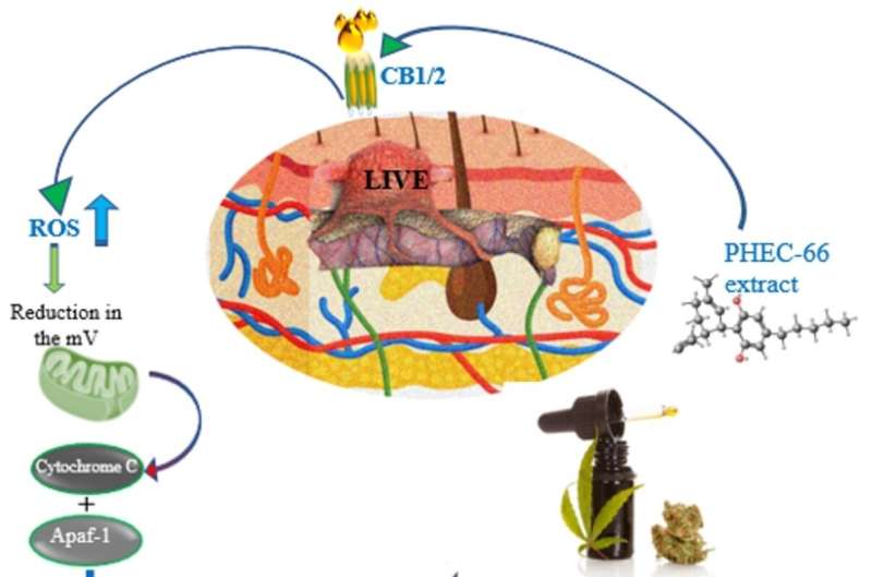
A cannabis extract has shown positive results in slowing down melanoma cell growth and increasing cell death rates, a new in-vitro study finds. Researchers from Charles Darwin University (CDU) and RMIT investigated programmed cell death caused by a specific cannabis extract (cannabinoid PHEC-66) from the cannabis sativa plant.

The study that was part of a Ph.D. project by RMIT’s Dr. Ava Bachari found that the extract binds to receptor sites on particular melanoma cells, then controls the growth of cells at two pivotal phases and increases the amount of damage to the cells. The paper was published in Cells.

CDU pharmaceutical lecturer and co-author Dr. Nazim Nassar said this damage effectively manipulates the cell into killing itself.

“The damage to the melanoma cell prevents it from dividing into new cells, and instead begins a programmed cell death, also known as apoptosis,” Dr. Nassar said.

“This is a growing area of important research because we need to understand cannabis extracts as much as possible, especially their potential to function as anticancer agents.

“If we know how they react to cancer cells, particularly in the cause of cell death, we can refine treatment techniques to be more specific, responsive and effective.”

Dr. Nassar said the next challenge was developing targeted delivery system development to the melanoma cells to get it ready for pre-clinical trials.

“Advanced delivery systems still need to be fully developed, underscoring the importance of ongoing efforts to ensure the proper and effective use of these agents at target sites,” he said.

Dr. Nassar specializes in cancer cell biology, pharmacology, drug delivery systems, and drug disposition and dynamics.

A practicing pharmacist and pharmacologist, he has co-authored several papers on applying cannabinoids in melanoma treatment, the therapeutic potential of cannabinoids in prostate cancer and an overview of current melanoma treatment.

He said that while the use of cannabis extracts to treat a variety of health conditions is stigmatized, future research into its application could revolutionize cancer treatment.

“Clinical uses of cannabis extracts include treatment for anxiety, cancer-related symptoms, epilepsy, and chronic pain. Intensive research into its potential for killing melanoma cells is only the start as we investigate how this knowledge can be applied to treating different types of cancers.”

Lead author and RMIT biotechnologist Professor Nitin Mantri emphasized the necessity for a long-term follow-up to ensure the sustained effectiveness and safety of the PHEC-66 extract in cancer treatment over extended periods.

He stressed the importance of testing the safety profile of the extract before its widespread adoption.

“The subsequent stage involves animal studies or pre-clinical trials to validate and further explore the efficacy of cannabinoid PHEC-66 in treating melanoma and other cancers,” Professor Mantri said.

He highlighted the critical collaboration with Dr. Nassar, emphasizing the need for support and sponsorship from pharmaceutical companies to qualify PHEC-66 as a registered medicine.

Oral interleukin-23-receptor antagonist peptide shows greater efficacy than placebo for plaque psoriasis.


JNJ-77242113 shows greater efficacy than placebo for plaque psoriasis

For patients with moderate-to-severe plaque psoriasis, the interleukin-23-receptor antagonist peptide JNJ-77242113 shows greater efficacy than placebo, according to a study published in the Feb. 8 issue of the New England Journal of Medicine.

Robert Bissonnette, M.D., from Innovaderm Research in Montreal, and colleagues conducted a phase 2 dose-finding trial involving 255 patients with moderate-to-severe plaque psoriasis who were randomly assigned to receive JNJ-77242113 at a dose of 25 mg once daily, 25 mg twice daily, 50 mg once daily, 100 mg once daily, or 100 mg twice daily or placebo for 16 weeks.

A reduction from baseline of at least 75 percent in the Psoriasis Area and Severity Index (PASI) score (PASI 75 response) at week 16 was the primary endpoint.

The researchers found that the percentage of patients with a PASI 75 response was higher among those receiving JNJ-77242113 versus placebo (37, 51, 58, 65, and 79 percent in the 25-mg once-daily, 25-mg twice-daily, 50-mg once-daily, 100-mg once-daily, and 100-mg twice-daily groups, respectively, versus 9 percent), with a significant dose-response relationship observed. COVID-19 and nasopharyngitis were the most common adverse events.

The percentages of patients with at least one adverse event was similar in the combined JNJ-77242113 dose group and the placebo group (52 and 51 percent, respectively).

“In this phase 2 trial, JNJ-77242113, an oral interleukin-23-receptor antagonist peptide, showed a dose-response relationship and greater efficacy than placebo,” the authors write.

Video game trauma can last for years, says researcher.


horror

“Cry of Fear” is a horror video game that explores a wide range of particularly sensitive topics, from depression to self-harm, murder, and suicide. Samuel Poirier-Poulin knows the game well.

A lecturer and doctoral student in the Department of Art History and Film Studies at Université de Montréal, Poirier-Poulin has spent time analyzing it—and come to a disturbing conclusion.

Games such as “Cry of Fear” can trigger intense emotions in players, a type of “gameplay trauma” that continues to resonate long after the game has ended, he found.

Fright is inherent

Studies of horror in movies and video games have often suggested that the pleasure that players get from being frightened is rooted in the games’ inherent safety. As players move through its virtual environment, they’re never in any real danger.

Bernard Perron, a professor in Poirier-Poulin’s department, has described this fear as “gameplay emotion.” But while the lecturer found that interpretation compelling, it didn’t fully describe his own experience of “Cry of Fear.” So to get a clearer picture, he turned to the field of trauma studies.

“Trauma studies have made significant headway since the ’90s,” Poirier-Poulin said. “We’re no longer just looking at trauma caused by horrifying events such as murder, car accidents or sexual assault. The research has shown that some people experience daily trauma from events such as cyclical family abuse or insidious forms of microaggression.”

It would be inaccurate, however, to lump gameplay trauma in with other forms of trauma, he added.

“Games such as ‘Cry of Fear’ can blur the line between fiction and reality,” Poirier-Poulin said. “When the two worlds ‘bleed’ into each other, the player can get hurt.”

‘Bleeding’ between worlds

The notion of spillover or “bleed” between worlds has been used in describing live-action role-play (LARP), a popular subject of study by researchers in Scandinavian countries.

In particular, they’ve looked at situations where the player embodies a fictional character. “For example, let’s say I’m LARPing,” Poirier-Poulin said, “and my character is in a relationship with another character. We’re not a couple in real life, but the fiction may lead me to start developing feelings for the other person.”

Some LARP games in Scandinavia deliberately introduce violence. Sensitive subjects such as rape may be verbalized, though they are not acted out. And because players physically embody their characters, extreme experiences such as these can leave a lasting mark on a player’s psyche, even though they are fictional.

Video game players don’t have quite the same experience, but emotions can still bleed from the character to the player.

This can be the case with particularly violent video games, such as “Spec Ops: The Line,” in which players must use white phosphorus, a highly controversial incendiary weapon, to destroy an enemy camp, only to find they have unwittingly scorched civilians. Poirier-Poulin quotes at length from Belgian researcher Tobi Smethurst’s description of the experience:

“I was the one who had pushed the buttons and dropped the bombs. Whether or not I knew I was doing it at the time, I had a hand in murdering those civilians. I did not experience the same trauma afterwards that the protagonist did, of course, but his reactions were certainly not too far from my own. I have taken thousands upon thousands of virtual lives, but these were the first for which I truly felt that I shared the burden of guilt.

“Rationally, I knew this was absurd. Afterwards I reminded myself that these were virtual civilians, not real ones; and besides, the game is programmed in such a way that if you want to finish it, you have to use the white phosphorus… But none of this rationalization changed how horrified I had felt, along with the protagonist, on discovering the charred bodies that he/we/I had produced. If trauma studies has taught me one thing, it is that fictional events can have real effects on one’s outlook and ethics… The fact that I felt the need to rationalize my guilt over ‘Spec Ops: The Line’ in the first place demonstrated just how ‘real’ of an effect the game had had on me.”

How does type 1 diabetes alter muscle structure and blood supply?


How Type 1 Diabetes Alters Muscle Structure and Blood Supply?
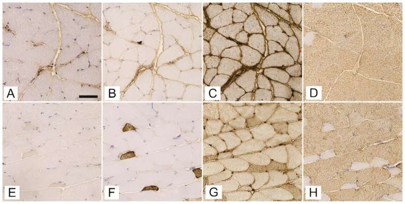
Expression of myosin heavy chain isoforms 1 (A, E), 2a (B, F), 2x/d (C, G), and 2b (D, H) in successive cross-sections of gluteus maximusmuscle of streptozotocin-induced diabetic mice (A–D) and age-matched non-diabetic mice (E–H). The scale bar indicates 50 μm.

In a recent study conducted by the Institute of Anatomy, Faculty of Medicine, University of Ljubljana, researchers have provided new insights into the detrimental effects of type 1 diabetes mellitus (T1DM) on skeletal muscle structure and capillary networks. Utilizing state-of-the-art 3D imaging technology, this comprehensive study marks a significant leap in understanding the multifaceted impact of T1DM on the body’s muscular system.

Diabetes mellitus disrupts the regulation of glucose levels, leading to high blood sugar and a myriad of related health issues. T1DM, characterized by the immune-mediated destruction of insulin-producing pancreatic β cells, has profound effects on various organs, especially skeletal muscles, which play a crucial role in glucose uptake and regulation.

This study, published in the journal Biomolecules and Biomedicine, aimed to explore the structural and functional adaptations of skeletal muscles to the metabolic disturbances caused by T1DM.

The hidden changes in muscle and blood vessels

Conducted on female C57BL/6J-OlaHsd mice using a streptozotocin (STZ)-induced model to simulate T1DM, the research focused on critical muscles like the soleus, gluteus maximus, and gastrocnemius. Researchers meticulously analyzed the expression of myosin heavy chain (MyHC) isoforms and the intricacies of the 3D capillary network.

“Our study provides a deeper understanding of how type 1 diabetes not only affects muscle fiber composition but also significantly alters the capillary networks that are essential for muscle health,” explained Nejc Umek, the study’s lead author.

How Type 1 Diabetes Alters Muscle Structure and Blood Supply?
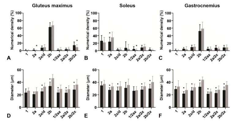
Numerical density and diameter of (A,D) gluteus maximus,(B,E) soleus, and (C,F) gastrocnemius muscle fibers. Comparison between type 1 diabetes mellitus mice (black columns; n=12) and non-diabetic mice (gray columns; n=12). Data are presented as mean±standard deviation. *P<0.05.

The research revealed that, despite the composition of fast-twitch type 2b fibers remaining consistent, notable differences were observed in the diabetic mice’s soleus muscle, which showed a reduced proportion of type 2a fibers and diminished fiber diameters across all muscles analyzed.

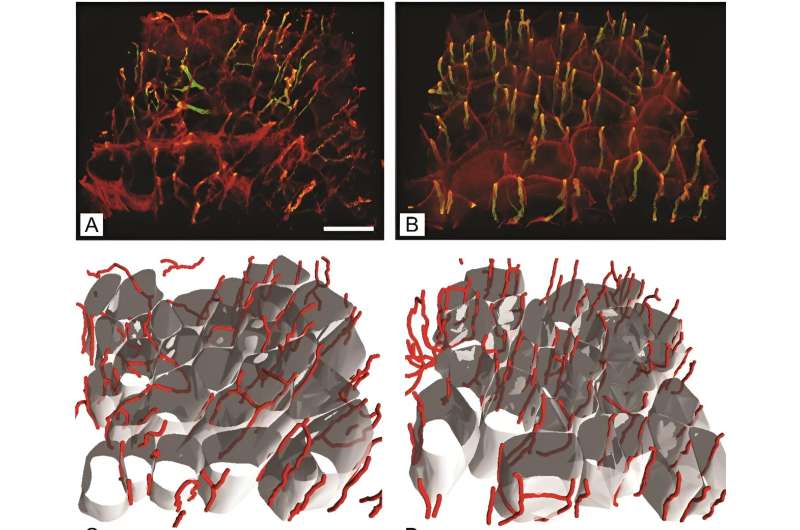
Additionally, an intriguing increase in capillary length per muscle volume was discovered in the gluteus maximus of diabetic mice, suggesting an adaptive mechanism to counterbalance muscle fiber atrophy induced by diabetes.

Methodological advances and key discoveries

The study utilized female mice, addressing a gap in diabetes research that often overlooks gender differences in disease progression and response to treatment. Through a single intraperitoneal administration of STZ, researchers successfully induced T1DM, confirmed by significantly elevated fasting glucose levels. This model allowed for an in-depth examination of diabetes-induced changes in a controlled environment.

By employing antibodies specific to different MyHC isoforms and cutting-edge 3D imaging, the team could precisely quantify changes in muscle fiber types and the capillary network. “The advanced 3D imaging techniques we used represent a significant improvement over traditional 2D analyses, offering a more detailed and accurate depiction of the capillary network changes in diabetic muscle tissue,” stated Erika Cvetko, the study’s senior author.

How Type 1 Diabetes Alters Muscle Structure and Blood Supply?
Capillaries and muscle fibers in gluteus maximus muscle of streptozotocin-induced diabetic mice (B, D) and age-matched non-diabeticmice (A, C).(A and B) Immunofluorescent staining with volume rendering of capillaries; (C and D) Reconstructed muscle fibers with supplying capillaries. The scale bar indicates 50μm.

Implications for diabetes management and future directions

The findings from this collaborative research effort highlight the necessity for comprehensive diabetes management plans that encompass not only glucose regulation but also the preservation of muscle structure and function. “Understanding the specific alterations in muscle tissue due to type 1 diabetes paves the way for developing targeted therapies that could significantly improve patient outcomes,” Cvetko added.

The study’s revelations about the increased capillary length per muscle volume in diabetic mice underscore the body’s potential compensatory responses to the structural changes induced by diabetes. These insights are crucial for designing interventions that aim to mitigate muscle deterioration and enhance overall diabetes care.

This novel study contributes significantly to the body of knowledge on diabetes and its systemic effects, particularly on skeletal muscle health. By highlighting the critical role of maintaining muscle integrity and vascular supply in the management of T1DM, the research opens new avenues for therapeutic strategies and underscores the importance of multidisciplinary approaches in tackling this complex disease.

New PET tracer detects inflammatory arthritis before symptoms appear


New PET tracer detects inflammatory arthritis before symptoms appear
(A) PET images of [68Ga]Ga-DOTA-ZCAM241 uptake at baseline and 3, 7, and 12 days after injection as inflammatory arthritis developed in single representative individual mouse. Images are normalized to SUV of 0.5 for direct comparison between time points. (B) CD69 immunofluorescence Sytox (Thermo Fisher Scientific) staining of joints of representative animals during matching time points.

A novel PET imaging technique can noninvasively detect active inflammation in the body before clinical symptoms arise, according to research published in the February issue of The Journal of Nuclear Medicine. Using a PET tracer that binds to proteins present on activated immune cells, the technique produces images of ongoing inflammation throughout the body, such as rheumatoid arthritis. This makes it easier for physicians to correctly diagnose and treat patients.

Rheumatoid arthritis is the most common type of inflammatory arthritis and affects 18 million people worldwide. It is a complex autoimmune disease characterized by chronic inflammation. This inflammation can cause the destruction of cartilage and bone, eventually leading to limitations, disabilities, loss of function, decreased quality of life, and possibly shortened life expectancy.

“A major interest of the rheumatology field is employing precision diagnostics to predict disease development in individuals with risk factors of rheumatoid arthritis,” said Fredrik Wermeling, Ph.D., associate professor and group leader at the Department of Medicine, Division of Rheumatology, Center for Molecular Medicine (CMM) at the Karolinska Institutet, in Solna, Sweden. “The hope is to find ways to identify such individuals even before they get sick, with the goal of being able to treat them so they never develop the disease.”

CD69 is one of the earliest cell surface markers seen on cells experiencing inflammation and is present in the tissue of patients with active rheumatoid arthritis. As such, researchers evaluated the performance of the CD69-targeting PET agent, 68Ga-DOTA-ZCAM241, for early disease detection in a mouse model of inflammatory arthritis.

In the study, mice were imaged with 68Ga-DOTA-ZCAM241 PET before and three, seven, and 12 days after induction of arthritis. Disease progression was monitored by clinical parameters, such as measuring body weight and scoring swelling in the paws. The uptake of 68Ga-DOTA-ZCAM241 in the paws was analyzed, and after the last PET scan, tissue biopsy samples were analyzed for CD69 expression. A second group of mice received PET scans with a nonspecific control peptide.

Increased uptake of the CD69-directed tracer 68Ga-DOTA-ZCAM241 was seen in the paws of mice with induced inflammatory arthritis three days after induction, which preceded the appearance of clinical symptoms five to seven days after induction. The uptake of 68Ga-DOTA-ZCAM241 also correlated with the clinical score and disease severity. The nonspecific control peptide demonstrated only low binding.

68Ga-DOTA-ZCAM241 is a potential candidate for PET imaging of activated immune cells during rheumatoid arthritis onset,” stated Olof Eriksson, Ph.D., associate professor and group leader of Translational PET Imaging at the Department of Medicinal Chemistry at Uppsala University, in Uppsala, Sweden. “We know that physicians are asking for better methods to image inflammation, for example in rheumatoid arthritis, and we hope this technology will be broadly used in many diseases that involve activated immune cells and inflammation.”