A controversial technique has produced detailed maps of the magnetic fields in colossal galaxy clusters. If confirmed, the approach could be used to reveal where cosmic magnetic fields come from.
Using radio waves, astronomers made a map of the magnetic field within the El Gordo cluster of galaxies, which spans 6 million light-years.
Introduction
By making maps of the magnetic fields hidden inside massive galaxy clusters, astronomers are getting closer to finding the origin of cosmic magnetism.
“These are the first maps of the detailed structure of magnetic fields on an unprecedentedly large scale,” said Alexandre Lazarian, an astronomer at the University of Wisconsin, Madison, and a co-author on the paper describing the maps, published today in Nature Communications.
Lazarian and his colleagues studied five galaxy clusters, each spanning millions of light-years. They made the maps using a technique he devised called synchrotron intensity gradient (SIG) mapping, which relies on radio observations to work out which way a cluster’s magnetic field points at a given location. By applying the same technique across an entire cluster, the researchers say they can construct a complete map of its magnetic fields. The results, if confirmed, would show that there’s a previously undetected order to magnetic fields in giant structures
Abstractions navigates promising ideas in science and mathematics. Journey with us and join the conversation.
Magnetism is ubiquitous in the universe. We see it from the smallest scales on Earth to the universe’s largest, where it sculpts cosmic structures such as stars and the interstellar medium. Magnetism is also crucial for life as we know it, influencing chirality at a molecular level and crafting the protective shield that encompasses Earth. But a big unanswered question has been where cosmic magnetism originated. Some scientists favor a primordial explanation, with magnetism arising in the first moments after the Big Bang with the other fundamental forces. Others favor a later arrival, with magnetism arising after hundreds of millions of years and growing from seed magnetic fields produced by objects such as stars and galaxies.
This new mapping technique might offer a solution by allowing astronomers to compare magnetic fields on the very largest scales. But the technique has its own limitations and remains somewhat controversial in the field of large-scale magnetism.
“If it works, it gives you a very observationally inexpensive way of mapping magnetic fields over very large areas of the sky,” said Kate Pattle, an astrophysicist at University College London.
Cosmic Cartography
Scientists typically find cosmic magnetic fields by studying synchrotron radiation — radio emissions produced as a magnetic field bends the path of electrons traveling close to the speed of light. Such observations can also use the orientation of those radio emissions — their polarization — to reveal the orientation of the magnetic fields. But polarization measurements are extremely time-consuming, and work best in denser and dustier regions of a galaxy cluster.
About seven years ago, Lazarian came up with a way to use synchrotron emission alone to reveal the direction of the magnetic field — no polarization needed. The technique uses observations of the changing strength of the radio emission as you move across space, or what researchers call the gradient.
“The gradient in the brightness, the direction in which the image becomes fainter or brighter, relates to the magnetic fields,” said Marcus Brüggen, a professor of astrophysics at the University of Hamburg in Germany who has previously studied large magnetic fields.
In preliminary observations of interstellar space, “everywhere we [looked], we revealed this magnetic field structure,” Lazarian said.
The team then turned to galaxy clusters, which grow as smaller groups of galaxies collide. When these mergers occur, they produce shock fronts that “plow through the [intracluster] medium,” Brüggen said. When magnetic fields interact with those turbulent shock fronts, they produce synchrotron emission. By observing the gradient of that emission, the researchers can infer the direction of the magnetic field, which in turn reflects the mergers that have built these clusters over time.
The method allows Lazarian to survey magnetic fields across the expanse of huge galaxy clusters, including the diffuse intergalactic space within the structure where polarization measurements are not possible. To make their maps, the team targeted five galaxy clusters, including El Gordo — a well-studied clump of hundreds of galaxies that stretches 6 million light-years across. They also looked at Abell 2345, 2 billion light-years away, Abell 3376, about a half-billion light-years away, and two others.
Not all scientists are convinced that the strategy accurately tracks the motion of magnetic fields, however. What look like shifts in magnetism-driven synchrotron gradients could just be changes in electron or gas density. The method also relies on a phenomenon known as turbulence in galaxy clusters, where magnetic fields twist and turn together — “a notoriously complex physical process,” said Andrea Botteon, an astrophysicist at the National Institute for Astrophysics in Italy.
Magnetic Life
In the future, Lazarian wants to use SIG — if the technique holds up — to map the magnetism in filaments between galaxies using a vast European radio network called the Low-Frequency Array. If the fields in those filaments are aligned with one another, as they are in clusters, it might suggest a primordial source of cosmic magnetic structure rather than a slow emergence from seed magnetic fields. Such an alignment would be “essentially impossible” for stars and galaxies to create during later cosmic epochs, Brüggen said.
“My hunch,” Brüggen said, “is we will find that magnetic fields were produced early in the universe.”
Divining the origin of magnetism may tell us something about the habitability of the cosmos. Life itself (at least as we know it on Earth) relies on magnetism and its influence on chirality to give the building blocks of life a right- or left-handedness. “If magnetic fields formed at the beginning of the universe, you can form molecules with chirality very early,” Lazarian said. Then, “we can ask the question [of] whether we should expect to see signals of civilizations which formed quite early in the history of the universe.”
He also noted that magnetic fields in galaxy clusters could be the source of some of the highest-energy cosmic rays known to pervade the universe, which still have a mysterious origin. “There’s a big question [about] whether these clusters of galaxies could be the sources of the cosmic rays of the highest energy,” he said, and mapping the fields within clusters could help resolve that question
The team’s next goal is to observe galaxy clusters that are farther away and further back in time. El Gordo, although immense, only stretches back to when the universe was 6.5 billion light-years old, about half its current age of 13.8 billion years. Upcoming radio telescopes such as the Square Kilometer Array, a vast array of antennas that will be spread across 1 million square meters in South Africa and Australia later this decade, could be powerful enough to apply this type of mapping to clusters that existed when the universe was just 3 billion years old.
“I would like to see what happened in the early universe,” said Yue Hu, a graduate student at the University of Wisconsin, Madison, and lead author on the paper.
But the origin of magnetism in the universe, and all the implications of that answer, will not be solved overnight using this method. “It’s one piece of the puzzle,” Brüggen said. “But it’s a very substantial piece.”
When your mind is wandering, your brain’s “default mode” network is active. Its discovery 20 years ago inspired a raft of research into networks of brain regions and how they interact with each other.
Introduction
Whenever you’re actively performing a task — say, lifting weights at the gym or taking a hard exam — the parts of your brain required to carry it out become “active” when neurons step up their electrical activity. But is your brain active even when you’re zoning out on the couch?
The answer, researchers have found, is yes. Over the past two decades they’ve defined what’s known as the default mode network, a collection of seemingly unrelated areas of the brain that activate when you’re not doing much at all. Its discovery has offered insights into how the brain functions outside of well-defined tasks and has also prompted research into the role of brain networks — not just brain regions — in managing our internal experience.
In the late 20th century, neuroscientists began using new techniques to take images of people’s brains as they performed tasks in scanning machines. As expected, activity in certain brain areas increased during tasks — and to the researchers’ surprise, activity in other brain areas declined simultaneously. The neuroscientists were intrigued that during a wide variety of tasks, the very same brain areas consistently dialed back their activity.
It was as if these areas had been active when the person wasn’t doing anything, and then turned off when the mind had to concentrate on something external.
Researchers called these areas “task negative.” When they were first identified, Marcus Raichle, a neurologist at the Washington University School of Medicine in St. Louis, suspected that these task-negative areas play an important role in the resting mind. “This raised the question of ‘What’s baseline brain activity?’” Raichle recalled. In an experiment, he asked people in scanners to close their eyes and simply let their minds wander while he measured their brain activity.
He found that during rest, when we turn mentally inward, task-negative areas use more energy than the rest of the brain. In a 2001 paper, he dubbed this activity “a default mode of brain function.” Two years later, after generating higher-resolution data, a team from the Stanford University School of Medicine discovered that this task-negative activity defines a coherent network of interacting brain regions, which they called the default mode network.
In a brain network, the individual parts interact to bring about effects that they can only produce together.
The discovery of the default mode network ignited curiosity among neuroscientists about what the brain is doing in the absence of an outward-focused task. Although some researchers believed that the network’s main function was to generate our experience of mind wandering or daydreaming, there were plenty of other conjectures. Maybe it controlled streams of consciousness or activated memories of past experiences. And dysfunction in the default mode network was floated as a potential feature of nearly every psychiatric and neurological disorder, including depression, schizophrenia and Alzheimer’s disease.
Since then, a flurry of research into the default mode has complicated that initial understanding. “It’s been very interesting to see the types of different tasks and paradigms that engage the default mode network in the last 20 years,” said Lucina Uddin, a neuroscientist at the University of California, Los Angeles.
The default mode was one of the first brain networks characterized by science. It consists of a handful of brain regions, including a few at the front of the brain, like the dorsal and ventral medial prefrontal cortices, and others scattered throughout the organ, like the posterior cingulate cortex, the precuneus and the angular gyrus. These regions are associated with memory, experience replay, prediction, action consideration, reward/punishment and information integration. (The colored highlighting in the following figure indicates some of the outer brain areas that become more active when the default network engages.)
Since its discovery, neuroscientists have loosely identified a handful of additional distinct networks that each activate seemingly disparate areas of the brain. These activated areas don’t act independently, but rather harmonize in synchrony with each other. “You can’t think about a symphony orchestra as just the violins or the oboes,” Raichle said. Similarly, in a brain network, the individual parts interact to bring about effects that they can only produce together.
According to research, the effects of the default mode network include mind wandering, remembering past experiences, thinking about others’ mental states, envisioning the future and processing language. While this may seem like a grab bag of unrelated aspects of cognition, Vinod Menon, the director of the Stanford Cognitive & Systems Neuroscience Laboratory, recently theorized that all of these functions may be helpful in constructing an internal narrative. In his view, the default mode network helps you think about who you are in relation to others, recall your past experiences and then wrap up all of that into a coherent self-narrative
Introduction
The default mode is clearly up to something complicated; it’s involved in many different processes that can’t be neatly described. “It’s kind of silly to think that we’re ever going to be like, ‘This one brain region or one brain network does one thing,’” Uddin said. “I don’t think that’s how it works.”
Uddin began investigating the default mode network because she was interested in self-recognition, and many self-recognition tasks, such as identifying your own face or voice, appeared to be associated with the network. In recent years, she has shifted her attention to interactions between brain networks. Just as different brain areas interact with each other to form networks, different networks interact with each other in meaningful ways, Uddin said. “Network interactions are more elucidating to study in some ways than just a network in isolation because they do work together and then come apart and then change what they’re doing over time.”
She’s particularly interested in how the default mode network interacts with the salience network, which seems to help us identify the most relevant piece of information at any given time. Her work suggests that the salience network detects when something is important to pay attention to and then acts as an off switch for the default mode network.
Researchers have also been examining whether mental health disorders like depression could be linked to problems with the default mode network. So far, the findings have been inconclusive. In people with depression, for example, some researchers have found that network nodes are overly connected, while others have found the opposite — that nodes are failing to connect. And in some studies, the default mode network itself isn’t abnormal, but its interactions with other networks are. These findings may appear incompatible, but they align with recent findings that depression is perhaps a cluster of different disorders that present with similar symptoms.
Meanwhile, Menon has developed what he calls the triple network theory. It posits that abnormal interactions between the default mode network, the salience network and a third one called the frontoparietal network could contribute to mental health disorders including schizophrenia, depression, anxiety, dementia and autism. Typically, the activity of the default mode network decreases when someone is paying attention to an external stimulus, while activity in the two other networks increases. This push and pull between networks may not work the same way in people with psychiatric or developmental disorders, Menon suspects
Deanna Barch, who studies the neurobiology of mental illnesses at Washington University in St. Louis, is intrigued by the triple network theory. Investigating how networks are wired up differently in people with mental health disorders can help researchers find underlying mechanisms and develop treatments, she said. However, she doesn’t think network interactions alone will fully explain mental illness. “I think of understanding connectivity differences as a starting point,” Barch said. “It’s not an endpoint.”
The current understanding of the default mode network is surely not its endpoint, either. Since its discovery, it has pushed neuroscientists to think beyond the responsibilities of single brain regions to the effects of interactions between brain networks. And it’s driven many people to appreciate the inward-focused activities of the mind — that even when we’re daydreaming or at rest, our brain is hard at work making it happen.
function Points to Potential Therapeutic Targets against Inflammation
Mitochondria are structures within cells that convert the energy from food into a form that cells can use. Although most DNA is packaged in chromosomes within the nucleus, mitochondria also have a small amount of their own DNA. Mitochondrial DNA (mtDNA) contains 37 genes, all of which are essential for normal mitochondrial function. When mtDNA remains where it belongs (inside of mitochondria), it sustains both mitochondrial and cellular health. However, when it goes where it doesn’t belong, it can initiate an immune response that promotes inflammation.
Now, Salk scientists and collaborators at the University of California, San Diego (UCSD), have discovered a novel mechanism used to remove improperly functioning mtDNA from inside to outside the mitochondria. When this happens, the mtDNA gets flagged as foreign DNA and activates a cellular pathway used to promote inflammation to rid the cell of pathogens.
In their study, the researchers noted “the relationship among aberrant mitochondria and nucleoid dynamics, mtDNA release and cGAS–STING activation remains unclear. Here we show that, under a variety of mtDNA replication stress conditions and during herpes simplex virus-1 infection, enlarged nucleoids that remain bound to TFAM exit mitochondria.”
“We knew that mtDNA was escaping mitochondria, but how was still unclear,” explained Gerald Shadel, PhD, senior and co-corresponding author of the study, professor, director of the San Diego-Nathan Shock Center of Excellence in the Basic Biology of Aging, and holder of the Audrey Geisel Chair in Biomedical Science at Salk. “Using imaging and cell biology approaches, we’re able to trace the steps of the pathway for moving mtDNA out of the mitochondria, which we can now try to target with therapeutic interventions to hopefully prevent the resulting inflammation.”
Scientists have been working to uncover how mtDNA leaves mitochondria and triggers the innate immune response, but the previously characterized pathways did not apply to the unique mtDNA stress conditions the Salk team was investigating.
“We had a huge breakthrough when we saw that mtDNA was inside of a mysterious membrane structure once it left mitochondria—after assembling all of the puzzle pieces, we realized that structure was an endosome,” said first author Laura Newman, PhD, former postdoctoral researcher in Shadel’s lab and current assistant professor at the University of Virginia. “That discovery eventually led us to the realization that the mtDNA was being disposed of and, in the process, some of it was leaking out.”
The team discovered a process beginning with a malfunction in mtDNA replication that caused mtDNA-containing protein masses called nucleoids to pile up inside of mitochondria. The cell then begins to remove the replication-halting nucleoids by transporting them to endosomes. The endosome gets overloaded with these nucleoids, springs a leak, and mtDNA is suddenly loose in the cell. The cell flags that mtDNA as foreign DNA and initiates the DNA-sensing cGAS-STING pathway to cause inflammation.
“Using our cutting-edge imaging tools for probing mitochondria dynamics and mtDNA release, we have discovered an entirely novel release mechanism for mtDNA,” added co-corresponding author Uri Manor, PhD, former director of the Waitt Advanced Biophotonics Core at Salk and current assistant professor at UCSD. “There are so many follow-up questions we cannot wait to ask, like how other interactions between organelles control innate immune pathways, how different cell types release mtDNA, and how we can target this new pathway to reduce inflammation during disease and aging.”
The researchers hope to further map out the mtDNA-disposal and immune-activation pathway, including what biological circumstances are required to initiate the pathway and what downstream effects there may be on human health. The findings offer new targets for therapeutics to disrupt the inflammatory pathway and mitigate inflammation during aging and diseases, like lupus or rheumatoid arthritis.
Exposure to even moderate concentrations of radon is associated with a significant increase in stroke risk, new research suggests.
An analysis of radon exposures in more than 150,000 postmenopausal women in the Women’s Health Initiative revealed a 14% higher stroke risk in those exposed to the highest concentrations compared with those exposed to the lowest concentrations. Even moderate concentrations of radon were associated with a 6% higher stroke risk.
Radon is the second leading cause of lung cancer, but little was known about how exposure to the gas might affect stroke risk in women.
“Our research found an increased risk of stroke among participants exposed to radon above — and as many as 2 picocuries per liter (pCi/L) below — concentrations that usually trigger Environmental Protection Agency recommendations to install a home radon mitigation system,” senior author Eric A. Whitsel, MD, MPH, professor of epidemiology and medicine, University of North Carolina, Chapel Hill, said in a news release.
The study was published online on January 31, 2024, in Neurology.
Women Particularly Affected
Radon is a naturally occurring odorless radioactive gas produced when uranium or radium break down in rocks and soil. Its presence is increasing as a result of climate change, and it is increasingly being found in people’s homes. When inhaled, this air pollutant releases ionizing radiation in the lungs and is seen as second only to smoking as an established cause of lung cancer.
The National Radon Action Plan of the US Environmental Protection Agency (EPA) lays out testing and mitigation guidelines based on the known role of radon in lung carcinogenesis. But radon testing and mitigation are less common than recommended, and the EPA’s action plan doesn’t cover diseases other than lung cancer.
Compared with men, women have a higher rate of stroke and, in the US, typically spend about 11% more hours per day indoors at home, which investigators note highlights a “potential role of the residential environment among other risk factors specific to women.”
Researchers examined longitudinal associations between home radon exposure and incident stroke in 158,910 women at baseline (mean age 63.2 years; 83% White) over a mean follow-up of 13.4 years. During this time, participants experienced a total of 6979 strokes.
Participants’ home addresses were linked to radon concentration data drawn from the US Geological Survey and the EPA, which recommends that average indoor radon concentrations not exceed 4 pCi/L.
The highest radon exposure group resided in areas where average radon concentrations were < 4 pCi/L; the middle exposure group lived in regions with average concentrations of 2-4 pCi/L; and the lowest exposure group lived in areas with average concentrations < 2 pCi/L.
The researchers adjusted for demographic, social, behavioral, and clinical characteristics.
Public Health Implications
The incidence rates of stroke per 100,000 women in the lowest, middle, and highest radon concentration areas were 333, 343, and 349, respectively.
Stroke risk was 6% higher among those in the middle exposure group (adjusted hazard ratio [aHR], 1.06; 95% CI, 0.99-1.13) and 14% higher in the highest exposure group (aHR, 1.14; 95% CI, 1.05-1.22) compared with the lowest exposure group.
Notably, stroke risk was significant even at concentrations ranging from 2 to 4 pCi/L (P = .0004) vs < 2 pCi/L, which is below the EPA’s Radon Action Level for mitigation.
The findings remained robust in sensitivity analyses, although the associations were slightly stronger for ischemic stroke (especially cardioembolic, small-vessel occlusive, and very large artery atherosclerotic) compared with hemorrhagic stroke.
“Radon is an indoor air pollutant that can only be detected through testing that measures concentrations of the gas in homes,” Whitsel said in the release. “More studies are needed to confirm our findings. Confirmation would present an opportunity to improve public health by addressing an emerging risk factor for stroke.”
The study lacked gender and racial/ethnic diversity, so the findings may not be generalizable to other populations.
“Replication studies of individual-level radon exposures are needed to confirm this positive radon-stroke association,” the authors write. “Confirmation would present a potential opportunity to affect public health by addressing a pervasive environmental risk factor for stroke and thereby merit reconsideration of extant radon policy.”
Amid a devastating epidemic stealing minds and memories, a new study suggests an astonishing 7-fold reduction in Alzheimer’s brain atrophy from a shockingly simple treatment: high-dose B vitamins.
With over 6 million Americans living with Alzheimer’s disease and no FDA-approved treatments that effectively slow its progression, there is a desperate need for new therapeutic strategies.1 Despite billions invested in developing medications targeting Alzheimer’s brain plaques and tangles, drug trial after drug trial has ended in disappointment.2 But an unlikely natural treatment – high doses of three humble B vitamins – may succeed where these drugs have failed.
A groundbreaking new study published in the Proceedings of the National Academy of Sciences (Douaud et al., 2013)3 suggests that high-dose B vitamin treatment could slow the progression of Alzheimer’s disease by as much as 7-fold. The double-blind randomized controlled trial included 156 elderly patients with mild cognitive impairment (MCI)4, an early Alzheimer’s risk state. Patients who took supplements of folic acid, vitamins B6 and B12 over two years had dramatically less brain atrophy detected via MRI scans compared to patients taking a placebo. Critically, the treatment was most effective in patients with elevated homocysteine levels.5 By lowering this amino acid, B vitamins reduced grey matter loss in brain regions vulnerable in Alzheimer’s.6
The results provide a glimmer of hope amid the litany of failed Alzheimer’s clinical trials.7 Unlike risky experimental drugs designed to remove plaques or tangles, vitamin B supplements are safe, natural, and already indicated for high homocysteine levels. If these unprecedented findings are confirmed in ongoing trials, vitamin supplementation could offer the first major advancement in Alzheimer’s treatment in over 15 years.8 With cases expected to triple in coming decades, this affordable regimen could have tremendous potential to alleviate the staggering burden of this epidemic.9
Brain fog is one of the most commo.n, persistent complaints in patients with long COVID. It affects as many as 46% of patients who also deal with other cognitive concerns like memory loss and difficulty concentrating.
Now, researchers believe they know why. A new study has found that these symptoms may be the result of a viral-borne brain injury that may cause cognitive and mental health issues that persist for years.
Researchers found that 351 patients hospitalized with severe COVID-19 had evidence of a long-term brain injury a year after contracting the SARS-CoV-2 virus. The findings were based on a series of cognitive tests, self-reported symptoms, brain scans, and biomarkers.
Brain Deficits Equal to 20 Years of Brain Aging
As part of the preprint study, participants took a cognition test with their scores age-matched to those who had not suffered a serious bout of COVID-19. Then a blood sample was taken to look for specific biomarkers, showing that elevated levels of certain biomarkers were consistent with a brain injury. Using brain scans, researchers also found that certain regions of the brain associated with attention were reduced in volume.
Patients who participated in the study were “less accurate and slower” in their cognition, and suffered from at least one mental health condition, such as depression, anxiety, or posttraumatic stress disorder, according to researchers.
The brain deficits found in COVID-19 patients were equivalent to 20 years of brain aging and provided proof of what doctors have feared: that this virus can damage the brain and result in ongoing mental health issues.
“We found global deficits across cognition,” said lead study author Benedict Michael, PhD, director of the Infection Neuroscience Lab at the University of Liverpool in Liverpool, England. “The cognitive and memory problems that patients complained of were associated with neuroanatomical changes to the brain.”
Proof That Symptoms Aren’t ‘Figment’ of Patients’ Imaginations
Cognitive deficits were common among all patients, but the researchers said they don’t yet know whether the brain damage causes permanent cognitive decline. But the research provides patients who have been overlooked by some clinicians with proof that their conditions aren’t a figment of their imaginations, said Karla L. Thompson, PhD, lead neuropsychologist at the University of North Carolina School of Medicine’s COVID Recovery Clinic.
“Even though we’re several years into this pandemic, there are still a lot of providers who don’t believe that their patients are experiencing these residual symptoms,” said Thompson, “That’s why the use of biomarkers is important, because it provides an objective indication that the brain has been compromised in some way.”
Some patients with long COVID have said that getting their doctors to believe they have a physical ailment has been a persistent problem throughout the pandemic and especially as it relates to the sometimes-vague collection of symptoms associated with brain fog. One study found that as many as 79% of study respondents reported negative interactions with their healthcare providers when they sought treatment for their long-COVID symptoms.
How Do COVID-Related Brain Injuries Happen?
Researchers are unsure what’s causing these brain injuries, though they have identified some clues. Previous research has suggested that such injuries might be the result of a lack of oxygen to the brain, especially in patients who were hospitalized, like those in this study, and were put on ventilators.
Brain scans have previously shown atrophy to the brain’s gray matter in COVID-19 patients, likely caused by inflammation from a heightened immune response rather than the virus itself. This inflammatory response seems to affect the central nervous system. As part of the new study, researchers found some neuroprotective effects of using steroids during hospitalization to reduce brain inflammation.
The results suggest that clinicians should overcome their skepticism and consider the possibility that their patients have suffered a brain injury and should be treated appropriately, said James C. Jackson, PsyD, a neuropsychiatrist at Vanderbilt University School of Medicine. “The old saying is that if it walks like a duck and talks like a duck, it’s a duck,” said Jackson.
He contends that treatments used for patients who have brain injuries have also been shown to be effective in treating long COVID–related brain fog symptoms. These may include speech, cognitive, and occupational therapy as well as meeting with a neuropsychiatrist for the treatment of related mental health concerns.
A New Path Forward
Treating long-COVID brain fog like a brain injury can help patients get back to some semblance of normalcy, researchers said. “What we’re seeing in terms of brain injury biomarkers and differences in brain scans correlates to real-life problems that these patients are dealing with on a daily basis,” said Jackson. These include problems at work and in life with multitasking, remembering details, meeting deadlines, synthesizing large amounts of information, and maintaining focus on the task at hand, he said.
There’s also a fear that even with treatment, the aging of the brain caused by the virus might have long-term repercussions and that this enduring injury may cause the early onset of dementia and Alzheimer’s disease in those who were already vulnerable to it. One study, from the National Institute of Neurological Disorders and Stroke (NINDS), found that in those infected with COVID-19 who already had dementia, the virus “rapidly accelerated structural and functional brain deterioration.”
“We already know the role that neuroinflammation plays in the brains of patients with Alzheimer’s disease,” said Thompson. “If long COVID is involved in prolonged inflammation of the brain, it goes a long way in explaining the mechanism underlying [the study’s reported] brain aging.”
Still More to Learn
In some ways, this study raises nearly as many questions as it does answers. While it provides concrete evidence around the damage the virus is doing to the brains of patients who contracted severe COVID-19, researchers don’t know about the impact on those who had less serious cases of the virus.
For Ziyad Al-Aly, MD, chief of research and development at the Veterans Affairs St. Louis Health Care System, the concern is that some long-COVID patients may be suffering from cognitive deficits that are more subtle but still impacting their daily lives, and that they’re not getting the help they need.
What’s more, said Al-Aly, it’s unclear whether the impacts of the brain damage are permanent or how to stop them from worsening. Researchers and clinicians need a better understanding of the mechanism that allows this virus to enter the brain and do structural damage. If it’s inflammation, will anti-inflammatory or antiviral medications work at preventing it? Will steroids help to offset the damage? “It’s critical we find some answers,” he said.
“SARS-CoV-2 isn’t going anywhere. It will continue to infect the population, so if this is indeed a virus that damages the brain in the long term or permanently, we need to figure out what can be done to stop it,” said Al-Aly.
X chromosome inactivation is a crucial biological process with some inadvertent autoimmune consequences.
Shaped by thousands of years of evolution, the body’s immune system is a vigilant guardian primed against external infectious threats. Too much vigilance, however, and this guardian of potent cells and chemicals instead becomes our own adversary. The result is a myriad of autoimmune diseases affecting around one in five Americans and around four percent worldwide.
While one’s risk for autoimmune disease is partly genetic, individuals who are disproportionately affected tend to be those assigned female at birth — a shocking 78 percent. Why there’s such a staggering difference has been a mystery, but scientists have been narrowing down suspects to risk factors like hormones, the gut microbiome, and now, the X chromosome itself.
Researchers led by Stanford University found that a routine biological process that silences one X chromosome in females (called X chromosome inactivation) may be a risk factor for autoimmune diseases. In particular, proteins that help with the silencing process might seem foreign to our bodies, and the body might create antibodies against them, setting the stage for autoimmune diseases.
When male mice prone to autoimmune disease were genetically engineered with the Xist gene, they also showed signs of autoimmunity similar to what’s seen in their female counterparts. In blood samples collected from human participants with autoimmune diseases, the researchers also found antibodies against the proteins involved in X chromosome inactivation.
“Scientists have a better idea now of pinpointing the autoimmune risk associated in different sexes, and this comes from the chromosomal basis, individual molecules we can now perhaps pin this [to],” Howard Chang, professor of cancer research and genetics at Stanford University of Medicine, who led the study, tells Inverse.
THE MASTER SILENCER
All humans and many other mammals have a pair of sex chromosomes: two Xs for biological females and one X and one Y for biological males. Because chromosomes are basically individual recipe books that a cell uses whenever it needs to make something, having duplicates of the same book means the cell would end up making more than it needs, like making twice as many cakes. Twice as many cakes may sound benign, but for a cell, having that many genes turned on and baking all their proteins and other molecules they code for would be lethal.
To balance out the books, each cell randomly chooses an X chromosome to muffle during early embryonic development. As you might guess, this doesn’t happen in biological males, as they only have one X chromosome.
This biological silencing is handled by a gene found on the X chromosome called Xist, which was first discovered in the early 1990s. Xist doesn’t code for any protein — almost 99 percent of DNA doesn’t; instead, this gene manufactures something called long noncoding RNA, RNA being a Xerox copy of DNA that’s free to float around in the cell, unlike DNA packed away in chromosomes and confined to the nucleus.
Paradoxically, Xist is turned on in whichever X chromosome gets the short stick, says Chang. Its long noncoding RNA recruits other proteins to join in on the silencing, the whole mass of RNA and proteins sticking together to form what scientists call a ribonucleoprotein complex. This complex then coats the X chromosome like some sort of form-fitting molecular bodysuit.
In 2015, Chang and a group of other researchers published a paper cataloging the proteins forming the ribonucleoprotein complex. They didn’t have autoimmune disease on their minds, per se, when conducting their study but discovered many of the proteins associated with the complex could become parts of autoantibodies, aka antibodies that respond to and attack your own cells and tissues.
This discovery had the researchers wondering: Is Xist, the master silencer, somehow nefariously involved in autoimmune disease?
GENETICS + ENVIRONMENT
To answer that question, Chang and his colleagues inserted the Xist gene into the genome of male mice, some of whom were susceptible to autoimmune disease and others resistant. The Xist gene was tweaked so it could be turned on and off at will by chemical means, and the portion of the gene that instructs its RNA to silence the X chromosome was deleted. However, the long noncoding RNA could still mingle with other proteins to make its ribonucleoprotein complex. The experiment also included male mice where Xist wasn’t turned on, female mice, and healthy, otherwise normal mice.
Turning on Xist itself didn’t trigger autoimmunity, but when the mice were injected with pristane, a compound known to cause a lupus-like disease in the animals, that’s where Chang and his colleagues noticed signs of autoimmune disease in the genetically vulnerable mice on par with some of the female mice injected with pristane.
“Because we made a male mouse get [autoimmune] disease just with this RNA, that really shows that you don’t need female hormones,” says Chang. “You don’t even need a second X chromosome, just this RNA can confer a lot of the risk.”
In mice with Xist who weren’t susceptible to autoimmune disease, nothing really happened to them, which Chang says highlights how not just Xist alone but one’s genetic susceptibility for conditions like lupus, inflammatory bowel disease, or multiple sclerosis (and exposures to potential environmental triggers) needs to be present to explain one’s risk for autoimmune disease.
These findings are just the first step in unraveling the complexity of autoimmune disease and identifying more precise diagnostics and treatments to better help patients, says Chang. However, there are some limitations, namely the fact this study was in mice using an engineered form of Xist, not the one normally found in the X chromosome.
“If you use a version [of Xist] that’s slightly different, that could lead to slightly different results,” he says.
In a separate but parallel study, the researcher analyzed blood samples obtained from anonymous donations to the Stanford Blood Center. In those whose blood demonstrated signs of autoimmunity with autoantibodies, those antibodies were reactive to 79 out of the 81 proteins part of Xist’s ribonucleoprotein complex. So there is a basis for Xist potentially mediating autoimmune disease in humans, although Chang says he would love to see these results replicated in a larger population.
For upcoming studies, the researchers would also like to suss out how exactly the immune system chances upon this ribonucleoprotein complex since it’s otherwise sequestered inside the cell’s nucleus, out of sight by immune cells.
“Understanding what different triggers do, whether they cause cell death in a way that leaks out small or large amounts of Xist, that would be a useful thing to think about in the future,” says Chang
The research could offer insight into metabolic diseases affecting pregnancy.
When it’s time to bring new life into the world, the human body transforms itself in remarkable ways. The immune system, which usually fights off anything that reads as foreign, learns to chill out so it doesn’t attack the baby; the uterus stretches and expands; the cardiovascular system grows stronger so it can pump more blood; and the kidneys and liver bulk up to process waste for two.
Yet, with all these amazing adaptations part and parcel of pregnancy, there’s still a lot of mystery surrounding the orchestra of changes happening. One group of researchers in China has created a metabolic treasure map that aims to demystify our understanding of this intricate process.
Comparing tissue samples collected from nonpregnant and pregnant crab-eating macaques, the researchers discovered that pregnancy rewires metabolic processes in contrasting yet dynamic ways throughout pregnancy across 23 different tissues from organs such as the heart, kidneys, and even skin. They identified 91 different metabolites — or chemicals produced by metabolism — involved in these changes; two standout stars were the steroid hormone corticosterone and a building block of fat called palmitoyl-carnitine.
The researchers say their findings, published last week in the journal Cell, illuminate the body’s tremendous metamorphosis during pregnancy. Moreover, their study may hold the key to improving maternal and fetal health by focusing a spotlight on these metabolic changes underpinning pregnancy-related diseases like preeclampsia and gestational diabetes.
METABOLIC PREGNANCY ATLAS
While scientists do have some idea of the metabolic changes the body undergoes to provide for the developing fetus, large gaps in knowledge remain because it’s simply impossible to investigate that without being deeply invasive, such as collecting tissue samples from various organs, such as the uterus, and even the placenta.
Analysis of human maternal blood samples has started to map out the intricate flux of metabolites over the course of pregnancy, some of which could be used like biological milestones to help predict crucial moments like how far along the pregnancy is, when the baby might arrive, and even the risk of preeclampsia, a serious pregnancy complication characterized by high blood pressure that can be life-threatening if left untreated.
But rodents aren’t the best models for a normal human pregnancy, so for their study, the researchers of the new paper turned to the crab-eating macaque, an invaluable non-human primate model used extensively in biomedical research that shares many similarities to humans in terms of how their bodies work (especially reproduction) and even how their metabolisms function.
The researchers obtained tissue samples from pregnant monkeys at three different time points: early, mid, and late pregnancy (crab-eating macaques have a gestational period of roughly 165 days). These samples were compared to their nonpregnant counterparts. The team collected samples from 23 different tissues across 10 organ systems, which included the uterus, ovaries, placenta, mammary glands, thymus (a small organ under the breastbone involved in the immune system), heart, pancreas, liver, kidneys, adrenal glands, spinal cord, skin, leg muscles, and blood serum.
They found 91 metabolites common to all the tissues across the different stages of pregnancy, although these varied in quantity. Some of these metabolites involved steroids, like progesterone, which were being produced and exerting their influence in unexpected places like the adrenal glands, pancreas, heart, and even the skin. This surprising finding suggests the production of steroids plays a broader role in adapting a person’s body for the challenges of pregnancy, potentially aiding in the growth of organs like the pancreas and heart, though further research is needed to fully understand these connections.
Another crucial finding the researchers uncovered was that corticosterone increases in many organ systems during pregnancy, which was a bit of a surprise because it’s usually not considered as important as the stress hormone cortisol. However, this research shows that corticosterone is key to helping the placenta grow and develop, overall keeping a pregnancy running smoothly.
There may also be a link with preeclampsia, a metabolic disorder affecting five to seven percent of pregnancies. Pregnant crab-eating macaques demonstrated higher levels of corticosterone, which contrasted with a separate study the researchers did that found the steroid hormone was lower in pregnant people with preeclampsia. This could mean that testing for targeting corticosterone might be used in the future to mitigate preeclampsia.
These findings are far from final and do have some limitations, namely being a non-human-based study. However, the researchers hope their efforts will “serve as a resource for future research into female metabolism,” paving the way to optimizing maternal and fetal health.
FDA DisclaimerThe information on this website has not been evaluated by the Food & Drug Administration or any other medical body. We do not aim to diagnose, treat, cure or prevent any illness or disease. Information is shared for educational purposes only. Learn More
Affliliate DisclosureIn compliance with the FTC guidelines, please assume the following about links and posts on this site: Many of the links on DrJockers.com are affiliate links of which I receive a small commission from sales of certain items, but the price is the same for you. If I post an affiliate link to a product, it is something that I personally use, support and would recommend without an affiliate link. Learn More
Privacy PolicyPlease read the Privacy Policy carefully before you start to use DrJockers.com. By using DrJockers.com or by clicking to accept or agree to Terms of Use when this option is made available to you, you accept and agree to be bound and abide by the Privacy Policy. Learn More
Cardiac Autophagy: Healing Damaged Heart Cells
Did you know that every 36 seconds someone dies from cardiovascular disease (1)? You don’t want to be one of them. Your heart is a very important organ that is working all the time, beating every second of your life to keep you healthy and alive. Your heart can’t stay healthy without help though. It needs your support to function optimally and support your health. This is where cardiac autophagy comes in: a way to repair your damaged heart health and support your overall health.
In this article, I will discuss the importance of cardiac health. You will understand how cardiac cells may become damaged. You will learn about what cardiac autophagy is and the importance of creating resilient cardiac mitochondria. I will share my top strategies to promote cardiac autophagy to support your health and well-being.
The Importance of Cardiac Health
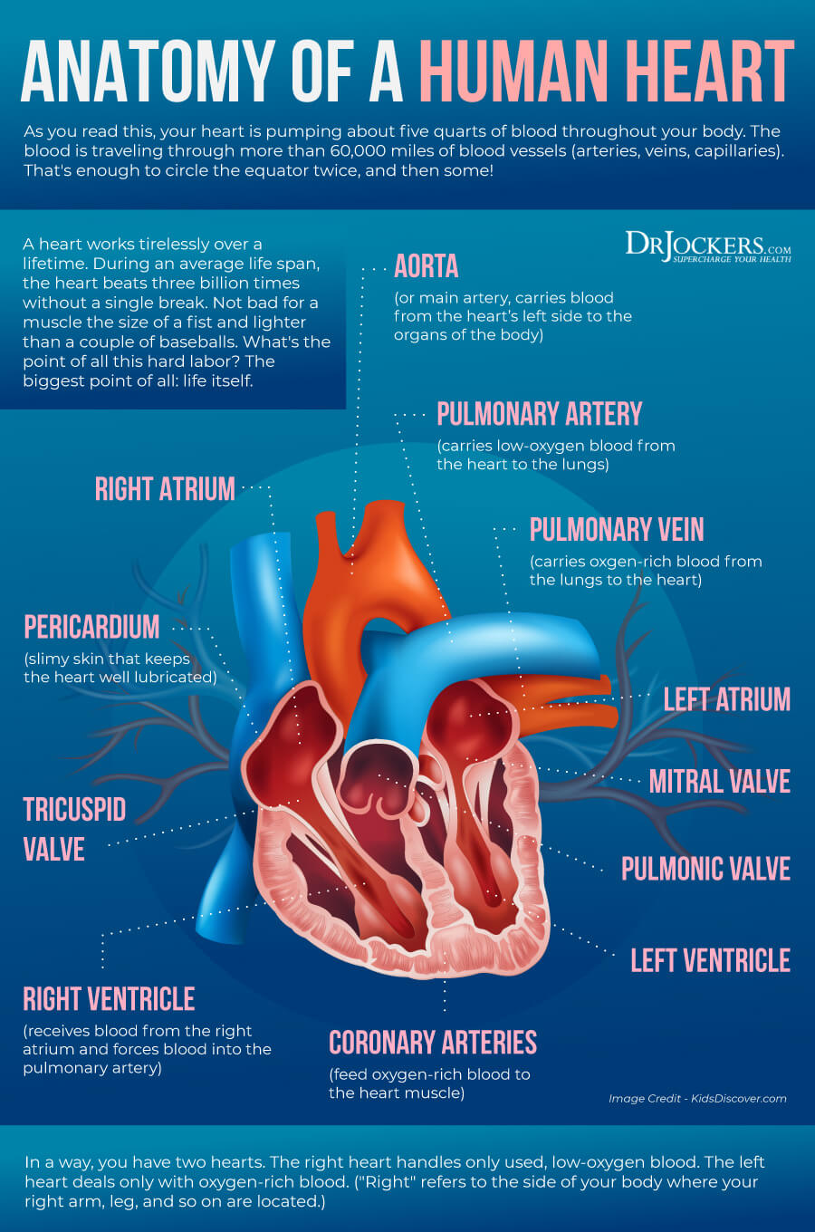
Cardiac health refers to your cardiovascular or heart health. Your heart is one of your most important organs and the center of your circulatory system. As the National Institute of Health (NIH), National Lung, Blood, and Heart Institute explains, your circulatory system is made up of a network of blood vessels, such as capillaries, veins, and arteries that carry blood full of oxygen and nutrients to your organs allowing healthy functioning. Your heart is working non-stop. It is beating all day long every minute of the day to make sure that the right amount of blood is flowing through your system at a healthy rate to keep your body’s systems healthy and functioning (2).
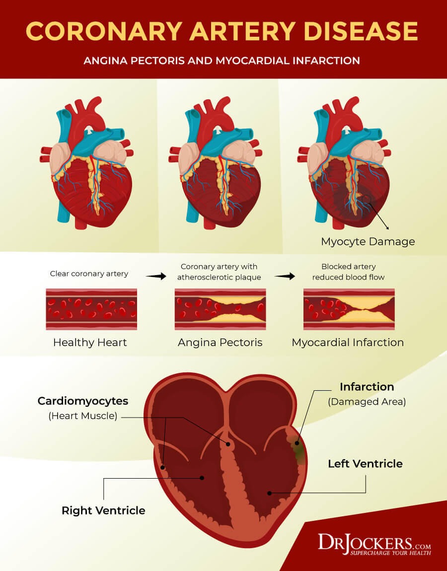
Your cardiac health is clearly important. If there is a problem, your heart will have a harder time to pump blood. If your heart experiences difficulties pumping blood, it can affect your entire body. Fat, cholesterol, calcium, and other substances can create plaques in your coronary arteries restricting blood flow and increasing the risk of heart attacks, strokes, and cardiac death.
According to the Centers for Disease Control and Prevention (CDC), heart disease is the leading cause of death in the United States killing one person every 36 seconds and 655,000 people a year. It is responsible for one in every four deaths in the country (1). It is clear that your cardiac health is important and non-negotiable if you want to live a healthy and long life.
How Cardiac Cells Become Damaged
Damage to your cardiac or heart cells and heart and circulatory system poses a major health risk and is a major cause of death and disability in our country. Cardiomyocytes are your cardiac or heart muscle cells that make up your cardiac muscle.
Damage to your cardiac cells may happen because of hypertension or high blood pressure, coronary artery disease, which leads to chronic blood supply inefficiency, or a heart attack, which is a sudden closing of a blood vessel that takes oxygen to the heart.
There are a variety of dietary, lifestyle, health, and environmental factors that may increase the risk of cardiac cell damage and cardiac health problems, including obesity, mitochondrial dysfunction, high levels of oxidative stress, chronic inflammation, insulin resistance, diabetes, unhealthy diet, sedentary lifestyle, smoking, family history of heart disease, and drinking too much alcohol (3, 4).
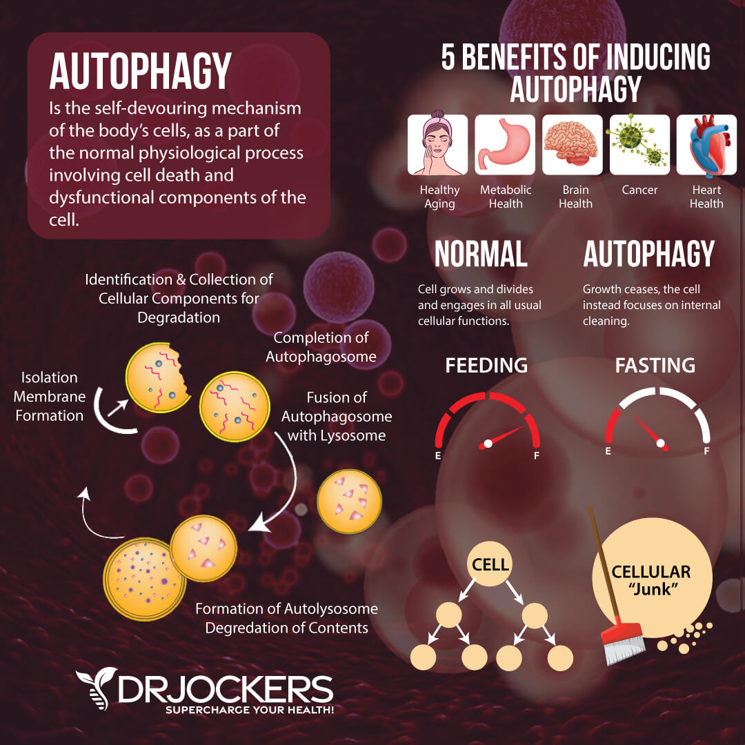
What is Cardiac Autophagy
To learn how to protect your cardiac cells, you need to understand cardiac autophagy first. The word autophagy comes from ‘auto-phlegein’, a Greek word for ‘self-eating’. Autophagy, or self-eating, refers to the process of cellular recycling in your body. During this process, a cell itself metabolizes various components in order to reuse them and create new and healthy cellular structures.
Your cells, including your cardiac cells, have a number of important components called organelles that act like organs in your body. When your body is exposed to stressors, such as nutrient deprivation, your cells make a phagophore, which is a double-membrane structure. This is a very flexible structure that is able to surround cellular components and take them to the lysosomes, which are unique organelles that can destroy particular components by releasing degrading enzymes upon them.
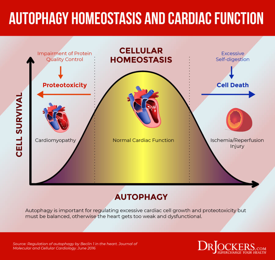
Cardiac Autophagy Homeostasis
Your body’s goal is always homeostasis and balance which is necessary or healthy. One of the major driving forces of autophagy is cellular stress. Nutrient deprivation from fasting, glucose deprivation from a low-carb diet, and exercise are great examples of this autophagy-driving stress.
When such stress happens, your body prepares for survival. To survive, it needs healthy cells. So it proceeds to break down damaged and old cells and cellular organelles to allow room for the creation of new and healthy cells for better energy efficiency, survival, and health.
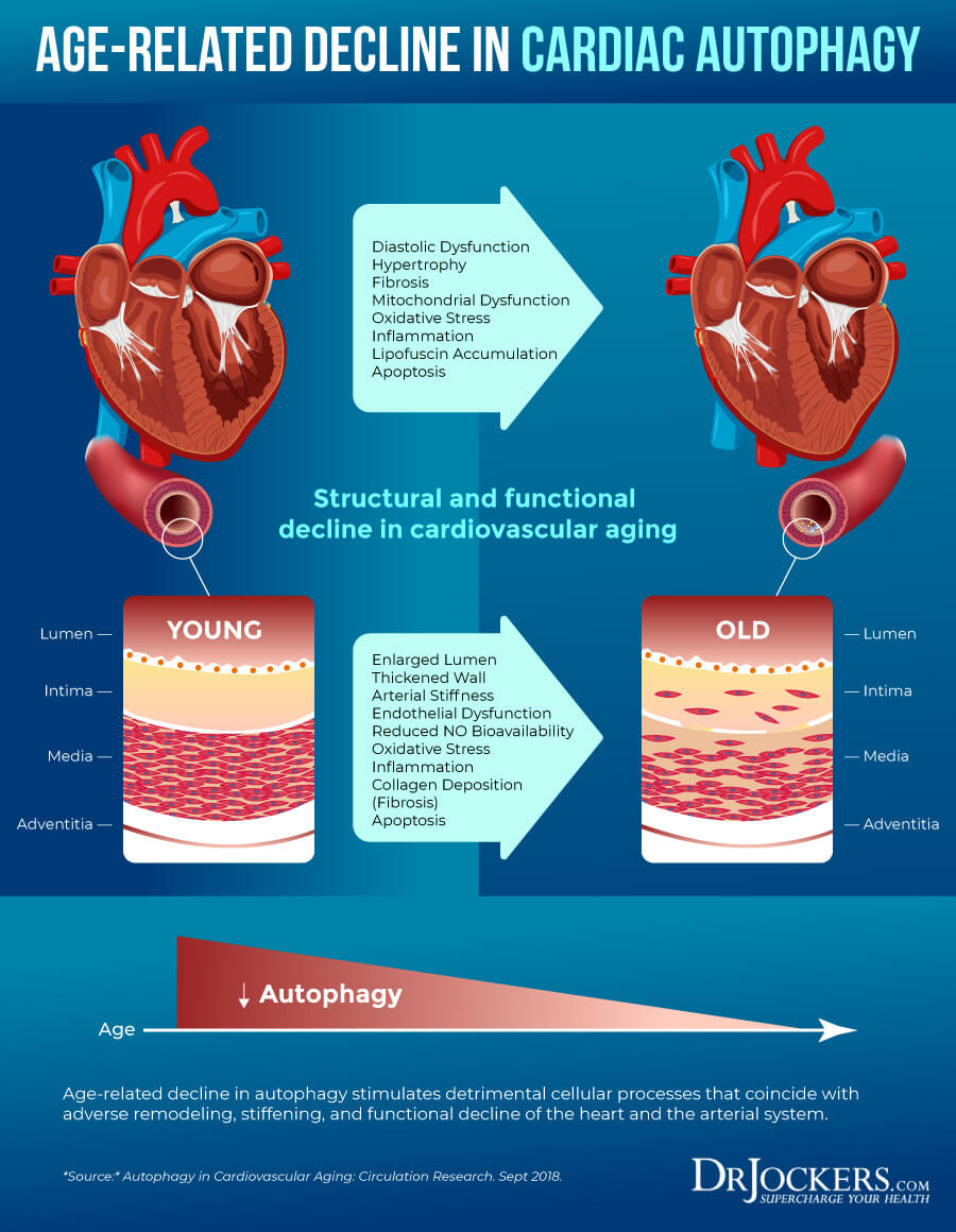
Autophagy is very important for your overall health and cardiac autophagy is beneficial for your cardiac health. According to a 2012 scientific review published in Heart Failure Reviews, autophagy plays a beneficial role in heart health, including in the setting of cardioprotection, hypertrophy, and heart failure (5).
According to a 2012 study published in the Journal of Cardiovascular Pharmacology, cardiac autophagy can improve the heart’s tolerance to myocardial ischemia, which happens when the blood flow to your heart is reduced and lowers your heart’s ability to receive enough oxygen (6). A 2018 study in Circulation Research showed that as we age, cardiac autophagy decreases and this increases the thickening of the extracellular matrix and results in endothelial dysfunction (7).
While we know that cardiac autophagy is very important for the healing process, too much autophagy can be problematic as well. A 2016 study out of The Journal of Molecular and Cellular Cardiology discussed the idea of autophagy homeostasis for optimal cardiovascular health and you can see the image below to better understand this (8). To learn more about the importance and benefits of autophagy, I recommend reading this article.
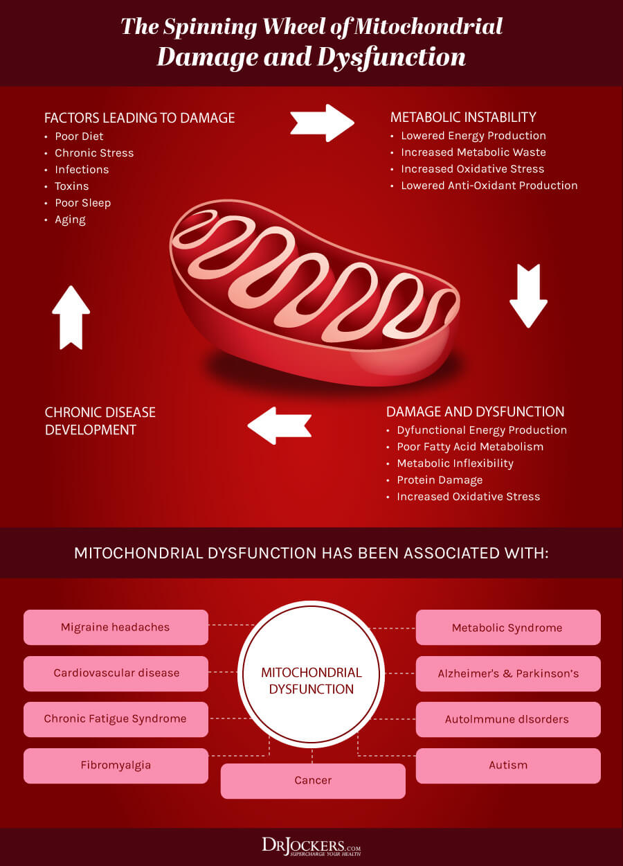
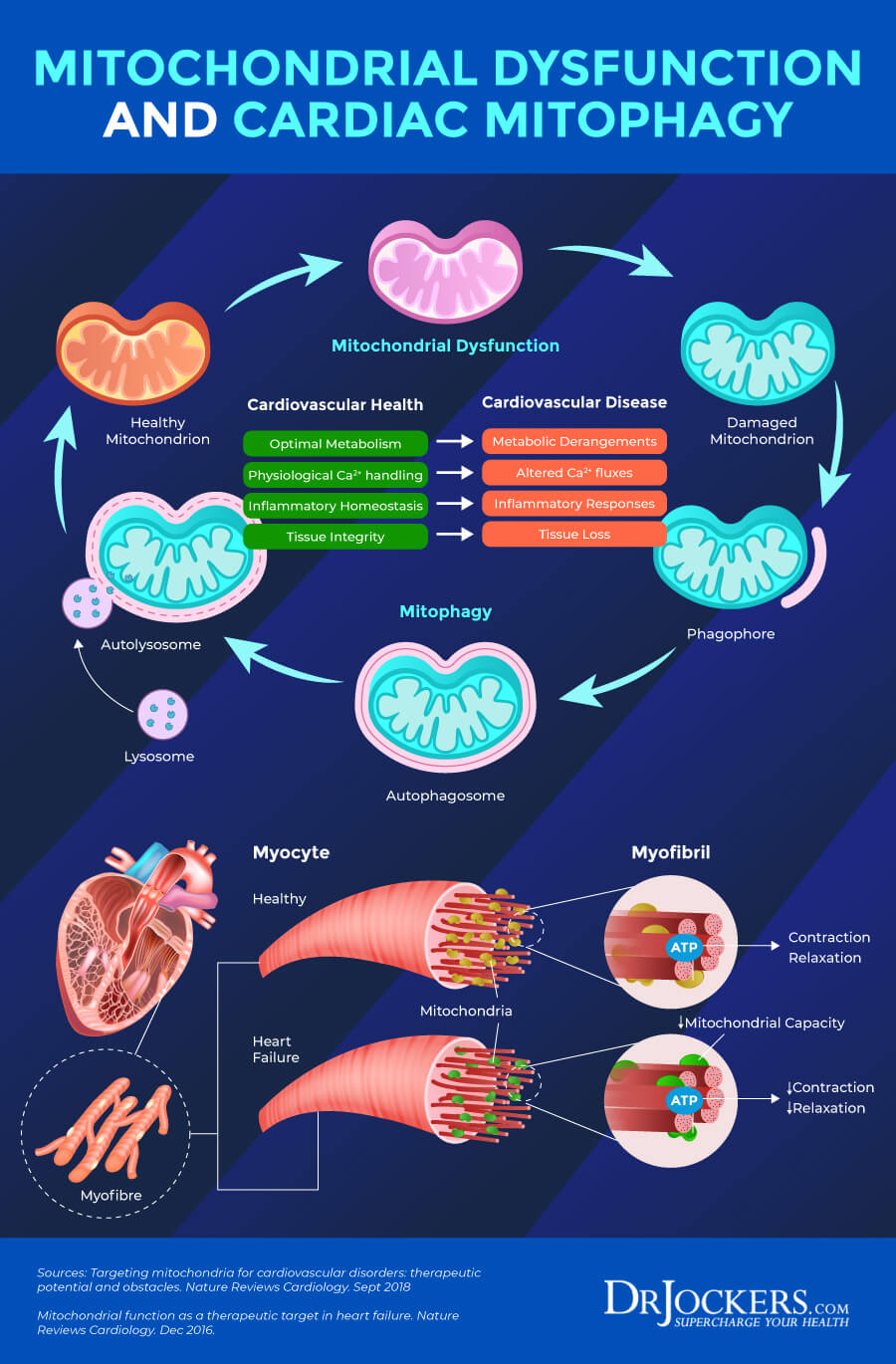
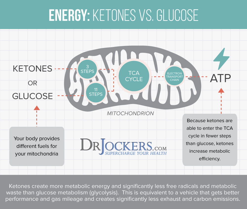
Cardiac Mitophagy
If there is one thing we’ve all learned in biology class is that the mitochondria are the powerhouse of your cells. They are specialized cells that are found in every single cell of your body, except for your red blood cells. Cardiac mitochondria are found in your cardiac cells.
Your mitochondria are responsible for generating about 90 percent of cellular energy in the form of adenosine triphosphate (ATP). They also help metabolic functions. Mitochondria are located at various concentrations in various tissues throughout your body serving various purposes. Your cardiac mitochondria are responsible for your heart’s health and functioning (9).
Creating Resilient Cardiac Mitochondria
There is a link between the performance and functioning of the mitochondria and the risk of developing a disease. Mitochondria become damaged from metabolically induced oxidative stress. Mitochondrial abnormalities include impaired mitochondrial electron transport chain activity, increased formation of reactive oxygen species, shifted metabolic substrate utilization, aberrant mitochondrial dynamics, and altered ion homeostasis.
Nature Reviews Cardiology has published a series of articles on how important it is to target mitochondrial autophagy, which is called mitophagy, as a means of improving heart and blood vessel tissue function and cardiovascular health (10, 11). In the image below, you can see in the cardiac muscle tissue and mitochondria and the importance of healthy mitophagy processes.
Chronic stress is a major risk factor for health issues and the development of stress-related chronic diseases. Heart disease is one of them. According to the book Stress Resilience published by Academic Press, improving mitochondrial function may be beneficial for improving stress resilience (12). According to a 2016 study published in the Impact Journal on Aging, aerobic exercise can improve cardiac mitochondrial resilience and heart health (13).
There are a variety of other ways that you can improve cardiac mitochondrial resilience. In the next section, you will learn the top strategies to benefit cardiac mitochondrial health. To learn more about how to improve mitochondrial health, I recommend reading this article.
Top Strategies to Promote Cardiac Autophagy
If you want to promote cardiac autophagy for cardiac health, there are a variety of simple strategies you can try. It is important to note that these strategies have not been FDA approved to prevent, mitigate, treat or cure heart disease or any other disease and should not be confused as such.
However, we do know that these can improve heart health and stimulate cardiac cell autophagy. Here are my top strategies to promote cardiac autophagy:
Consume Nutrient-Dense Foods
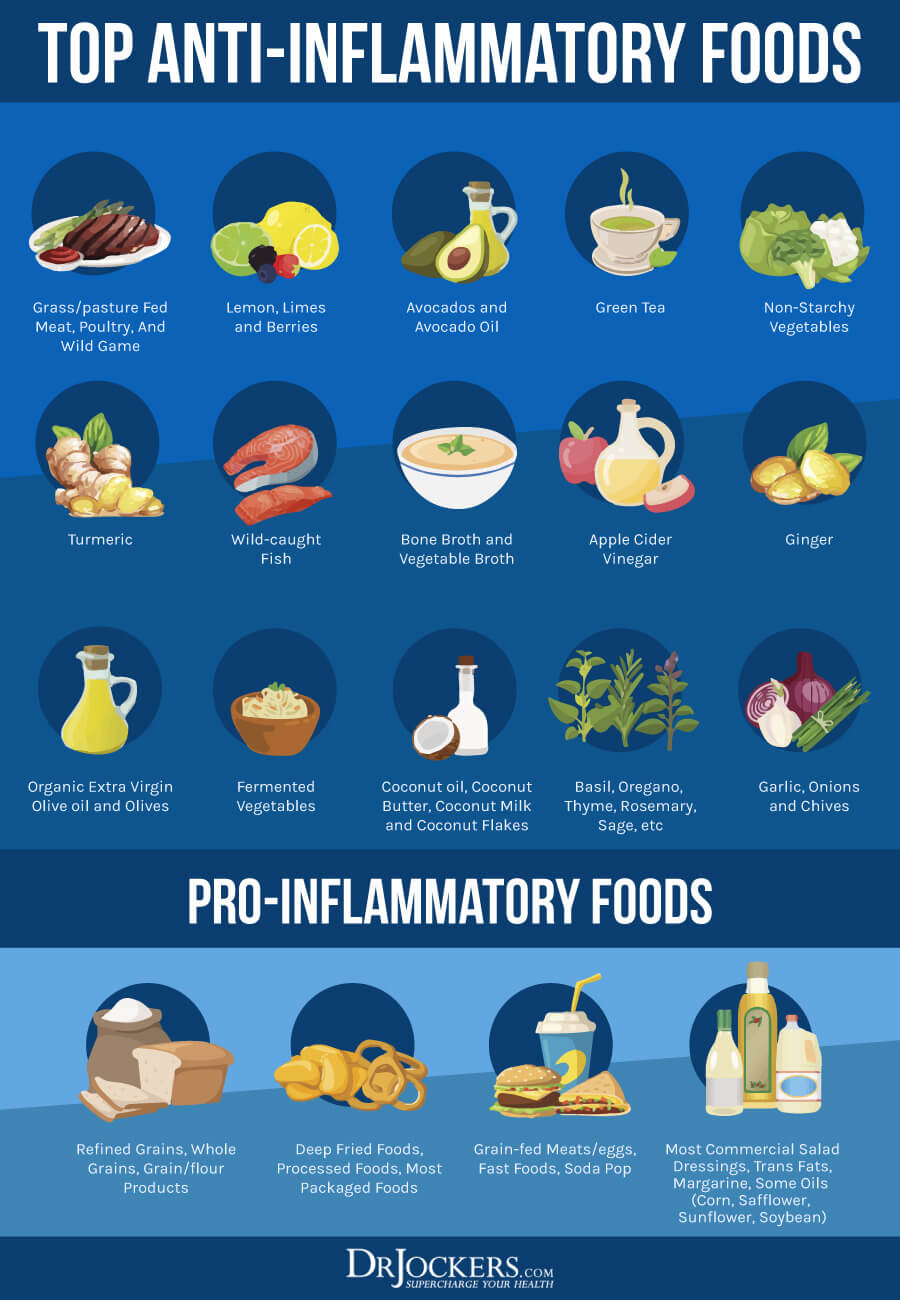
Eating an inflammatory, unhealthy diet can seriously compromise your cardiac and overall health. I recommend that you remove all refined sugar, refined oils, processed foods, deep-fried food, junk food, and artificial ingredients from your diet, and consume nutrient-dense foods instead.
I recommend eating a nutrient-dense, anti-inflammatory, low-carb ketogenic diet rich in greens, vegetables, low-glycemic index fruits, herbs, spices, fermented foods, healthy fats, such as coconut oil, avocados, and organic butter and ghee, and clean protein, such as grass-fed beef, pasture-raised poultry and eggs, wild-caught fish, and wild game.
According to a 2010 research published in Human Molecular Genetics, a ketogenic diet may slow down the progression of mitochondrial myopathy (14). According to a 2014 randomized trial published in the Annals for Internal Medicine, which followed 119 participants for 12 months and aimed to compare the benefits of a low-fat versus a low-carb diet on cardiac, has found that a low-carb diet leads to greater weight loss and better cardiovascular markers than a low-fat diet (15). To learn more about the diet that I recommend, read this article.
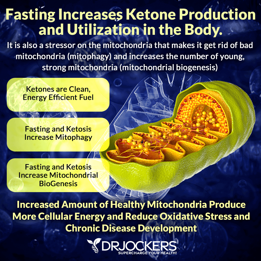
Intermittent Fasting
Intermittent fasting is a form of fasting that cycles between a period of not eating (fasting) and a period of eating (feasting) within a day. As I explained in detail in this article, intermittent fasting has a number of health benefits, including increased autophagy, improved genetic repair, improved insulin sensitivity, improved immune regulation, and a lower risk of disease. It is also great for your cardiac health.
According to a 2019 review published in the Journal of American Heart Association, intermittent fasting may help to reverse advanced cardiomyopathy. The paper discussed that intermittent fasting has cell protective benefits. It can reduce high blood pressure, aging‐induced cardiac hypertrophy, and other cardiovascular conditions, benefit insulin resistance and may benefit cardiomyopathy (16). To learn more about how intermittent fasting may benefit your heart health, I recommend reading this article.
Extended Fasting
It is not only intermittent fasting but also extended fasting that may benefit your cardiac health. According to a 2014 interview with Dr. Alan Goldhamer published in Integrative Medicine: a Clinician’s Journal, water fasting can benefit all kinds of health conditions, including cardiovascular health, diabetes, and autoimmune conditions (17).
Extended fasting can last for a couple of days or weeks. During your extended fast, you will only be consuming water, and in some cases, other non-calorie liquids, such as herbal tea. It is important that before you practice intermittent fasting for a while and make sure that you do well on more advanced forms of intermittent fasting before you embark on an extended fast. To learn more about extended fasting or water fasting and how to prepare for it, I recommend reading this article.
Keep Stress Under Control
Chronic stress is a major contributing factor to inflammation, pain, and disease. According to a 2008 study published in the Journal of American College of Cardiology, psychological stressors, including job loss, the difficulties of caregiving, and marital unhappiness put major stress of cardiac health and can contribute to heart health issues, and may even trigger serious cardiac issues and events including myocardial ischemia, myocardial infarction, wall motion abnormalities, and death (18).
Keeping your stress levels under control is clearly important for your cardiac health. Reduce events, activities, and situations that stress you out. Learn better coping mechanisms to deal with stress better.
Practice breathwork, gratitude, mindfulness, meditation, journaling, yoga, grounding, and laughter to lower your stress levels. Spend time with uplifting and supportive people. Spend time in nature. Have some regular me-time and practice a positive mindset.
Prioritize Restorative Sleep
Getting regular restorative sleep is critical for your cardiac and overall health. Poor or not enough sleep can contribute to stress, increase inflammation, and increase the risk of disease. According to a 2020 study in the Journal of American College of Cardiology, irregular sleep can increase your risk of cardiac issues (19).
Make sure to get 7 to 9 hours of restorative sleep each night. Support your natural circadian rhythms by going to bed and waking up around the same time. Make sure that your bed and bedding are comfortable and your bedroom is a peaceful sanctuary. Avoid food, caffeine, stress, and electronics several hours before bed.
Develop a nighttime routine and engage in relaxing activities that wind you down. Sipping on herbal tea, stretching, reading, meditation, breathwork, prayer, and journaling are great ideas for the evening.
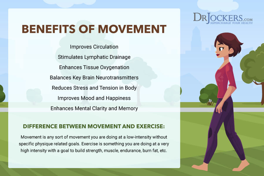
Regular Movement & Exercise
Regular movement and exercise are important for your health and well-being, including your cardiac health. According to a 2018 study in Frontiers in Cardiovascular Medicine, regular exercise is associated with lower blood pressure, higher insulin sensitivity, lower risk of cardiovascular health issues, and lower risk of cardiac death (20).
Moving your body regularly is important. Start your day with light stretching, yoga, or a short walk. Take a stroll at lunch. Stretch regularly throughout the day. Do some light movement in the evening, such as stretching, dancing, yoga, or walking. Take the stairs, park far from the grocery store and walk to the door, walk or bike instead of driving short distances, garden, dance to your favorite song, walk your dog, or play with your kids to add other simple movements to your day. Exercise at least two days a week and up to five days a week for 20 to 30 minutes each day.
Cardio workouts, such as biking, dancing, trampolines, aerobics classes, jogging, or swimming are important for your heart health. However, make sure to add strength and resistance training, such as weight lifting, kettlebells workouts, or bodyweight training, and low-impact exercises, such as yoga, pilates, or Barre workouts to the mix.
High-intensity interval training (HIIT) and Tabata workouts are a great way to get your cardio, strength workout, and resistance training within a short session. If you are new to working out, try different forms of exercise until you find the combination that you love and works for you.
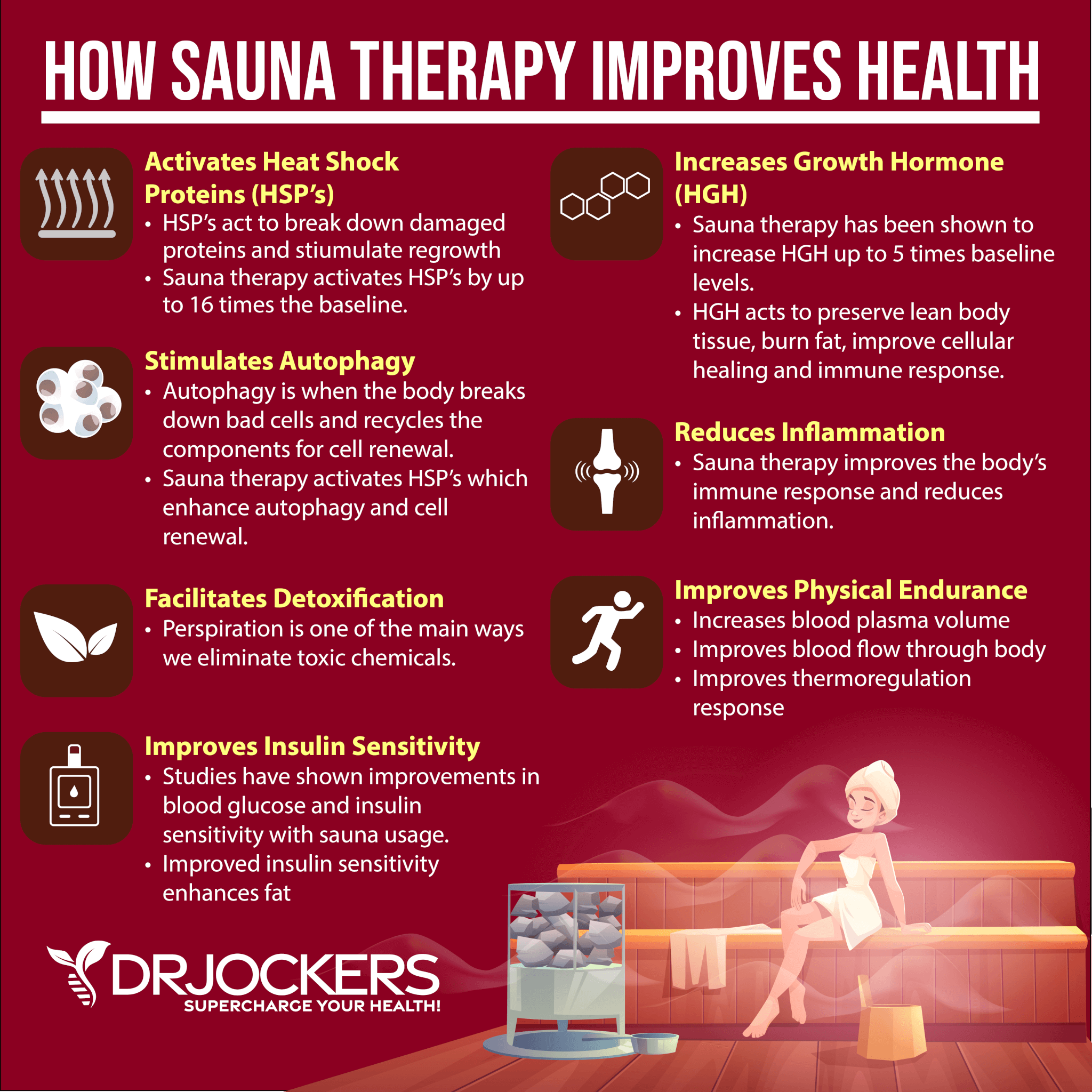
Use Heat Therapy
Heat therapy is a great way to activate cardiac autophagy. According to a 2018 study published in the Journal of Molecular Sciences, heat shock is great for autophagy (21). Experiencing heat for a short period of time can activate autophagy by putting major stressors on your body through extreme temperature change. Your body will want to return to homeostasis quickly which can support autophagy and improve your health.
To experience the benefits of heat therapy, I recommend using an infrared sauna regularly. I recommend and personally love the High Tech Health Sauna for infrared sauna therapy because it uses non-toxic poplar wood, is low EMF, has the highest level certifications and works wonderfully!
For a portable, lower cost option, I really like the HigherDOSE Infrared Sauna Blanket here. This is also low EMF and is designed by expert Sauna maker Dr Raleigh Duncan. Your head stays out of this which most people find really comfortable and their heat tolerance increases so they are able to stay in longer and get more benefits from the sauna therapy. Use the coupon code DRJOCKERS to save 15% off on HigherDOSE products including the Sauna Blanket.
I personally try to do do infrared sauna therapy at least 3 times per week for roughly 15-30 minutes per session which is a clinically effective dose to help enhance healing. To learn more about infrared sauna therapy, I recommend reading this article.
Use Cold Therapy
Just like heat therapy, cold therapy is also great for cardiac autophagy. According to a 2017 study published in Neural Regeneration Research, cold exposure induces autophagy (22). Experiencing cold for a short period of time can activate autophagy by putting unexpected stressors on your body through extreme temperature change. Your body will want to return to balance homeostasis which supports autophagy.
To experience the benefits of cold therapy, I recommend taking cold showers or finishing your shower with cold water. You may take ice baths or jump into a cold pool. I also recommend cryotherapy.
Using the combination of hot and cold therapy by jumping into the cold pool or taking a cold shower after an infrared sauna session or alternating between hot and cold water when taking a shower is the best way to get the benefits of both worlds.
Use Autophagy-Enhancing Herbs
To further support cardiac autophagy and cardiac resistance, you can try taking some autophagy-enhancing herbs. Resveratrol is a polyphenol found in the skin of red grapes, peanuts, and berries. According to a 2019 study in the International Journal of Molecular Sciences, resveratrol may help to reduce cardiovascular disease and heart failure (23).
Quercetin is a flavonol with similar and complimentary benefits to resveratrol. According to a 2013 study published in the International Journal of Preventive Medicine, quercetin may improve cardiovascular biomarkers in those with diabetes (24).
Curcumin is the potent compound found in the spice, turmeric. According to a 2009 study published in the International Journal of Cardiology, curcumin has anti-inflammatory and cardioprotective benefits (25). Epigallocatechin gallate (EGCG), a powerful polyphenol found in green tea, offers important health benefits. According to a 2007 study in the Journal of American College of Nutrition, regular consumption of green tea and EGCG has shown cardiovascular and metabolic health benefits (26).
I recommend taking Resveratrol Power, a fantastic supplement powered by resveratrol and quercetin, daily to support autophagy and cardiac resistance. Resveratrol Power helps to improve mitochondrial function, improve circulation, support optimal immune function, reduce oxidative stress, and supports skin function. Take one capsule once a day for optimal results.
Final Thoughts
Cardiovascular disease is the leading cause of death in the United States. It doesn’t have to be this way and you certainly don’t have to become part of the statistics. You can support your heart health naturally through a healthy diet and lifestyle and some simple strategies. Follow my tips to improve cardiac resilience, support your health, and live a long heart-healthy life.
Antioxidants are substances that can help protect cells from damage caused by free radicals, or unstable molecules. That damage, called “oxidative stress,” is linked to the kind of damage in DNA mutations that can contribute to the risk of certain cancers, as well as diabetes, Alzheimer’s disease, and Parkinson’s disease.
In most cases, you can find all the antioxidants you need in a healthy, plant-based diet.
Some studies have shown that taking antioxidant supplements – such as vitamins A, C, E, folic acid, and beta-carotene – might help reduce the risk for certain diseases related to oxidative stress, including cancer. A 1993 clinical trial from China found “significantly reduced” stomach cancer mortality rates in participants who took beta-carotene, vitamin E, and selenium over a five-year period.
However, another study found that elevated antioxidant levels might heighten the risk for certain types of cancer. In a 1994 Swedish trial, male smokers who took beta-carotene regularly were more likely to develop lung cancer than male smokers who did not take the supplement. Researchers concluded that the supplements they studied could possibly have both harmful and beneficial effects.
To date, nine randomized controlled clinical trials of dietary antioxidant supplements for cancer prevention have been conducted worldwide to study the effects of antioxidant supplements and cancer. The different studies reached varying conclusions about the efficacy and safety of taking antioxidant supplements to help prevent cancer or taking them during cancer treatment. “Additional large randomized controlled trials are needed to provide clear scientific evidence about the potential benefits or harms of taking antioxidant supplements during cancer treatment,” the National Cancer Institute (NCI), reports.
While the Chinese and Swedish studies found that antioxidants could decrease or increase cancer risk, respectively, other studies found that antioxidant supplements had no effect on cancer risk. The 2005 Women’s Health Study concluded that there was “no benefit or harm associated with two years of beta-carotene supplementation” in regards to cancer and cardiovascular disease risk in women 45 or older
Antioxidant supplements have also been shown to decrease the effectiveness of certain cancer treatments. Some treatments, such as radiation and certain forms of chemotherapy, use free radicals to kill cancer cells. But because antioxidant supplements work by combating free radicals, taking supplements as a cancer patient may lessen the effectiveness of certain cancer treatments, some studies have stated. However, getting antioxidants from foods like fruits, vegetables, and grains, which are rich sources of dietary antioxidants, does not reduce the potency of treatment. They also help provide nutrients that support a healthy immune system.
It’s important to discuss all supplements with your doctor, including antioxidants, before beginning a regimen, especially during active cancer treatment.
In most cases, you can find all the antioxidants you need in a healthy, plant-based diet. Eating plenty of differently colored fruits and vegetables can help ensure you get important antioxidants like lycopene, beta-carotene, and anthocyanins.