A new study illustrates how parts of the brain need to work together to focus on important information while filtering out distractions.
Imagine a busy restaurant: dishes clattering, music playing, people talking loudly over one another. It’s a wonder that anyone in that kind of environment can focus enough to have a conversation.
The new research from researchers at Brown University’s Carney Institute for Brain Science provides some of the most detailed insights yet into the brain mechanisms that help people pay attention amid such distraction, as well as what’s happening when they can’t focus.
In an earlier psychology study, the researchers established that people can separately control how much they focus (by enhancing relevant information) and how much they filter (by tuning out distraction). The team’s new research in Nature Human Behaviour unveils the process by which the brain coordinates these two critical functions.
Lead author and neuroscientist Harrison Ritz likens the process to how humans coordinate muscle activity to perform complex physical tasks.
“In the same way that we bring together more than 50 muscles to perform a physical task like using chopsticks, our study found that we can coordinate multiple different forms of attention in order to perform acts of mental dexterity,” says Ritz, who conducted the study while a PhD student at Brown.
The findings provide insight into how people use their powers of attention as well as what makes attention fail, says coauthor Amitai Shenhav, an associate professor in Brown’s cognitive, linguistic, and psychological sciences department.
“These findings can help us to understand how we as humans are able to exhibit such tremendous cognitive flexibility—to pay attention to what we want, when we want to,” Shenhav says. “They can also help us better understand limitations on that flexibility, and how limitations might manifest in certain attention-related disorders such as ADHD.”
To conduct the study, Ritz administered a cognitive task to participants while measuring their brain activity in an fMRI machine. Participants saw a swirling mass of green and purple dots moving left and right, like a swarm of fireflies. The tasks, which varied in difficulty, involved distinguishing between the movement and colors of the dots. For example, participants in one exercise were instructed to select which color was in the majority for the rapidly moving dots when the ratio of purple to green was almost 50/50.
Ritz and Shenhav then analyzed participants’ brain activity in response to the tasks.
Ritz, who is now a postdoctoral fellow at the Princeton Neuroscience Institute, explains how the two brain regions work together during these types of tasks.
“You can think about the intraparietal sulcus as having two knobs on a radio dial: one that adjusts focusing and one that adjusts filtering,” Ritz says. “In our study, the anterior cingulate cortex tracks what’s going on with the dots. When the anterior cingulate cortex recognizes that, for instance, motion is making the task more difficult, it directs the intraparietal sulcus to adjust the filtering knob in order to reduce the sensitivity to motion.
“In the scenario where the purple and green dots are almost at 50/50, it might also direct the intraparietal sulcus to adjust the focusing knob in order to increase the sensitivity to color. Now the relevant brain regions are less sensitive to motion and more sensitive to the appropriate color, so the participant is better able to make the correct selection.”
Ritz’s description highlights the importance of mental coordination over mental capacity, revealing an often-expressed idea to be a misconception.
“When people talk about the limitations of the mind, they often put it in terms of, ‘humans just don’t have the mental capacity’ or ‘humans lack computing power,’” Ritz says. “These findings support a different perspective on why we’re not focused all the time. It’s not that our brains are too simple, but instead that our brains are really complicated, and it’s the coordination that’s hard.”
Ongoing research projects are building on these study findings. A partnership with physician-scientists at Brown University and Baylor College of Medicine is investigating focus-and-filter strategies in patients with treatment-resistant depression. Researchers in Shenhav’s lab are looking at the way motivation drives attention; one study co-led by Ritz and Brown PhD student Xiamin Leng examines the impact of financial rewards and penalties on focus-and-filter strategies.
Our fear response is a survival mechanism that signals us to remain alert and avoid dangerous situations. Those who have suffered episodes of severe or life-threatening stress can later experience intense feelings of fear, even during situations that lack a real threat. Experiencing this generalization of fear is psychologically damaging and can result in conditions such as post-traumatic stress disorder (PTSD). The stress-induced mechanisms that cause our brain to produce feelings of fear in the absence of threats has not been fully understood. Now, neurobiologists at the University of California (UC), San Diego, have identified the changes in brain biochemistry and mapped the neural circuitry that cause such a generalized fear experience.
“Overgeneralization of fear to harmless situations is a core feature of anxiety disorders resulting from acute stress, yet the mechanisms by which fear becomes generalized are poorly understood,” the researchers wrote. “In this study, we show that generalized fear in mice results from a transmitter switch from glutamate to γ-aminobutyric acid (GABA) in serotonergic neurons of the lateral wings of the dorsal raphe. A similar change in transmitter identity was found in the postmortem brains of individuals with post-traumatic stress disorder (PTSD). Overriding the transmitter switch in mice prevented the acquisition of generalized fear.”
In their research, former UC San Diego assistant project scientist Hui-quan Li, PhD, (now a senior scientist at Neurocrine Biosciences), Nick Spitzer, PhD, the Atkinson Family Distinguished Professor of the School of Biological Sciences, and their colleagues described the research behind their discovery of the neurotransmitters—the chemical messengers that allow the brain’s neurons to communicate with one another—at the root of stress-induced generalized fear.
The researchers studied the brains of mice in an area known as the dorsal raphe, and discovered that acute stress induced a switch in the chemical signals in the neurons, flipping from excitatory “glutamate” to inhibitory “GABA” neurotransmitters, which led to generalized fear responses.
“Our results provide important insights into the mechanisms involved in fear generalization,” said Spitzer, who is also a member of UC San Diego’s department of neurobiology and the Kavli Institute for Brain and Mind. “The benefit of understanding these processes at this level of molecular detail—what is going on and where it’s going on—allows an intervention that is specific to the mechanism that drives related disorders.”
The researchers then examined the postmortem human brains of individuals who had suffered from PTSD. A similar glutamate-to-GABA neurotransmitter switch was confirmed in their brains as well.
The researchers then found a way to block the production of generalized fear. Prior to the experience of acute stress, they injected the dorsal raphe of the mice with an adeno-associated virus (AAV) to suppress the gene responsible for synthesis of GABA. This method prevented the mice from acquiring generalized fear.
Further, when mice were treated with the antidepressant fluoxetine (branded as Prozac) immediately after a stressful event, the transmitter switch and subsequent onset of generalized fear were prevented.
Not only did the researchers identify the location of the neurons that switched their transmitter, but they demonstrated the connections of these neurons to the central amygdala and lateral hypothalamus, brain regions that were previously linked to the generation of other fear responses.
“Now that we have a handle on the core of the mechanism by which stress-induced fear happens and the circuitry that implements this fear, interventions can be targeted and specific,” said Spitzer.
When your mind is wandering, your brain’s “default mode” network is active. Its discovery 20 years ago inspired a raft of research into networks of brain regions and how they interact with each other.
Introduction
Whenever you’re actively performing a task — say, lifting weights at the gym or taking a hard exam — the parts of your brain required to carry it out become “active” when neurons step up their electrical activity. But is your brain active even when you’re zoning out on the couch?
The answer, researchers have found, is yes. Over the past two decades they’ve defined what’s known as the default mode network, a collection of seemingly unrelated areas of the brain that activate when you’re not doing much at all. Its discovery has offered insights into how the brain functions outside of well-defined tasks and has also prompted research into the role of brain networks — not just brain regions — in managing our internal experience.
In the late 20th century, neuroscientists began using new techniques to take images of people’s brains as they performed tasks in scanning machines. As expected, activity in certain brain areas increased during tasks — and to the researchers’ surprise, activity in other brain areas declined simultaneously. The neuroscientists were intrigued that during a wide variety of tasks, the very same brain areas consistently dialed back their activity.
It was as if these areas had been active when the person wasn’t doing anything, and then turned off when the mind had to concentrate on something external.
Researchers called these areas “task negative.” When they were first identified, Marcus Raichle, a neurologist at the Washington University School of Medicine in St. Louis, suspected that these task-negative areas play an important role in the resting mind. “This raised the question of ‘What’s baseline brain activity?’” Raichle recalled. In an experiment, he asked people in scanners to close their eyes and simply let their minds wander while he measured their brain activity.
He found that during rest, when we turn mentally inward, task-negative areas use more energy than the rest of the brain. In a 2001 paper, he dubbed this activity “a default mode of brain function.” Two years later, after generating higher-resolution data, a team from the Stanford University School of Medicine discovered that this task-negative activity defines a coherent network of interacting brain regions, which they called the default mode network.
In a brain network, the individual parts interact to bring about effects that they can only produce together.
The discovery of the default mode network ignited curiosity among neuroscientists about what the brain is doing in the absence of an outward-focused task. Although some researchers believed that the network’s main function was to generate our experience of mind wandering or daydreaming, there were plenty of other conjectures. Maybe it controlled streams of consciousness or activated memories of past experiences. And dysfunction in the default mode network was floated as a potential feature of nearly every psychiatric and neurological disorder, including depression, schizophrenia and Alzheimer’s disease.
Since then, a flurry of research into the default mode has complicated that initial understanding. “It’s been very interesting to see the types of different tasks and paradigms that engage the default mode network in the last 20 years,” said Lucina Uddin, a neuroscientist at the University of California, Los Angeles.
The default mode was one of the first brain networks characterized by science. It consists of a handful of brain regions, including a few at the front of the brain, like the dorsal and ventral medial prefrontal cortices, and others scattered throughout the organ, like the posterior cingulate cortex, the precuneus and the angular gyrus. These regions are associated with memory, experience replay, prediction, action consideration, reward/punishment and information integration. (The colored highlighting in the following figure indicates some of the outer brain areas that become more active when the default network engages.)
Since its discovery, neuroscientists have loosely identified a handful of additional distinct networks that each activate seemingly disparate areas of the brain. These activated areas don’t act independently, but rather harmonize in synchrony with each other. “You can’t think about a symphony orchestra as just the violins or the oboes,” Raichle said. Similarly, in a brain network, the individual parts interact to bring about effects that they can only produce together.
According to research, the effects of the default mode network include mind wandering, remembering past experiences, thinking about others’ mental states, envisioning the future and processing language. While this may seem like a grab bag of unrelated aspects of cognition, Vinod Menon, the director of the Stanford Cognitive & Systems Neuroscience Laboratory, recently theorized that all of these functions may be helpful in constructing an internal narrative. In his view, the default mode network helps you think about who you are in relation to others, recall your past experiences and then wrap up all of that into a coherent self-narrative
Introduction
The default mode is clearly up to something complicated; it’s involved in many different processes that can’t be neatly described. “It’s kind of silly to think that we’re ever going to be like, ‘This one brain region or one brain network does one thing,’” Uddin said. “I don’t think that’s how it works.”
Uddin began investigating the default mode network because she was interested in self-recognition, and many self-recognition tasks, such as identifying your own face or voice, appeared to be associated with the network. In recent years, she has shifted her attention to interactions between brain networks. Just as different brain areas interact with each other to form networks, different networks interact with each other in meaningful ways, Uddin said. “Network interactions are more elucidating to study in some ways than just a network in isolation because they do work together and then come apart and then change what they’re doing over time.”
She’s particularly interested in how the default mode network interacts with the salience network, which seems to help us identify the most relevant piece of information at any given time. Her work suggests that the salience network detects when something is important to pay attention to and then acts as an off switch for the default mode network.
Researchers have also been examining whether mental health disorders like depression could be linked to problems with the default mode network. So far, the findings have been inconclusive. In people with depression, for example, some researchers have found that network nodes are overly connected, while others have found the opposite — that nodes are failing to connect. And in some studies, the default mode network itself isn’t abnormal, but its interactions with other networks are. These findings may appear incompatible, but they align with recent findings that depression is perhaps a cluster of different disorders that present with similar symptoms.
Meanwhile, Menon has developed what he calls the triple network theory. It posits that abnormal interactions between the default mode network, the salience network and a third one called the frontoparietal network could contribute to mental health disorders including schizophrenia, depression, anxiety, dementia and autism. Typically, the activity of the default mode network decreases when someone is paying attention to an external stimulus, while activity in the two other networks increases. This push and pull between networks may not work the same way in people with psychiatric or developmental disorders, Menon suspects
Deanna Barch, who studies the neurobiology of mental illnesses at Washington University in St. Louis, is intrigued by the triple network theory. Investigating how networks are wired up differently in people with mental health disorders can help researchers find underlying mechanisms and develop treatments, she said. However, she doesn’t think network interactions alone will fully explain mental illness. “I think of understanding connectivity differences as a starting point,” Barch said. “It’s not an endpoint.”
The current understanding of the default mode network is surely not its endpoint, either. Since its discovery, it has pushed neuroscientists to think beyond the responsibilities of single brain regions to the effects of interactions between brain networks. And it’s driven many people to appreciate the inward-focused activities of the mind — that even when we’re daydreaming or at rest, our brain is hard at work making it happen.
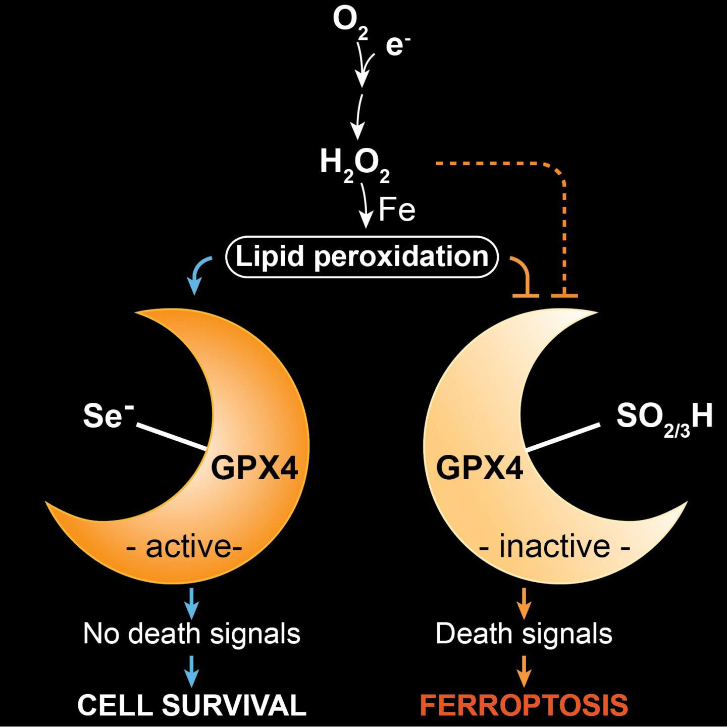
Selenium protects a specific type of interneurons in the brain.
Exactly 200 years ago, the Swedish scientist Jöns Jacob Berzelius discovered the trace element selenium, which he named after the goddess of the moon, Selene. Besides its industrial applications (chemical industry, production of semiconductors and toners), selenium is an essential trace element and indispensable for humans, many animals, and some bacteria. A team led by Dr. Marcus Conrad, research group leader at the Institute of Developmental Genetics (IDG) at Helmholtz Zentrum München, showed for the first time why selenium is a limiting factor for mammals.
Scientific ‘by-catch‘ solves decades-old mystery
The scientists have been investigating for years the processes of a novel type of cell death, known as ferroptosis. In this context, the enzyme GPX4, which normally contains selenium in the form of the amino acid selenocysteine, plays an important role.
In order to better understand the role of GPX4 in this death process, we established and studied mouse models in which the enzyme was modified,” said study leader Conrad. “In one of these models, we observed that mice with a replacement of selenium to sulfur in GPX4 did not survive for longer than three weeks due to neurological complications.”
In their search for the underlying reasons, the researchers identified a distinct subpopulation of specialized neurons in the brain, which were absent when selenium-containing GPX4 was lacking. “In further studies, we were able to show that these neurons were lost during postnatal development, when sulfur- instead of selenium-containing GPX4 was present,” stated first author of the study, Irina Ingold.
Furthermore, the scientists were able to show that ferroptosis is triggered by oxidative stress, which is known to occur for instance during high metabolic activity of cells and high neuronal activity. “Our study demonstrates for the first time that selenium is an essential factor for the postnatal development of a specific type of interneurons,” said Dr. José Pedro Friedmann Angeli, a scientist at the IDG, describing the results. “Selenium-containing GPX4 protects these specialized neurons from oxidative stress and from ferroptotic cell death.”
Thus, the study explains why certain selenoenzymes are essential in some organisms, including mammals, whereas they are dispensable in other organisms, such as fungi and higher plants. In future investigations, study leader Marcus Conrad and his team aim to investigate how ferroptosis is triggered in cells. As a long-term goal, he wants to elucidate the role of ferroptosis in various disease conditions in order to be able to alleviate diseases, such as cancer or neurodegeneration, which are currently difficult to tackle.
Further Information
GPX4 stands for the enzyme glutathione peroxidase 4, one of 25 selenoproteins in humans. In the enzyme, selenium is an integral part of the 21st amino acid selenocysteine. The enzyme plays a decisive role in ferroptosis. The word ferroptosis, which means a type of programmed cell death dependent on iron, is derived from the Greek ptosis: fall and Latin ferrum: iron. Ferroptosis has not yet been completely elucidated, but the importance of cellular suicide has already been impressively confirmed, for example, by apoptosis, which has been more extensively studied.
New ways of thinking about how addictions form have the potential to change how we approach treatment.
Many people are wired to seek and respond to rewards. Your brain interprets food as rewarding when you are hungry and water as rewarding when you are thirsty. However, addictive substances like alcohol and drugs of abuse can overwhelm the natural reward pathways in your brain, resulting in intolerable cravings and reduced impulse control.
A popular misconception is that addiction is a result of low willpower. However, an explosion of knowledge and technology in the field of molecular genetics has changed our basic understanding of addiction drastically over the past decade. The general consensus among scientists and healthcare professionals is that there is a strong neurobiological and genetic basis for addiction.
As a behavioral neurogeneticistleading a team investigating the molecular mechanisms of addiction, I combine neuroscience with genetics to understand how alcohol and drugs influence the brain. In the past decade, I have seen changes in our understanding of the molecular mechanisms of addiction, largely due to a better understanding of how genes are dynamically regulated in the brain. New ways of thinking about how addictions form have the potential to change how we approach treatment.
ALCOHOL AND DRUGS AFFECT BRAIN GENE ACTIVITY
Each of your brain cells has your genetic code stored in long strands of DNA. For all that DNA to fit into a cell, it needs to be packed tightly. This is achieved by winding the DNA around “spools” of proteins called histones. Areas where DNA is unwound contain active genes coding for proteins that serve important functions within the cell.
When gene activity changes, the proteins your cells produce also change. Such changes can range from a single neuronal connection in your brain to how you behave. This genetic choreography suggests that while your genes affect how your brain develops, which genes are turned on or off when you are learning new things is dynamic and adapts to suit your daily needs.
Recent data from animal models suggests that alcohol and drug abuse directly influence changes in gene expression in areas of the brain that help drive memory and reward responses.
There are many ways addictive substances can change gene expression. They can alter which proteins bind to DNA to turn genes on and off and which segments of DNA are unwound. They can change the process of how DNA is read and translated into proteins, as well as alter the proteins that determine how cells use energy to function.
For example, alcohol can cause an alternative form of a gene to be expressed in the memory circuits in fliesand people, resulting in changes in dopamine receptors and transcription factors involved in reward signaling and neuronal function. Similarly, cocaine can cause an alternative form of a gene to be expressed in the reward centersof mice, leading them to seek out more cocaine.
Exactly how these drugs cause changes in gene regulation is unknown. However, a direct link between alcohol consumption and changes in gene expression in mice provides a clue. A byproduct of alcohol being broken down in the liver called acetate can cross the blood-brain barrier and unwind DNA from histones in mouse memory circuits.
Alcohol, nicotine, cocaine, and opioids also all activate important signaling pathways that are central regulators of metabolism. This suggests they can also affect many aspects of neuronal function and consequently affect which genes are expressed.
CHANGING BRAIN GENE ACTIVITY WITH LIFESTYLE
How addictive substances change cell function is complex. The version of a gene you’re born with can be modified in many ways before it becomes a functional protein, including exposure to alcohol and drugs. Rather than discouraging researchers, this complexity is empowering because it provides evidence that changes to gene expression in your brain aren’t permanent. They can also be altered by medications and lifestyle choices.
Many commonly prescribed medications for mental health disorders also affect gene expression. Antidepressants andmood stabilizers can change how DNA is modified and which genes are expressed. For example, a commonly prescribed drug for depression called escitalopram affects how tightly wound DNA is and can change the expression of genes important to brain plasticity.
Additionally, mRNA-based therapies can specifically change which genes are expressed to treat diseases like cancer. In the future, we may discover similar therapies for alcohol and substance use disorders. These treatments could potentially target important signaling pathways linked to addiction, altering how brain circuits function and how alcohol and drugs affect them.
Lifestyle choices can also affect gene expression in your brain, though researchers don’t yet know whether they can alter the changes induced by addictive substances.
Like alcohol and drugs, dietary changes can affect gene expression in many ways. In flies, a high-sugar diet can reprogram the ability to taste sweetness by tapping into a gene expression network involved in development.
Work in animal models has also shown that exercise changes gene expression by altering both histonesand themolecular tags directly attached to DNA. This increases the activity of genes important to the activity and plasticity of neurons, supporting the idea that exercise improves learning and memory and can decrease the risk of dementia.
From Dry January and beyond, many factors can have profound effects on your brain biology. Taking steps to reduce your consumption of alcohol and drugs and picking up healthy lifestyle practices can help stabilize and bring long-lasting benefits to your physical and mental health.
Researchers have developed a novel synaptic transistor that mimics the human brain’s integrated processing and memory capabilities. This device operates at room temperature, is energy-efficient, and can perform complex cognitive tasks such as associative learning, making it a significant advancement in the field of artificial intelligence. Credit: Xiaodong Yan/Northwestern University
A transistor conducts energy-efficient associative learning at room temperature.
This advanced device not only processes but also stores information, mirroring the multifunctional nature of the human brain. Recent experiments by the team have shown that this transistor goes beyond simple machine-learning tasks to categorize data and is capable of performing associative learning.
Although previous studies have leveraged similar strategies to develop brain-like computing devices, those transistors cannot function outside cryogenic temperatures. The new device, by contrast, is stable at room temperatures. It also operates at fast speeds, consumes very little energy and retains stored information even when power is removed, making it ideal for real-world applications.
The study was recently published in the journal Nature.
Mimicking the Brain’s Efficiency
“The brain has a fundamentally different architecture than a digital computer,” said Northwestern’s Mark C. Hersam, who co-led the research. “In a digital computer, data move back and forth between a microprocessor and memory, which consumes a lot of energy and creates a bottleneck when attempting to perform multiple tasks at the same time. On the other hand, in the brain, memory and information processing are co-located and fully integrated, resulting in orders of magnitude higher energy efficiency. Our synaptic transistor similarly achieves concurrent memory and information processing functionality to more faithfully mimic the brain.”
Hersam is the Walter P. Murphy Professor of Materials Science and Engineering at Northwestern’s McCormick School of Engineering. He also is chair of the department of materials science and engineering, director of the Materials Research Science and Engineering Center, and member of the International Institute for Nanotechnology. Hersam co-led the research with Qiong Ma of Boston College and Pablo Jarillo-Herrero of MIT.
Driving Forces Behind the Development
Recent advances in artificial intelligence (AI) have motivated researchers to develop computers that operate more like the human brain. Conventional, digital computing systems have separate processing and storage units, causing data-intensive tasks to devour large amounts of energy. With smart devices continuously collecting vast quantities of data, researchers are scrambling to uncover new ways to process it all without consuming an increasing amount of power. Currently, the memory resistor, or “memristor,” is the most well-developed technology that can perform combined processing and memory functions. But memristors still suffer from energy-costly switching.
“For several decades, the paradigm in electronics has been to build everything out of transistors and use the same silicon architecture,” Hersam said. “Significant progress has been made by simply packing more and more transistors into integrated circuits. You cannot deny the success of that strategy, but it comes at the cost of high power consumption, especially in the current era of big data where digital computing is on track to overwhelm the grid. We have to rethink computing hardware, especially for AI and machine-learning tasks.”
Innovative Design Using Moiré Patterns
To rethink this paradigm, Hersam and his team explored new advances in the physics of moiré patterns, a type of geometrical design that arises when two patterns are layered on top of one another. When two-dimensional materials are stacked, new properties emerge that do not exist in one layer alone. And when those layers are twisted to form a moiré pattern, unprecedented tunability of electronic properties becomes possible.
For the new device, the researchers combined two different types of atomically thin materials: bilayer graphene and hexagonal boron nitride. When stacked and purposefully twisted, the materials formed a moiré pattern. By rotating one layer relative to the other, the researchers could achieve different electronic properties in each graphene layer even though they are separated by only atomic-scale dimensions. With the right choice of twist, researchers harnessed moiré physics for neuromorphic functionality at room temperature.
“With twist as a new design parameter, the number of permutations is vast,” Hersam said. “Graphene and hexagonal boron nitride are very similar structurally but just different enough that you get exceptionally strong moiré effects.”
Advanced Capabilities and Testing
To test the transistor, Hersam and his team trained it to recognize similar — but not identical — patterns. Just earlier this month, Hersam introduced a new nanoelectronic device capable of analyzing and categorizing data in an energy-efficient manner, but his new synaptic transistor takes machine learning and AI one leap further.
“If AI is meant to mimic human thought, one of the lowest-level tasks would be to classify data, which is simply sorting into bins,” Hersam said. “Our goal is to advance AI technology in the direction of higher-level thinking. Real-world conditions are often more complicated than current AI algorithms can handle, so we tested our new devices under more complicated conditions to verify their advanced capabilities.”
First, the researchers showed the device one pattern: 000 (three zeros in a row). Then, they asked the AI to identify similar patterns, such as 111 or 101. “If we trained it to detect 000 and then gave it 111 and 101, it knows 111 is more similar to 000 than 101,” Hersam explained. “000 and 111 are not exactly the same, but both are three digits in a row. Recognizing that similarity is a higher-level form of cognition known as associative learning.”
In experiments, the new synaptic transistor successfully recognized similar patterns, displaying its associative memory. Even when the researchers threw curveballs — like giving it incomplete patterns — it still successfully demonstrated associative learning.
“Current AI can be easy to confuse, which can cause major problems in certain contexts,” Hersam said. “Imagine if you are using a self-driving vehicle, and the weather conditions deteriorate. The vehicle might not be able to interpret the more complicated sensor data as well as a human driver could. But even when we gave our transistor imperfect input, it could still identify the correct response.”
The cerebellum is responsible for far more than coordinating movement. New techniques reveal that it is, in fact, a hub of sensory and emotional processing in the brain.
Introduction
In recent decades, neuroscience has seen some stunning advances, and yet a critical part of the brain remains a mystery. I am referring to the cerebellum, so named for the Latin for “little brain,” which is situated like a bun at the back of the brain. This is no small oversight: The cerebellum contains three-quarters of all the brain’s neurons, which are organized in an almost crystalline arrangement, in contrast to the tangled thicket of neurons found elsewhere.
Encyclopedia articles and textbooks underscore the fact that the cerebellum’s function is to control body movement. There is no question that the cerebellum has this function. But scientists now suspect that this long-standing view is myopic.
Or so I learned in November in Washington, D.C., while attending the Society for Neuroscience annual meeting, the largest meeting of neuroscientists in the world. There, a pair of neuroscientists organized a symposium on newly discovered functions of the cerebellum unrelated to motor control. New experimental techniques are showing that in addition to controlling movement, the cerebellum regulates complex behaviors, social interactions, aggression, working memory, learning, emotion and more.
A Crack in Dominant Wisdom
The connection between the cerebellum and movement has been known since the 19th century. Patients suffering trauma to the brain region had obvious difficulties with balance and movement, leaving no doubt that it was critical for coordinating motion. Over the decades, neuroscientists developed a detailed understanding of how the cerebellum’s unique neural circuitry controls motor function. The explanation of how the cerebellum worked seemed watertight.
Then, in 1998, in the journal Brain, neurologists reported on wide-ranging emotional and cognitive disabilities in patients with damage to the cerebellum. For example, in 1991, a 22-year-old female college student had fallen while ice skating; a CT scan revealed a tumor in her cerebellum. After it was removed surgically, she was a completely different person. The bright college student had lost her ability to write with proficiency, do mental arithmetic, name common objects or copy a simple diagram. Her mood flattened. She hid under covers and behaved inappropriately, undressing in the corridors and speaking in baby talk. Her social interactions, including recognizing familiar faces, were also impaired.
This and similar cases puzzled the authors. These high-level cognitive and emotional functions were understood to reside in the cerebral cortex and limbic system. “Precisely what that cerebellar role is, and how the cerebellum accomplishes it, is yet to be established,” they concluded.
Despite these clues from clinical studies that conventional wisdom was on the wrong track, leading authorities still insisted that the function of the cerebellum was to control movement and nothing more. “It is kind of sad because it has been 20 years [since these cases were reported],” said Diasynou Fioravante, a neurophysiologist at the University of California, Davis, who co-organized the conference symposium.
Other neurologists have noticed neuropsychiatric deficits in their patients all along, said the neuroscientist Stephanie Rudolph of Albert Einstein College of Medicine, who co-organized the symposium with Fioravante. However, there was no hard anatomical evidence for how the cerebellum’s unique neural circuitry could possibly regulate the reported psychological and emotional functions, so the clinical reports were overlooked.
Now, a better understanding of the cerebellum’s circuitry is proving those case studies right and dominant wisdom wrong.
Precision Wiring
The wiring pattern in the cerebellum is precisely organized and compacted to concentrate three-quarters of the brain’s neurons into a 4-inch lobe. The principal type of neuron in the cerebellum, called the Purkinje cell, is widely branching like a fan coral, yet flattened and nearly two-dimensional. The fan’s blades are the neuron’s dendrites, which receive incoming signals. These flat neurons are arranged in parallel, as if millions of fan corals were stacked atop each other in a tight bundle. Thousands of tiny neurons run axons — the brain’s transmission cables for electrical impulses — perpendicularly through the stack of dendrites, like threads in a loom. Each axon connects with the dendrites of tens of thousands of Purkinje cells
This level of interconnectivity gives the cerebellum’s 50 billion neurons an astonishing capacity for integration. This circuitry, unique to the cerebellum, can crunch enormous amounts of incoming data from the senses to regulate body movement. The fluid movement of a ballerina leaping across the stage requires the cerebellum to rapidly process information from all senses while tracking the changing positions of limbs, maintaining balance, and mapping the space through which the body is moving. The cerebellum uses that dynamic information to control muscles with precise timing, and to do so in the right social context, driven by emotion and motivation.
Fioravante and Rudolph told me that neuroscientists are now realizing that the powerful neural circuitry in the cerebellum that integrates information for body movement also equips it to handle complex mental processes and behaviors.
“For example, right now,” Rudolph explained as we talked before the symposium began, “you ask questions, and we give answers. That is a complex behavior.” She needed to comprehend my speech, formulate a response, and then use muscles to produce words. She also had to take in my body language and other subtle signals. “You are nodding right now, for example, so from this I can conclude that you are listening and interested,” she said.
I had not fully appreciated the complexity of the motor control required for speech before. The physicality includes not only the intricate gymnastics of tongue and lips — to produce sound as well as adjust pitch and volume — but also gesticulation. Our words are timed so we don’t talk over the other person, and they are regulated for the social context: infused with the proper emotion and driven by motivation, thought, anticipation and mood.
Coordinating these diverse functions requires tapping into nearly everything the brain does — from regulation of heart rate and blood pressure, performed in deep brain regions, to the processing of sensory and emotional information, performed by the limbic system. It also requires engaging with the highest-level cognitive functions of comprehension, inhibition and decision-making in the prefrontal cerebral cortex.
For the cerebellum to do that, it would have to have connections that span the entire brain. Until now, evidence for that was lacking, but new techniques are uncovering these pathways.
A Hub of Sensory Input
Mere decades ago, when neuroanatomists mapped the brain, they couldn’t find any direct connections from the cerebellum to brain regions that control emotion and cognition, such as the limbic system and the prefrontal cortex. That led them to believe that the cerebellum was somewhat isolated and uninvolved in these higher cognitive functions. But just as bandits might evade a tracker by changing vehicles, neural signals can leap from one neuron to the next. This undercover action threw neuroanatomists off the cerebellum’s trail.
New methods have enabled neuroanatomists to trace those pathways from the cerebellum across relay points, following them across the entire brain. Researchers can, for example, plant rabies viruses in neurons to see precisely which other neurons they contact. They’ve genetically engineered fluorescent proteins to flash when a neural impulse fires so they can see the flow of traffic in neural circuits. They can also track footprints left behind by neuronal traffic: The appearance of proteins produced when a neuron fires can help identify all the cells communicating in a neural network when a specific behavior is performed.
At the symposium, researchers shared a flurry of fascinating new findings revealed by these new methods that demonstrate their evolving understanding of the cerebellum.
Jessica Verpeut of Arizona State University reported data describing the intricate and expansive network of cerebellar connections that are activated throughout the brain in mice when they socialize or learn to negotiate a maze.
Rudolph shared experiments showing that maternal behavior, studied in female mice caring for their pups, was affected by hormones acting on the cerebellum, especially the hormone oxytocin, which promotes maternal bonding. When this mechanism was disrupted experimentally, the mother no longer cared for her pups
Yi-Mei Yang of the University of Minnesota showed that when she disrupted certain cerebellar neurons, mice lost interest in engaging with unfamiliar mice introduced into their cage. However, they had no difficulties interacting with and remembering novel inanimate objects. This indicated a deficit in complex social-recognition memory, similar to what autistic people experience.
In fact, the cerebellum is often smaller in autistic people, and Aleksandra Badura from Erasmus University Medical Center in Rotterdam presented new data suggesting that the cerebellum is involved in autism because it is a hub of sensory input, especially for signals related to social contexts.
This new research goes beyond mouse studies. Andreas Thieme from University Hospital Essen in Germany presented a new clinical test used to accurately diagnose the emotional and cognitive impairments caused by cerebellar damage.
These new, groundbreaking studies show that in addition to controlling movement, the cerebellum regulates complex social and emotional behavior. To achieve this global influence, the cerebellum must be a data-crunching hub with connections throughout the brain. No wonder it has so many neurons. To accomplish this high-order command and control on its own, it must be, in fact, a little brain.
Observations in mice hint at role of daydreams in remodeling the brain
At a glance:
During quiet waking, brain activity in mice suggests the animals are daydreaming about a recent image.
Having daydreams about a recently viewed image predicted how the brain would respond to the image in the future.
The findings provide a clue that daydreams may play a role in brain plasticity.
You are sitting quietly, and suddenly your brain tunes out the world and wanders to something else entirely — perhaps a recent experience, or an old memory. You just had a daydream.
Yet despite the ubiquity of this experience, what is happening in the brain while daydreaming is a question that has largely eluded neuroscientists.
Now, a study in mice, published Dec. 13 in Nature, has brought a team led by researchers at Harvard Medical School one step closer to figuring it out.
The researchers tracked the activity of neurons in the visual cortex of the brains of mice while the animals remained in a quiet waking state. They found that occasionally these neurons fired in a pattern similar to one that occurred when a mouse looked at an actual image, suggesting that the mouse was thinking — or daydreaming — about the image. Moreover, the patterns of activity during a mouse’s first few daydreams of the day predicted how the brain’s response to the image would change over time.
The research provides tantalizing, if preliminary, evidence that daydreams can shape the brain’s future response to what it sees. This causal relationship needs to be confirmed in further research, the team cautioned, but the results offer an intriguing clue that daydreams during quiet waking may play a role in brain plasticity — the brain’s ability to remodel itself in response to new experiences.
“We wanted to know how this daydreaming process occurred on a neurobiological level, and whether these moments of quiet reflection could be important for learning and memory,” said lead author Nghia Nguyen, a PhD student in neurobiology in the Blavatnik Institute at HMS.
An overlooked brain region
Scientists have spent considerable time studying how neurons replay past events to form memories and map the physical environment in the hippocampus, a seahorse-shaped brain region that plays a key role in memory and spatial navigation.
By contrast, there has been little research on the replay of neurons in other brain regions, including the visual cortex. Such efforts would provide valuable insights about how visual memories are formed.
“My lab became interested in whether we could record from enough neurons in the visual cortex to understand what exactly the mouse is remembering — and then connect that information to brain plasticity,” said senior author Mark Andermann, professor of medicine at Beth Israel Deaconess Medical Center, and professor of neurobiology at HMS.
During the experiments, mice repeatedly looked at one of two images, shown here, with one-minute breaks in between. The images were selected based on their ability to elicit a strong response from neurons in the visual cortex.
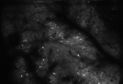
In the new study, the researchers repeatedly showed mice one of two images, each consisting of a different checkerboard pattern of gray and dappled black and white squares. Between images, the mice spent a minute looking at a gray screen. The team simultaneously recorded activity from around 7,000 neurons in the visual cortex.
The researchers found that when a mouse looked at an image, the neurons fired in a specific pattern, and the patterns were different enough to discern image one from image two. More important, when a mouse looked at the gray screen between images, the neurons sometimes fired in a similar, but not identical, pattern, as when the mouse looked at the image, a sign that it was daydreaming about the image. These daydreams occurred only when mice were relaxed, characterized by calm behavior and small pupils.
Unsurprisingly, mice daydreamed more about the most recent image — and they had more daydreams at the beginning of the day than at the end, when they had already seen each image dozens of times.
Between images, mice spent a minute looking at a gray screen. During this time, neurons in the visual cortex of the brain, shown here, occasionally fired in a pattern similar to one seen when the mice were looking at an image, suggesting that mice were daydreaming about the image.
But what the researchers found next was completely unexpected.
Throughout the day, and across days, the activity patterns seen when the mice looked at the images changed — what neuroscientists call “representational drift.” Yet this drift wasn’t random. Over time, the patterns associated with the images became even more different from each other, until each involved an almost entirely separate set of neurons. Notably, the pattern seen during a mouse’s first few daydreams about an image predicted what the pattern would become when the mouse looked at the image later.
“There’s drift in how the brain responds to the same image over time, and these early daydreams can predict where the drift is going,” Andermann said.
Finally, the researchers found that the visual cortex daydreams occurred at the same time as replay activity occurred in the hippocampus, suggesting that the two brain regions were communicating during these daydreams.
To sit, perchance to daydream
Based on the results of the study, the researchers suspect that these daydreams may be actively involved in brain plasticity.
“When you see two different images many times, it becomes important to discriminate between them. Our findings suggest that daydreaming may guide this process by steering the neural patterns associated with the two images away from each other,” Nguyen said, while noting that this relationship needs to be confirmed.
Nguyen added that learning to differentiate between the images should help the mouse respond to each image with more specificity in the future.
These observations align with a growing body of evidence in rodents and humans that entering a state of quiet wakefulness after an experience can improve learning and memory.
Next, the researchers plan to use their imaging tools to visualize the connections between individual neurons in the visual cortex and to examine how these connections change when the brain “sees” an image.
“We were chasing this 99 percent of unexplored brain activity and discovered that there’s so much richness in the visual cortex that nobody knew anything about,” Andermann said.
Whether daydreams in people involve similar activity patterns in the visual cortex is an open question, and the answer will require additional experiments. However, there is preliminary evidence that an analogous process occurs in humans when they recall visual imagery.
For the researchers, the results of their study and others suggest that it may be important to make space for moments of quiet waking that lead to daydreams. For a mouse, this may mean taking a pause from looking at a series of images and, for a human, this could mean taking a break from scrolling on a smartphone.
“We feel pretty confident that if you never give yourself any awake downtime, you’re not going to have as many of these daydream events, which may be important for brain plasticity,” Andermann said.
The brain is the most energy-demanding organ, which uses about half of all the sugar energy in the body.
This is Part 1 in the series “The Ultimate Guide to Kicking Sugar”
In this series, we will explore the good and bad sweeteners, uncover the unexpected outcomes of cutting out sugar, and discover the ultimate way to achieve this.
Our brains often instinctively crave sugar. It could be a slice of cake during times of stress, a bar of chocolate when bored, or a sweetened coffee when needing a pick-me-up. The inability to quit sugar may not stem from a lack of willpower but rather from not fully grasping the nature of sugar and not finding the most effective methods to quit.
Sweet Cravings: The Instinct for Survival and Growth
“Sugar is very important for our body and our brain. And I think this is where a lot of the difficulty (in cutting out sugar) lies,” Jessica Russo, a clinical psychologist from Philadelphia, told The Epoch Times during an interview.
Sugar serves as the primary energy source for every cell in our body and much of the food we eat is broken down into various sugars.
“The brain is the most energy-demanding organ, which uses about half of all the sugar energy in the body.”
“We’re biologically driven toward sweet foods,” as this is a survival mechanism, Ms. Russo said, explaining that in nature, sweet-tasting foods are generally healthy, while toxic foods may taste bitter, and spoiled or rotten foods may taste sour, both of which lack sweetness.
Therefore, when we taste something sweet, our brains signal, “Oh, this is good!”
Besides helping us identify safe food, sweetness also plays a role in human survival and growth
“We see babies being born with the ability to detect sweet taste and to prefer it,” Julie A. Mennella, a scientist at the Monell Chemical Senses Center in Philadelphia, during an interview. It indicates that sweetness is associated with the quality of breast milk, which can attract infants to suckle.
There is evidence to suggest that children’s preference for sweet foods may be linked to their higher caloric needs during the growth stage, which typically continues from infancy until the end of puberty, she told The Epoch Times.
The Effect of Sugar on Our Brains
When we consume sugar, the receptors on our tongues send sweet signals to the brain, triggering the release of dopamine, which can induce feelings of joy and happiness.
“We taste with our brains,” Ms. Mennella explained. Sweetness makes us feel good because these signals are sent to various parts of the brain, many of which are associated with rewards.
“The brain pathways used are significant for pleasure, memory, and reward,” she said.
This means that when we engage in activities that trigger dopamine release, we experience joy, form memories, and look forward to doing it again.
“In the brain, it has very specific actions and is the most important molecule in the brain that’s involved in bringing about well-being,” said Kenneth Blum, a renowned scientist with a doctorate in neuropharmacology.
Dopamine can also counteract stress, said Mr. Blum, who is a professor at the Western University of Health Sciences’ Graduate School of Biomedical Science, and a part-time professor at the University of Vermont and Wright University.
“When you have stress, the dopamine is released 100 times above the normal rate.” It can block the action of stress hormones such as adrenaline.
However, Mr. Blum emphasized the importance of maintaining a balance for this crucial molecule; otherwise, the brain could suffer severe negative consequences.
Many people are unaware that excessive sugar consumption can lead to consequences very similar to drug abuse.
Mr. Blum explained that excessive sugar consumption can trigger acute dopamine release.
“It’s like abusing alcohol or other drugs of abuse,” he said.
Over time, that can result in a chronic decrease in dopamine levels. Consequently, individuals may seek larger quantities of sugar to experience the same level of pleasure, eventually leading to an addictive state where they consume more and more.
When you consume a large amount of refined sugar, “your brain lights up like a pinball machine due to the intense release of dopamine,” said James DiNicolantonio, a cardiovascular research scientist and doctor of pharmacy at Saint Luke’s Mid America Heart Institute in Kansas City, Missouri.
When ingesting refined sugar, the sweet receptors signal the brain’s reward system more effectively than when eating fruit. The levels of dopamine released by the brain far exceed what we can handle.
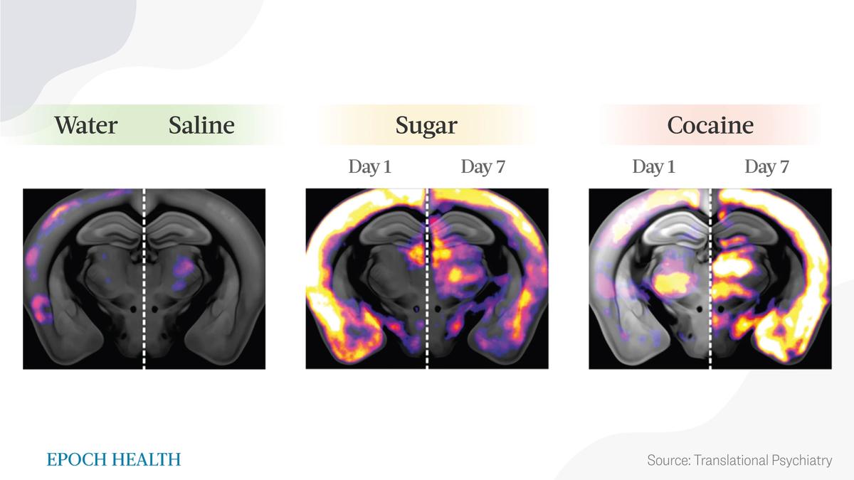
In 2023, a study published in the journal Translational Psychiatry revealed that when a mouse drinks water or is injected with saline, the brain remains relatively calm. However, when administered a sucrose solution or injected with cocaine, multiple regions of the brain’s neurons become activated (highlighted in the image). Multiple brain regions that respond to sugar signals also exhibit responses to cocaine signals.
Activation of brain neurons in specific regions upon sucrose and cocaine administration.
“Our research shows how similarly both additive and nonadditive rewards are processed by our brains, both on the whole-brain scale and on a cellular level,” said Anna Beroun, the study’s lead author and the head of the Laboratory of Neuronal Plasticity at the Nencki-EMBL Center of Excellence for Neural Plasticity and Brain Disorders (BRAINCITY) of the Nencki Institute of Experimental Biology of the Polish Academy of Sciences in Warsaw, Poland.
“Sugar/food becomes addictive if we value it over other rewards.”
Is Sugar More Addictive Than Drugs?
Sugar is irresistibly alluring, not only because it stimulates the brain to produce dopamine, which brings joy, but also because it triggers the production of endogenous opioids in the brain, which can lead to addiction and dependence.
Mr. Blum said that the brain has glucose receptors, and when they are stimulated by sugar, it triggers a series of signaling pathways that ultimately lead to the production of addictive substances. This mechanism is inherently present “so that if you abuse sugar, you’re going to order the brain–reward circuitry in a negative way, as if you use heroin.”
An experiment revealed that mice fed large amounts of sugar intermittently exhibited withdrawal symptoms when injected with a drug that blocks opioids. These symptoms included teeth chattering, forepaw tremors, and headshakes.
Sugar’s effect on the brain not only shares similarities with drugs but also, in certain circumstances, is even more alluring.
Over the years, French researchers have conducted a series of animal experiments, with the results revealing that when given the choice between cocaine and sucrose, rodents consistently preferred sucrose over cocaine. This preference held even for mice previously addicted to cocaine before the experiments.
“When we over-consume sugar, there is a release of dopamine and endogenous opioids that cause a ‘high,’ but then we get a ‘low.’ If we do this over a prolonged period of time, this can lead to dependency on sugar, especially in those who are vulnerable,” said Mr. DiNicolantonio, summarizing the addictive mechanism of sugar.
When there is a deficiency of dopamine and endogenous opioids, one may feel sad, confused, sluggish, and unable to concentrate, all of which can further drive the desire to consume more sugar.
Additionally, numerous human experiments have demonstrated the link between sugar and addiction.
For example, a prospective observational study published in Addiction Biology in 2021 revealed that a significant proportion of individuals with alcohol use disorder (40 percent) experienced an increased craving for sugar during their inpatient alcohol detoxification.
Additionally, a study published in the journal Addiction showed that children with a family history of alcoholism and depression were likelier to prefer intense sweetness. On average, these children opted for water with a sucrose concentration of 24 percent, equivalent to about 14 teaspoons of sugar in a glass of water—more than twice the sugar concentration found in regular soda water.
In contrast, children without such familial backgrounds preferred water with a sucrose concentration of 18 percent.
More from this series
Know Your Sugars: The Key to Overcoming Addiction
Sugar and the brain share an innate strong connection. Unfortunately, modern diets are filled with highly refined sugars that evoke drug-like allure. In fact, the sweetness we consume today differs significantly from what our ancestors once had.
Ms. Russo vividly described the body and brain’s conflicting views on sugar with a lively scene, noting that our bodies resist certain sugars while are more receptive to others.
She says, “The brain says, ‘We need sugar; we must have sugar; we can’t survive without it.’ On the other hand, the body disagrees, saying, ‘We don’t like all types of sugar.’”
There is an ancient Chinese saying: “If you know the enemy and know yourself, you need not fear the result of a hundred battles.” To quit sugar, one must first understand sugar. However, the truth is some sugars and sweet substances are natural and even beneficial to the body.
Obesity is a widespread health issue, affecting more than two billion people globally. Studies continue to show that too much weight is one of the leading causes of chronic health problems — from diabetes to heart disease. Now, a new study finds obesity may have more to do with the brain than it does with someone’s diet.
Researchers from the Baylor College of Medicine say that molecular mechanisms of early brain development likely play a key role in who ends up becoming obese. They add that previous studies have suggested that genes connected to obesity start expressing themselves in the developing brain after childbirth.
The new study looked at epigenetic development in mice, which is the system of molecular bookmarking that determines which genes are active in different cells.
“Decades of research in humans and animal models have shown that environmental influences during critical periods of development have a major long-term impact on health and disease,” says corresponding author Dr. Robert Waterland, a professor of pediatrics-nutrition and a member of the USDA Children’s Nutrition Research Center at Baylor, in a university release. “Body weight regulation is very sensitive to such ‘developmental programming,’ but exactly how this works remains unknown.”
“In this study we focused on a brain region called the arcuate nucleus of the hypothalamus, which is a master regulator of food intake, physical activity and metabolism,” adds first author Dr. Harry MacKay. “We discovered that the arcuate nucleus undergoes extensive epigenetic maturation during early postnatal life. This period is also exquisitely sensitive to developmental programming of body weight regulation, suggesting that these effects could be a consequence of dysregulated epigenetic maturation.”
Male and female brains develop differently
To uncover obesity’s link to the growing brain, the team conducted a genome-wide analyses of both DNA methylation (an important epigenetic tag) and gene expression. They studied these processes both before and after a critical window in developmental programming for body weight among postnatal mice — meaning right after birth.
“One of our study’s biggest strengths is that we studied the two major classes of brain cells, neurons and glia,” MacKays explains. “It turns out that epigenetic maturation is very different between these two cell types.”
“Our study is the first to compare this epigenetic development in males and females,” Waterland says. “We were surprised to find extensive sex differences. In fact, in terms of these postnatal epigenetic changes, males and females are more different than they are similar. And, many of the changes occurred earlier in females than in males, indicating that females are precocious in this regard.”
To their surprise, the team discovered clear similarities between epigenetic data in mice and human genetic changes linked to obesity. Genomic regions targeted for epigenetic maturation in the mouse arcuate nucleus stunningly matched up with human brain regions connected to body mass index.
“These associations suggest that obesity risk in humans is determined in part by epigenetic development in the arcuate nucleus,” MacKay reports. “Our results provide new evidence that developmental epigenetics is likely involved in both early environmental and genetic influences on obesity risk. Accordingly, prevention efforts targeting these developmental processes could be the key to stopping the worldwide obesity epidemic.”


