
A groundbreaking development in bone regeneration has been achieved with the creation of a piezoelectric scaffold by a KAIST-led research team. Utilizing hydroxyapatite (HAp) to generate electrical signals under pressure, this scaffold mimics the natural environment of bone tissue, showing promising results in promoting bone growth through both laboratory and animal studies. This advancement opens new pathways for biomaterial design and the understanding of bone regeneration processes.
Bone regeneration is a complicated procedure, and the current approaches for facilitating this regeneration, such as grafts and the application of growth factors, encounter challenges like elevated expenses. However, a breakthrough has been made with the creation of a piezoelectric material that has the capability to enhance bone tissue development.
A KAIST research team led by Professor Seungbum Hong from the Department of Materials Science and Engineering (DMSE) announced on January 25 the development of a biomimetic scaffold that generates electrical signals upon the application of pressure by utilizing the unique osteogenic ability of hydroxyapatite (HAp). This research was conducted in collaboration with a team led by Professor Jangho Kim from the Department of Convergence Biosystems Engineering at Chonnam National University.
HAp is a basic calcium phosphate material found in bones and teeth. This biocompatible mineral substance is also known to prevent tooth decay and is often used in toothpaste.
Breakthrough in Bone Regeneration
Previous studies on piezoelectric scaffolds confirmed the effects of piezoelectricity on promoting bone regeneration and improving bone fusion in various polymer-based materials, but were limited in simulating the complex cellular environment required for optimal bone tissue regeneration. However, this research suggests a new method for utilizing the unique osteogenic abilities of HAp to develop a material that mimics the environment for bone tissue in a living body.
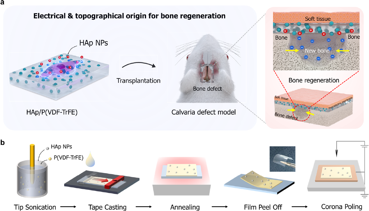
Figure 1. Design and characterization of piezoelectrically and topographically originated biomimetic scaffolds. (a) Schematic representation of the enhanced bone regeneration mechanism through electrical and topographical cues provided by HAp-incorporated P(VDF-TrFE) scaffolds. (b) Schematic diagram of the fabrication process. Credit: KAIST Materials Imaging and Integration Laboratory
The research team developed a manufacturing process that fuses HAp with a polymer film. The flexible and free-standing scaffold developed through this process demonstrated its remarkable potential for promoting bone regeneration through in-vitro and in-vivo experiments in rats.
Understanding the Principles of Bone Regeneration
The team also identified the principles of bone regeneration that their scaffold is based on. Using atomic force microscopy (AFM), they analyzed the electrical properties of the scaffold and evaluated the detailed surface properties related to cell shape and cell skeletal protein formation. They also investigated the effects of piezoelectricity and surface properties on the expression of growth factors.
Figure 1. Design and characterization of piezoelectrically and topographically originated biomimetic scaffolds. (a) Schematic representation of the enhanced bone regeneration mechanism through electrical and topographical cues provided by HAp-incorporated P(VDF-TrFE) scaffolds. (b) Schematic diagram of the fabrication process. Credit: KAIST Materials Imaging and Integration Laboratory
Professor Hong from KAIST’s DMSE said, “We have developed a HAp-based piezoelectric composite material that can act like a ‘bone bandage’ through its ability to accelerate bone regeneration.” He added, “This research not only suggests a new direction for designing biomaterials, but is also significant in having explored the effects of piezoelectricity and surface properties on bone regeneration.”
. DOI: 10.1038/s41589-023-01525-w")

