Unlocking Bacterial Self-Destruction to Combat Infections


A team of researchers has discovered a method to activate a bacterial defense system, known as CBASS, to self-destruct and prevent the spread of viruses among bacteria, potentially offering a new way to manage bacterial infections and combat antibiotic resistance. Credit: SciTechDaily.com

Researchers unveil how the self-killing activity of bacteria can be harnessed in the fight against antibiotic resistance.

Scientists at the Icahn School of Medicine at Mount Sinai have identified a new approach to controlling bacterial infections. The findings were described in the February 6 online issue of Nature Structural & Molecular Biology.

The team found a way to turn on a vital bacterial defense mechanism to fight and manage bacterial infections. The defense system, called cyclic oligonucleotide-based antiphage signaling system (CBASS), is a natural mechanism used by certain bacteria to protect themselves from viral attacks. Bacteria self-destruct as a means to prevent the spread of virus to other bacterial cells in the population.

CBASS Defense Mechanism Explored

“We wanted to see how the bacterial self-killing CBASS system is activated and whether it can be leveraged to limit bacterial infections,” says co-senior author Aneel Aggarwal, PhD, Professor of Pharmacological Sciences at Icahn Mount Sinai. “This is a fresh approach to tackling bacterial infections, a significant concern in hospitals and other settings. It’s essential to find new tools for fighting antibiotic resistance. In the war against superbugs, we need to constantly innovate and expand our toolkit to stay ahead of evolving drug resistance.”

According to a 2019 report by the Centers for Disease Control and Preventionmore than 2.8 million antimicrobial-resistant infections occur in the United States each year, with over 35,000 people dying as a result.

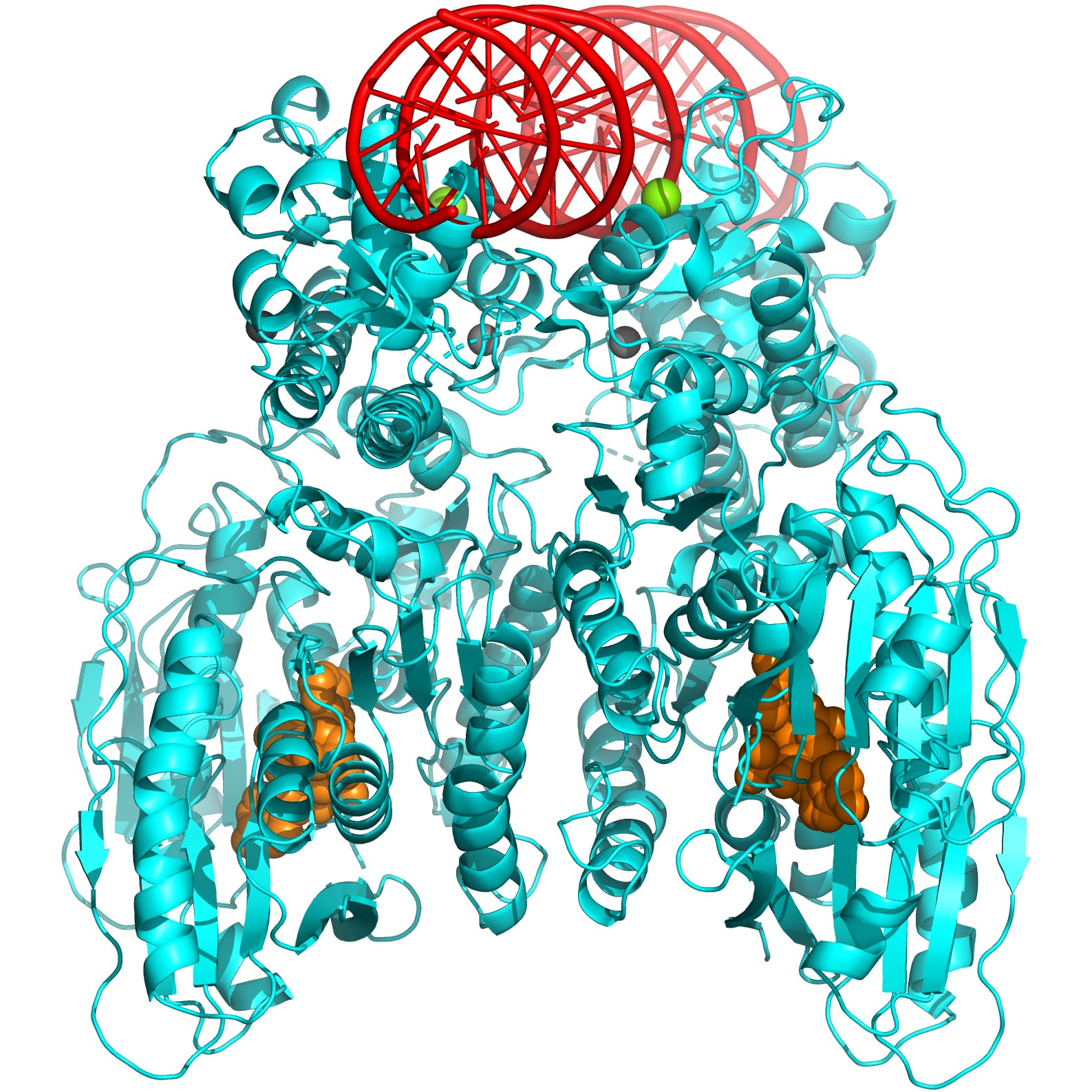
Icahn Mount Sinai researchers unveil how the self-killing activity of bacteria can be used in the fight against antibiotic resistance. Above: 3-D structure of CBASS Cap5 protein tetramer (shown in cyan) formed upon binding to the cyclic dinucleotide (shown in orange) to destroy bacteria’s own DNA (model, shown in red). Essential magnesium ions for DNA cleavage are shown in green.

Innovative Strategies Against Superbugs

As part of the experiments, the researchers studied how “Cap5,” or CBASS-associated protein 5, is activated for DNA degradation and how it could be used to control bacterial infections through a combination of structural analysis and various biophysical, biochemical, and cellular assays. Cap5 is a key protein that becomes activated by cyclic nucleotides (small signaling molecules) to destroy the bacterial cell’s own DNA.

“In our study, we started by identifying which of the many cyclic nucleotides could activate the effector Cap5 of the CBASS system,” says co-senior author Olga Rechkoblit, PhD, Assistant Professor of Pharmacological Sciences at Icahn Mount Sinai. “Once we figured that out, we looked closely at the structure of Cap5 when it’s bound to these small signaling molecules. Then, with expert help from Daniela Sciaky, PhD, a researcher at Icahn Mount Sinai, we showed that by adding these special molecules to the bacteria’s environment, these molecules could potentially be used to eliminate the bacteria.”

Overcoming Technical Challenges

The researchers found that determining the structure of Cap5 with cyclic nucleotides posed a technical challenge, requiring expert help from Dale F. Kreitler, PhD, AMX Beamline Scientist at Brookhaven National Laboratory. It was achieved by using micro-focused synchrotron X-ray radiation at the same facility. Micro-focused synchrotron X-ray radiation is a type of X-ray radiation that is not only produced using a specific type of particle accelerator (synchrotron) but is also carefully concentrated or focused on a tiny area for more detailed imaging or analysis.

Future Directions

Next, the researchers will explore how their discoveries apply to other types of bacteria and assess whether their method can be used to manage infections caused by various harmful bacteria

Unlocking the Genetic Puzzle of Obesity Across Sexes and Ages


A study has identified genes that influence obesity risk differently across sexes and age groups, offering new insights into the biological pathways of obesity. These discoveries underscore the importance of considering sex and age in obesity research and could lead to new treatments.

Researchers have discovered genes that impact obesity risk differently in men and women and across various ages, revealing potential new pathways for understanding and treating obesity.

From influencing how our body stores fat to how our brain regulates appetite, hundreds of genes, along with environmental factors, collectively determine our weight and body size. Now, researchers add several genes, that appear to affect obesity risk in certain sexes and ages, to that list. The study, published in the journal Cell Genomics, may shed light on new biological pathways that underlie obesity and highlight how sex and age contribute to health and disease.

The Impact of Sex and Age on Obesity Risk

“There are a million and one reasons why we should be thinking about sex, age, and other specific mechanisms rather than just lumping everyone together and assuming that disease mechanism works the same way for everyone,” says senior author John Perry, a geneticist and professor at the Wellcome-MRC Institute of Metabolic Science, University of Cambridge, U.K. “We’re not expecting people to have completely different biology, but you can imagine things like hormones and physiology can contribute to specific risks.”

To untangle sex’s role in obesity risk, the research team sequenced the exome—the protein-coding part of the genome—of 414,032 adults from the UK Biobank study. They looked at variants, or mutations, within genes associated with body mass index (BMI) in men and women, respectively. Based on height and weight, BMI is an estimated measurement of obesity. The search turned up five genes influencing BMI in women and two in men.

Researchers identified age-specific and sex-specific obesity genes by looking into the genome of 414,032 people from the UK.

Among them, faulty variants of three genes—DIDO1, PTPRG, and SLC12A5—are linked to higher BMI in women, up to nearly 8 kg/m² more, while having no effect on men. Over 80% of the women with DIDO1 and SLC12A5 variants had obesity, as approximated by their BMI. Individuals carrying DIDO1 variants had stronger associations with higher testosterone levels and increased waist-to-hip ratio, both risk indicators for obesity-related complications like diabetes and heart disease. Others with SLC12A5 variants had higher odds of having type 2 diabetes compared with non-carriers. These findings highlight previously unexplored genes that are implicated in the development of obesity in women but not men.

Gene Variants and Their Specific Effects

Perry and his colleague then repeated their method to look for age-specific factors by searching for gene variants associated with childhood body size based on participants’ recollections. They identified two genes, OBSCN and MADD, that were not previously linked to childhood body size and fat. While carriers of OBSCN variants had higher odds of having higher weight as a child, MADD variant carriers were associated with smaller body sizes. In addition, the genetic variants acting on MADD had no association with adult obesity risk, highlighting age-specific effects on body size.

“What’s quite surprising is that if you look at the function of some of these genes that we identified, several are clearly involved in DNA damage response and cell death,” says Perry. Obesity is a brain-related disorder, whereas biological and environmental factors act to influence appetite. “There’s currently no well-understood biological paradigm for how DNA damage response would influence body size. These findings have given us a signpost to suggest variation in this important biological process may play a role in the etiology of obesity.”

Implications for Future Research

Next, the research team hopes to replicate the study in a larger and more diverse population. They also plan to study the genes in animals to peer into their function and relationship with obesity.

“We’re at the very earliest stages of identifying interesting biology,” says Perry. “We hope the study can reveal new biological pathways that may one day pave the way to new drug discovery for obesity.”

Circadian Protein Helps Plants Manage Short- and Long-Term Stresses


Bioluminescent image of Arabidopsis seedlings expressing circadian clock reporter genes in response to water stress.”

Researchers at the Keck School of Medicine of the University of Southern California, have uncovered new insights into how plants regulate their responses to stress, which may aid future efforts to engineer crops that are better able to thrive in the face of drought or high soil salinity levels associated with climate change.

The investigators found that plants use their circadian clocks to respond to changes in external water and salt levels throughout the day. That same circuitry—an elegant feedback loop controlled by a protein known as ABF3—also helps plants adapt to extreme conditions such as drought.

“The bottom line is plants are stuck in place,” said research lead Steve A. Kay, PhD, University and Provost Professor of Neurology, biomedical engineering and quantitative Computational Biology at the Keck School of Medicine and director of the USC Michelson Center for Convergent Bioscience. “They can’t run around and grab a drink of water. They can’t move into the shade when they want to or away from soil that has excess salt. Because of that, they’ve evolved to use their circadian clocks to exquisitely measure and adapt to their environment.”

Kay and colleagues reported on their results in the journal Proceedings of the National Academy of Sciences (PNAS), in a paper titled “The interplay between the circadian clock and abiotic stress responses mediated by ABF3 and CCA1/LHY,” in which they concluded that their findings “… offer valuable insights for developing genetic and molecular approaches to enhance plant resilience in the face of climate change.”

Climate change is a global concern for all life on our planet, including humans and plants, the authors noted. “As sessile organisms, plants must constantly adapt to the environment and cope with various stresses, such as drought, salinity, and extreme temperatures,” they wrote. “Understanding how plants respond to abiotic stresses is crucial for addressing this challenge.” One of the most crucial mechanisms in plants is the ABA signaling pathway, the team further explained. “ABA is a plant hormone that plays a key role in regulating responses to abiotic stress such as drought, high salinity, and high temperatures.”

The newly reported study builds on a long line of research from Kay’s lab on the role of circadian clock proteins in both plants and animals. “As the internal timekeeping machinery, the circadian clock enables plants to synchronize with daily and seasonal environmental changes and directly controls many developmental processes throughout the life cycle,” the authors further explained. In the model plant organism Arabidopsis, the circadian clock includes what the researchers described as “… a plethora of oscillating proteins” that are expressed and function in an orchestrated manner. “Studies in crops also revealed the important role of the circadian clock in regulating stress responses,” they added.

Clock proteins regulate biological changes over the course of the day, and may provide a shrewd solution to an ongoing challenge in crop engineering. Creating drought-resistant plants is difficult because plants respond to stress by slowing their own growth and development—an overblown stress response means an underperforming plant. “There’s a delicate balance between boosting a plant’s stress tolerance while maximizing its growth and yield,” Kay said. “Solving this challenge is made all the more urgent by climate change.”

Previous plant biology research showed that clock proteins regulate about 90% of genes in plants and are central to their responses to temperature, light intensity and day length, including seasonal changes that determine when they flower. But one big outstanding question was whether and how clock proteins control the way plants handle changing water and soil salinity levels.

To explore the link, Kay and his team studied Arabidopsis, which is commonly used in research because it is small, has a rapid life cycle, a relatively simple genome and shares common traits and genes with many agricultural crops. They created a library of all of the more than 2000 Arabidopsis transcription factors, which are proteins that control the way genes are expressed under different circumstances. Transcription factors can provide key insights about regulation of biological processes. The researchers then built a data analysis pipeline to analyze each transcription factor and search for associations.

“We got a really big surprise: that many of the genes the clock was regulating were associated with drought responses,” Kay said, particularly those controlling the hormone abscisic acid, a type of stress hormone that plants produce when water levels are very high or very low. The analysis revealed that abscisic acid levels are controlled by clock proteins as well as the transcription factor ABF3 in what Kay calls a “homeostatic feedback loop.”

The authors wrote, “We demonstrate how the circadian clock influences ABF3 expression, which in turn delivers stress signals to core clock genes and adjusts the circadian period in response to stress … Our study reveals a unique mechanism of the interplay between the circadian clock and abiotic stress responses mediated by ABF3 and CCA1/LHY.”

The findings indicated that during the course of a day, clock proteins regulate ABF3 to help plants respond to changing water levels, then ABF3 feeds information back to clock proteins to keep the stress response in check. That same loop helps plants adapt when conditions become extreme, for instance during a drought. Genetic data also revealed a similar process for handling changes in soil salinity levels. “Specifically, we found that CCA1 and LHY regulate the expression of ABF3 under diel conditions, as well as seed germination under salinity. Conversely, ABF3 controls the expression of core clock genes and orchestrates the circadian period in a stress-responsive manner,” they stated. “Our research also uncovers a unique function of ABF3 in modulating the circadian clock. ABF3 controls the expression of core clock genes and modulates the circadian period in a stress-responsive manner.”

Kay added, “What’s really special about this circuit is that it allows the plant to respond to external stress while keeping its stress response under control, so that it can continue to grow and develop.”

The authors claim that elucidating the regulatory pathways and feedback loops involving CCA1, LHY, and ABF3, has offered up a deeper understanding of how plants coordinate their physiological and developmental responses to environmental challenges. “Understanding the intricate relationship between the circadian clock and abiotic stress response at the molecular level may aid in the development of targeted strategies to enhance plant resilience in the face of climate change,” they concluded.

The findings point to two new approaches that may help boost crop resilience. For one, agricultural breeders can search and select for naturally occurring genetic diversity in the circadian ABF3 circuit that gives plants a slight edge in responding to water and salinity stress. Even a small increase in resilience could substantially improve crop yield on a large scale. Kay and colleagues also plan to explore a genetic modification approach, using CRISPR to engineer genes that promote ABF3 in order to design highly drought-resistant plants. “This could be a significant breakthrough in thinking about how to modulate crop plants to be more drought resistant,” Kay said.

Immune Response and Not Acute Viral Infections Found Responsible for Neurological Damage.


Researchers using a mouse model of the Zika virus (ZIKV) infection discovered that the severity of neurological disease did not correlate with brain ZIKV titers, but rather with infiltration of bystander activated NKG2D+CD8+ T cells. [Westend61/Getty Images]

Scientists at McMaster University report that they have found that it is the immune system response, and not acute viral infections like Zika or COVID-19, that is directly responsive for neurological damage. They published their study “Bystander activated CD8+ T cells mediate neuropathology during viral infection via antigen-independent cytotoxicity” in Nature Communications.

“Although many viral infections are linked to the development of neurological disorders, the mechanism governing virus-induced neuropathology remains poorly understood, particularly when the virus is not directly neuropathic. Using a mouse model of Zika virus (ZIKV) infection, we found that the severity of neurological disease did not correlate with brain ZIKV titers, but rather with infiltration of bystander activated NKG2D+CD8+ T cells,” wrote the investigators.

“Antibody depletion of CD8 or blockade of NKG2D prevented ZIKV-associated paralysis, suggesting that CD8+ T cells induce neurological disease independent of TCR signaling. Furthermore, spleen and brain CD8+ T cells exhibited antigen-independent cytotoxicity that correlated with NKG2D expression. Finally, viral infection and inflammation in the brain was necessary but not sufficient to induce neurological damage.

“We demonstrate that CD8+T cells mediate virus-induced neuropathology via antigen-independent, NKG2D-mediated cytotoxicity, which may serve as a therapeutic target for treatment of virus-induced neurological disease.”

“We were interested in trying to understand why so many viral infections are associated with neurological diseases,” said Elizabeth Balint, a PhD student at McMaster. “Our evidence suggests that it’s not the virus itself that causes the damage, but a unique population of T cells, which are part of the immune system, that are actually responsible for the damage.”

A team focused on Zika

To come to this conclusion, the McMaster team focused on Zika virus. During laboratory testing, researchers, as expected, found T cells that were specific for Zika and designed to eliminate infected cells. They found something else, too.

“What was interesting in our study is that although we did find some T cells specific for Zika, we identified cells that weren’t functioning like a normal T cell and were killing cells that weren’t infected with Zika,” added Balint.

According to the researchers, this aggressive behavior of these NKG2D+CD8+ T cells is responsible for neurological damage suffered from infections beyond just Zika, like COVID-19 and even septic shock. The response is the result of the body producing large amounts of cytokines, which in moderation help to coordinate the body’s response in battling an infection or injury by telling immune cells where to go and what to do when they arrive.

“If our body’s immune cells overreact and over produce inflammatory cytokines, this condition will lead to non-specific activation of our immune cells which in turn leads to collateral damage. This can have severe consequences if it happens in the brain,” explained, Ali Ashkar, PhD, DVM, a professor with the department of medicine and the Canada Research Chair in Natural Immunity and NK Cell Function.

The discovery offers scientists a new target for treatments of neurological diseases sparked by acute viral infections. In fact, Balint has already found a treatment that holds promise.

“Elizabeth has experimented with an antibody that can completely block and treat devastating neurotoxicity in the animal model, which is already in clinical trials for different uses in humans,” pointed out Ashkar.

Balint hopes to continue her work towards finding a treatment that would be effective in humans.

“There are a few different other viruses we’re interested in studying, which will aid us in creating the best treatment options,” Balint said.

Warning Issued After Researchers Link Energy Drinks to Suicidal Thoughts in Children


Energy drinks could pose a risk to young brains, said UK researchers.

New research revealed that energy drinks could pose a greater risk to children’s and younger people’s brains than previously thought.

Those who consumed energy drinks were shown to have a higher risk of mental health problems such as depression, suicidal thoughts, attention-deficit hyperactivity disorder (ADHD), and anxiety, according to a study from Fuse, the Centre for Translational Research in Public Health at Teesside University, and Newcastle University in the UK. It was published in the Public Health journal last month.

Researchers said they looked at data from 57 studies of more than 1.2 million children and younger people from more than 21 countries to come up with their conclusions.

It found that boys consumed more energy drinks than girls, while “many studies” reported an association between energy drink consumption and alcohol use, binge drinking, and smoking, as well as other substance use.

“Additional health effects noted in the updated review included increased risk of suicide, psychological distress, attention-deficit hyperactivity disorder symptoms, depressive and panic behaviors, allergic diseases, insulin resistance, dental caries, and erosive tooth wear,” an abstract of the paper said.

Regarding the impacts on mental health, it found that “frequent” drinking of energy drinks “was associated with suicide attempts and severe stress,” while there “were also higher rates of suicide ideation and attempts with [energy drink] intake greater than once per day.

“Longitudinal analysis reported that [energy drink] consumption was related to increased ADHD inattention, conduct disorder, depressive,  and panic symptoms,” it continued to say.

A co-author, Shelina Visram, with Newcastle University, said in a news release that she is “deeply concerned about the findings that energy drinks can lead to psychological distress and issues with mental health.”

“These are important public health concerns that need to be addressed,” she added. “There has been policy inaction on this area despite [UK] government concern and public consultations. It is time that we have action on the fastest growing sector of the soft drink market.

The researchers, who are based in the UK, also called on the government to either ban or restrict the energy drinks for younger people and children.

“This evidence suggests that energy drinks have no place in the diets of children and young people,” author Amelia Lake, professor of public health nutrition at Teesside University, told Fox News on Thursday. “Policymakers should follow the example from countries that have placed age restrictions on their sales to children.”

It’s because, their study shows, the researchers have “found an even greater list of mental and physical health outcomes associated with children and young people consuming energy drinks,” she said.

“We repeated [the review] only to find an ever-growing evident space that suggests the consumption of these drinks is associated with negative health outcomes,” Ms. Lake continued.

Several countries have already tried to regulate energy drinks, including bans on sales to minors in Latvia and Lithuania. Other countries such as Finland and Poland are also reportedly looking to ban the products from being sold to people under the age of 18.

https://www.ganjingworld.com/embed/1fi193kb1cd30rvSOhrgtYGQQ1r11c

The study, meanwhile, drew a response from UK officials. A spokesperson for the UK Department of Health and Social Care told the BBC that “we consulted on a proposal to end the sale of energy drinks to children under 16 in England, and will set out our full response in due course” and that “in the meantime, many larger retailers and supermarkets have voluntarily introduced a ban on the sale of energy drinks to children under 16.”

But several years ago, Christopher Snowdon, the head of Lifestyle Economics at the UK-based Institute of Economic Affairs, found that such bans unfairly target teenagers and said there is a lack of evidence to link the drinks to negative behaviors.

“The current scientific evidence alone is not sufficient to justify a measure as prohibitive as a statutory ban on the sale of energy drinks to children,” he wrote in an article published in 2020.

Breakthrough Molecule RvT4 Reverses Inflammation and Clears Arterial Blockages in Rheumatoid Arthritis Patients.


Researchers have discovered that RvT4, a molecule, significantly boosts the body’s defenses against atherosclerosis in rheumatoid arthritis patients by enhancing macrophages’ ability to reduce inflammation and clear vascular blockages. This finding opens up new avenues for treating individuals with rheumatoid arthritis, who are at a heightened risk for cardiovascular diseases.

A new study reveals that RvT4 improves macrophage function to fight atherosclerosis in rheumatoid arthritis, suggesting new treatment pathways.

Scientists from Queen Mary University of London have found that the molecule RvT4 enhances the body’s natural defenses against atherosclerosis (hardening of the arteries) in patients with rheumatoid arthritis.

Studies in mice undertaken by researchers from Queen Mary University of London’s William Harvey Research Institute and Centre for Inflammation and Therapeutic Innovation, and funded by the European Research Council (ERC) and Barts Charity, shows that increasing levels of the RvT4 molecule in the body improves the ability of the body’s own defense mechanisms [macrophages] to reduce local inflammation and remove blockages in blood vessels.

This breakthrough in understanding the processes involved could lead to better treatments for people who have rheumatoid arthritis (RA), and who are at higher risk of developing cardiovascular disease.

The Link Between Rheumatoid Arthritis and Cardiovascular Disease

Rheumatoid arthritis (RA) is the most common form of inflammatory arthritis in the UK and affects around 1% of the population. Approximately 10,000 people receive a diagnosis of RA every year. Alongside the more widely-known symptoms of joint inflammation, people with the condition are also twice as likely as others to develop blood vessel disease. This can lead to serious complications and an increased risk of premature death.

One type of blood vessel disease seen in people with RA is atherosclerosis, which is caused by a build-up of fatty material called ‘plaque’ along the artery walls. This build-up causes the arteries to harden and narrow, making it more difficult to circulate blood around the body. These blockages can also break free, causing heart attacks and strokes.

Understanding the reasons why RA patients are at increased risk of these cardiovascular problems is critical in developing better treatments for this group and others.

Insights Into RvT4’s Role in Combatting Atherosclerosis

To gain a better understanding of the causes of blood vessel disease in patients with RA, researchers explored the role of a group of molecules called 13-series resolvins (RvTs). In experimental arthritis the levels of one of these molecules, RvT4, are markedly reduced, a phenomenon that associates with a higher degree of blood vessel disease. This study was designed to explore why this might be the case.

The study found that treating arthritic mice with RvT4 reduced blood vessel inflammation by re-programming macrophages — a group of white blood cells that accumulate in the diseased vessels — to release stored lipids. Researchers observed that these lipids were preventing the macrophage from carrying out their usual work of clearing dead cells and reducing localized inflammation in blood vessels.

Once freed of their lipid burden, the macrophages were able to move and work much more effectively to reduce the causes of atherosclerosis. The observation that RvT4 restores protective macrophage biological activities is an exciting finding.

RA patients also often present with metabolic dysfunction and this is thought to exacerbate vascular disease. The study found that administration of RvT4 to mice engineered to develop characteristics of metabolic dysfunction, advanced atherosclerosis, and arthritis led to an overall decrease in lipoprotein-associated cholesterol in plasma and an increase in the ratio of HDL-associated cholesterol to total cholesterol.

Jesmond Dalli, Professor in Molecular Pharmacology and Lipid Mediator Unit Director at the William Harvey Institute, Queen Mary University of London, said: “The study is important because it identifies for the first time the loss of RvT4 production as a potential new cause of blood vessel inflammation in the context of arthritis, offering a mechanistic explanation on the cause of this important disease in RA patients. It also showed that RvT4 restores the biological activities of lipid-loaded macrophages by promoting lipid breakdown and efflux from the cells, an observation that can guide the development of new treatments to limit the incidence and/or severity of cardiovascular disease in patients with RA.”

Victoria King, Director of Funding and Impact at Barts Charity said: “This exciting new discovery helps to explain why certain patients with rheumatoid arthritis are more likely to develop blood vessel disease. This could pave the way for the development of new treatments for these patients to help them live longer and healthier lives.”

Potential Implications for Other Conditions

Dysregulation of macrophage biological responses by lipid accumulation is also involved in the onset and development of many other conditions, including obesity. Medicines derived from RvT4 or RvT4-based compounds may therefore be useful to limit inflammation and promote the release of accumulated lipids out of macrophages in patients with a number of other medical conditions.

Unmasking the Immune System’s Secret Role in Brain Damage From Viral Infections.


Recent research has revealed that neurological damage from acute viral infections, like Zika and COVID-19, is caused by the immune system’s response, particularly by a unique population of T cells. This discovery shifts the focus from the viruses to the immune system, offering new avenues for treatment.

A new study discovered that T cells, not viruses, are responsible for neurological damage in diseases like Zika and COVID-19, suggesting new treatment strategies.

For years, there has been a long-held belief that acute viral infections like Zika or COVID-19 are directly responsible for neurological damage, but researchers from McMaster University have now discovered that it’s the immune system’s response that is behind it.

The research, published today (February 5, 2024) in Nature Communications, was led by Elizabeth Balint, a PhD student at McMaster, and Ali Ashkar, a professor with the Department of Medicine and the Canada Research Chair in Natural Immunity and NK Cell Function.

The Role of T Cells in Neurological Diseases

“We were interested in trying to understand why so many viral infections are associated with neurological diseases,” says Balint. “Our evidence suggests that it’s not the virus itself that causes the damage, but a unique population of T cells, which are part of the immune system, that are actually responsible for the damage.”

To come to this conclusion, the McMaster team focused on Zika virus. During laboratory testing, researchers, as expected, found T cells that were specific for Zika and designed to eliminate infected cells. They found something else, too.

“What was interesting in our study is that although we did find some T cells specific for Zika, we identified cells that weren’t functioning like a normal T cell and were killing lots of cells that weren’t infected with Zika.”

These cells are called NKG2D+CD8+ T cells and researchers say their aggressive response is responsible for neurological damage suffered from infections beyond just Zika, like COVID-19 and even septic shock.

Immune Response and Potential Treatments

The aggressive response is the result of the body producing large amounts of inflammatory proteins called cytokines, which in moderation help to coordinate the body’s response in battling an infection or injury by telling immune cells where to go and what to do when they arrive.

“If our body’s immune cells overreact and overproduce inflammatory cytokines, this condition will lead to non-specific activation of our immune cells which in turn leads to collateral damage. This can have severe consequences if it happens in the brain,” Ashkar says.

The discovery offers researchers and scientists a new target for treatments of neurological diseases sparked by acute viral infections. In fact, Balint has already found a treatment that holds promise.

“Elizabeth has experimented with an antibody that can completely block and treat devastating neurotoxicity in the animal model, which is already in clinical trials for different uses in humans,” says Ashkar.

Balint hopes to continue her work towards finding a treatment that would be effective in humans.

“There are a few different other viruses we’re interested in studying, which will aid us in creating the best treatment options,” Balint says.

The Future of Weight Loss? New Vibrating Pill Developed by MIT Reduces Food Intake by 40%


MIT engineers have developed an ingestible capsule that vibrates in the stomach, simulating fullness by activating stretch receptors, which in animal studies reduced food intake by about 40%. This non-invasive approach, potentially useful for weight control, is seen as a cost-effective alternative to current obesity treatments.

Ingesting the device prior to eating may induce a sensation of satiety, deceiving the brain into believing that it’s time to stop eating.

When you eat a large meal, your stomach sends signals to your brain that create a feeling of fullness, which helps you realize it’s time to stop eating. A stomach full of liquid can also send these messages, which is why dieters are often advised to drink a glass of water before eating.

MIT engineers have now come up with a new way to take advantage of that phenomenon, using an ingestible capsule that vibrates within the stomach. These vibrations activate the same stretch receptors that sense when the stomach is distended, creating an illusory sense of fullness.

In animals who were given this pill 20 minutes before eating, the researchers found that this treatment not only stimulated the release of hormones that signal satiety, but also reduced the animals’ food intake by about 40 percent. Scientists have much more to learn about the mechanisms that influence human body weight, but if further research suggests this technology could be safely used in humans, such a pill might offer a minimally invasive way to treat obesity, the researchers say.

“For somebody who wants to lose weight or control their appetite, it could be taken before each meal,” says Shriya Srinivasan PhD ’20, a former MIT graduate student and postdoc who is now an assistant professor of bioengineering at Harvard University. “This could be really interesting in that it would provide an option that could minimize the side effects that we see with the other pharmacological treatments out there.”

Srinivasan is the lead author of the new study, which appears today in Science Advances. Giovanni Traverso, an associate professor of mechanical engineering at MIT and a gastroenterologist at Brigham and Women’s Hospital, is the senior author of the paper.

A sense of fullness

When the stomach becomes distended, specialized cells called mechanoreceptors sense that stretching and send signals to the brain via the vagus nerve. As a result, the brain stimulates the production of insulin, as well as hormones such as C-peptide, Pyy, and GLP-1. All of these hormones work together to help people digest their food, feel full, and stop eating. At the same time, levels of ghrelin, a hunger-promoting hormone, go down.

While a graduate student at MIT, Srinivasan became interested in the idea of controlling this process by artificially stretching the mechanoreceptors that line the stomach, through vibration. Previous research has shown that vibration applied to a muscle can induce a sense that the muscle has stretched farther than it actually has.

“I wondered if we could activate stretch receptors in the stomach by vibrating them and having them perceive that the entire stomach has been expanded, to create an illusory sense of distension that could modulate hormones and eating patterns,” Srinivasan says.

As a postdoc in MIT’s Koch Institute for Integrative Cancer Research, Srinivasan worked closely with Traverso’s lab, which has developed many novel approaches to oral delivery of drugs and electronic devices. For this study, Srinivasan, Traverso, and a team of researchers designed a capsule about the size of a multivitamin, that includes a vibrating element. When the pill, which is powered by a small silver oxide battery, reaches the stomach, acidic gastric fluids dissolve a gelatinous membrane that covers the capsule, completing the electronic circuit that activates the vibrating motor.

MIT engineers designed an ingestible capsule that vibrates within the stomach. These vibrations activate the same stretch receptors that sense when the stomach is distended, creating an illusory sense of fullness and reducing appetite. Such a pill could offer a minimally invasive, cost-effective way to treat obesity. Credit: Courtesy of Shriya Srinivasan, Giovanni Traverso, MIT News

In a study in animals, the researchers showed that once the pill begins vibrating, it activates mechanoreceptors, which send signals to the brain through stimulation of the vagus nerve. The researchers tracked hormone levels during the periods when the device was vibrating and found that they mirrored the hormone release patterns seen following a meal, even when the animals had fasted.

The researchers then tested the effects of this stimulation on the animals’ appetite. They found that when the pill was activated for about 20 minutes, before the animals were offered food, they consumed 40 percent less, on average, than they did when the pill was not activated. The animals also gained weight more slowly during periods when they were treated with the vibrating pill.

“The behavioral change is profound, and that’s using the endogenous system rather than any exogenous therapeutic. We have the potential to overcome some of the challenges and costs associated with the delivery of biologic drugs by modulating the enteric nervous system,” Traverso says.

The current version of the pill is designed to vibrate for about 30 minutes after arriving in the stomach, but the researchers plan to explore the possibility of adapting it to remain in the stomach for longer periods of time, where it could be turned on and off wirelessly as needed. In the animal studies, the pills passed through the digestive tract within four or five days.

The study also found that the animals did not show any signs of obstruction, perforation, or other negative impacts while the pill was in their digestive tract.

An alternative approach

This type of pill could offer an alternative to the current approaches to treating obesity, the researchers say. Nonmedical interventions such as diet and exercise don’t always work, and many of the existing medical interventions are fairly invasive. These include gastric bypass surgery, as well as gastric balloons, which are no longer used widely in the United States due to safety concerns.

Drugs such as GLP-1 agonists can also aid weight loss, but most of them have to be injected, and they are unaffordable for many people. According to Srinivasan, the MIT capsules could be manufactured at a cost that would make them available to people who don’t have access to more expensive treatment options.

“For a lot of populations, some of the more effective therapies for obesity are very costly. At scale, our device could be manufactured at a pretty cost-effective price point,” she says. “I’d love to see how this would transform care and therapy for people in global health settings who may not have access to some of the more sophisticated or expensive options that are available today.”

The researchers now plan to explore ways to scale up the manufacturing of the capsules, which could enable clinical trials in humans. Such studies would be important to learn more about the devices’ safety, as well as determine the best time to swallow the capsule before a meal and how often it would need to be administered

Shattering Galactic Beliefs: Astronomers Uncover Surprising Magnetic Field Structures in Milky Way


Researchers have mapped the magnetic fields in a Milky Way spiral arm, discovering significant variations from previous galactic models. This groundbreaking study, leveraging advanced telescopes and the Gaia satellite, shows that galactic magnetic fields, particularly in the Sagittarius arm, are more complex and influential in star formation than previously thought, offering new insights into the evolution of galaxies.

A team of astronomers including those from the University of Tokyo created the first-ever map of magnetic field structures within a spiral arm of our Milky Way galaxy. Earlier research provided only a broad overview of galactic magnetic fields. However, this novel study uncovers that the magnetic fields within the galaxy’s spiral arms deviate markedly from this broad overview, displaying a significant tilt from the galactic average. These discoveries indicate that magnetic fields have a substantial influence on regions where stars are formed, implying their role in the formation of our solar system.

It might come as a surprise to some that magnetic fields can exist on scales larger than a planet. Most of our daily experience with magnetic fields involves either sticking things to our refrigerator, or perhaps using a compass to point north. The latter shows the existence of magnetic fields generated by our planet. Our sun also creates a vast magnetic field, and this can affect phenomena like solar flares. But magnetic fields that span the galaxy are almost too large to comprehend, and yet they likely have a role in the formation of stars and planets.

New Insights into the Milky Way’s Magnetic Structure

“Until now, all observations of magnetic fields within the Milky Way led to a very limited model that was uniform all over and largely matched the disc shape of the galaxy itself,” said Assistant Professor Yasuo Doi from the Department of Earth Science and Astronomy. “Thanks in part to telescope facilities at Hiroshima University capable of measuring polarized light to help us ascertain magnetic signatures, and the Gaia satellite launched by the European Space Agency in 2013, which specialized in measuring the distances to stars, we are able to build a better model with finer details in three dimensions. We focused on a specific area, the Sagittarius arm of our spiral galaxy (we are in the neighboring Orion arm), and found the dominant magnetic field there breaks away from the plane of the galaxy significantly.”

The white lines superimposed on this image of the Sagittarius arm of the Milky Way show the polarization, or orientation, of light. This correlates with the orientation of local magnetic field lines. Combined, this information builds a detailed map of the magnetic field in that arm of the galaxy. Credit: 2023 Doi et al.

Rethinking Galactic Magnetic Field Models

Previous models and observations could only imagine a smooth and largely homogeneous magnetic field in our galaxy; whereas the new data show that although magnetic field lines in the spiral arms do roughly align with the galaxy at large, at small scales the lines are actually spread out across a range of distances due to various astrophysical phenomena such as supernovae and stellar winds.

The galactic magnetic fields are also incredibly weak, around 100,000 times weaker than Earth’s own magnetic field. Despite this, however, over long time spans, gas and dust in interstellar space are accelerated by these fields which explains the presence of some stellar nurseries — star-forming regions — that cannot be explained by gravity alone. This finding implies further mapping of the magnetic fields within our galaxy could help better explain the nature and evolution of the Milky Way and other galaxies too.

7.5 Magnitude Earthquake Lifts Parts of Japan Up to 13 Feet.


Ground displacement on the Noto Peninsula in northwestern Honshu, Japan caused by the Earthquake on January 1, 2024.

Some parts of the peninsula rose up to 4 meters (13 feet), shifting the position of coastlines and leaving some ports dry.

The first day of 2024 brought catastrophe to parts of Japan. At 4:10 p.m. Japan Standard Time (07:10 Universal Time), the land on the Noto Peninsula in northwestern Honshu began to lurch, shaking violently for about 50 seconds. The 7.5 magnitude mainshock was followed by dozens of strong aftershocks in the following minutes, hours, and days.

The earthquake on January 1, 2024, was the strongest to hit Ishikawa Prefecture since 1885 and mainland Japan since the 2011 Tohoku earthquake. Shaking was felt across much of Honshu including Tokyo, located about 300 kilometers southeast of the earthquake’s epicenter. Shaking was most intense in the towns of Suzu, Noto, Wajima, and Anamizu, close to the epicenter on the northern Noto Peninsula.

Damage to infrastructure ignited fires that burned through communities. Heavy snow that fell after the quake complicated emergency response efforts, making it difficult for aid to reach some communities.

Scientific Analysis of the Earthquake

As first responders reacted to the disaster from the ground, several teams of scientists tracked the situation using satellites. The map above shows the amount of ground displacement—the shifting of the land—caused by the earthquake. Red areas were pushed upward and toward the northwest. The scattered dark blue and red areas around the airport and other cleared areas and settlements throughout the peninsula are likely false signals caused by how the shapes of buildings or other features reflect radar signals.

“The surface moved upward as much as 4 meters (13 feet) on some parts of the north coast of the Noto Peninsula,” said Eric Fielding, a geophysicist at NASA’s Jet Propulsion Laboratory (JPL). “The uplift is large because the fault ruptured close to the surface—at a depth of about 10 kilometers (6 miles). It occurred on a fault with a steep dip angle, and the south side of the fault moved upward—what we call a thrust earthquake.”

Earthquakes occur at a variety of depths. Those that occur between 0 to 70 kilometers are shallow, between 70 and 300 kilometers are intermediate, and between 300 and 700 kilometers are deep. Earthquakes that occur at shallow depths, like this one, tend to be more destructive because the seismic waves generated have less time to lose energy as they travel from the source of a quake to the surface.

Advanced Satellite Data and Coastal Changes

The map is based on data from the Advanced Rapid Imaging and Analysis (ARIA) team at JPL and the California Institute of Technology’s Seismological Laboratory, a team that develops state-of-the-art deformation measurements, change detection methods, and physical models for use in hazards science and response. The ARIA team used synthetic aperture radar data from the PALSAR-2 sensor on the Japan Aerospace Exploration Agency’s ALOS-2 (Advanced Land Observing Satellite-2) and a pixel offset tracking technique to measure surface displacement in the line-of-sight between the ground and the satellite.

Additional analysis of ALOS-2 observations by scientists from the Geospatial Information Authority of Japan indicates that the earthquake uplifted land along 85 kilometers (52 miles) of coastline. It shifted the location of the coastline roughly 200 meters seaward at Minazuki Bay, one of the areas that saw the most uplift. They also reported a large amount of uplift and new land in Waijma and Nafune.

Goto Hideaki, a geomorphologist from Hiroshima University, with colleagues from the Association for Japanese Geographers, used aerial photographs and satellite data to estimate that the quake exposed a total of 4.4 square kilometers of land along the coasts of the Nota Peninsula.

Satellite image of the coastline around Minazuki Bay, Japan captured by the Operational Land Imager-2 on Landsat 9 on January 10, 2022.

Satellite image of the coastline around Minazuki Bay, Japan captured by the Operational Land Imager on Landsat 8 on January 17, 2024.

Some of the coastline changes around Minazuki Bay are visible in the pair of Landsat images above. The upper image, from the OLI-2 (Operational Land Imager-2) on Landsat 9 was acquired on January 10, 2022, before the earthquake. The lower image, from the OLI (Operational Land Imager) on Landsat 8 was acquired on January 17, 2024, after the event. The bay hosts two small fishing ports that were left much higher and drier than usual. More than 15 fishing ports in Ishikawa Prefecture reported uplift, according to The Asashi Shimbun.

Satellite data often proves useful for emergency aid organizations assisting with disaster response immediately after an event because it can be used to rapidly locate the most severely damaged areas. Over longer time spans, satellite data can also help authorities make more informed decisions about recovery and rebuilding as they prepare for the possibility of future events.