Researchers say their approach could lead to robotic hands with fingertip sensitivity comparable to humans
Researchers have overcome a major challenge in biomimetic robotics by developing a sensor that, assisted by AI, can slide over braille text, accurately reading it at twice human speed. The tech could be incorporated into robot hands and prosthetics, providing fingertip sensitivity comparable to humans.
Human fingertips are incredibly sensitive. They can communicate details of an object as small as about half the width of a human hair, discern subtle differences in surface textures, and apply the right amount of force to grip an egg or a 20-lb (9 kg) bag of dog food without slipping.
As cutting-edge electronic skins begin to incorporate more and more biomimetic functionalities, the need for human-like dynamic interactions like sliding becomes more essential. However, reproducing the human fingertip’s sensitivity in a robotic equivalent has proven difficult despite advances in soft robotics.
Researchers at the University of Cambridge in the UK have brought it a step closer to reality by adopting an approach that uses vision-based tactile sensors combined with AI to detect features at high resolutions and speeds.
“The softness of human fingertips is one of the reasons we’re able to grip things with the right amount of pressure,” said Parth Potdar, the study’s lead author. “For robotics, softness is a useful characteristic, but you also need lots of sensor information, and it’s tricky to have both at once, especially when dealing with flexible or deformable surfaces.”
The researchers set themselves a challenging task: to develop a robotic ‘fingertip’ sensor that can read braille by sliding along it like a human’s finger would. It’s an ideal test. The sensor needs to be highly sensitive because the dots in each representative letter are placed so closely together.
“There are existing robotic braille readers, but they only read one letter at a time, which is not how humans read,” said study co-author David Hardman. “Existing robotic braille readers work in a static way: they touch one letter pattern, read it, pull up from the surface, move over, lower onto the next letter pattern, and so on. We want something that’s more realistic and far more efficient.”
So, the researchers created a robotic sensor with a camera in its ‘fingertip’. Aware that the sensor’s sliding action results in motion blurring, the researchers used a machine-learning algorithm trained on a set of real static images that had been synthetically blurred to ‘de-blur’ the images. Once the motion blur had been removed, a computer vision model detected and classified each letter.
“This is a hard problem for roboticists as there’s a lot of image processing that needs to be done to remove motion blur, which is time- and energy-consuming,” Potdar said.
Incorporating the trained machine learning algorithm meant the robotic sensor could read braille at 315 words per minute with 87.5% accuracy, twice the speed of a human reader and about as accurate. The researchers say that’s significantly faster than previous research, and the approach can be scaled with more data and more complex model architectures to achieve better performance at even higher speeds.
“Considering that we used fake blur to train the algorithm, it was surprising how accurate it was at reading braille,” said Hardman. “We found a nice trade-off between speed and accuracy, which is also the case with human readers.”
Although the sensor was not designed to be an assistive technology, the researchers say that its ability to read braille quickly and accurately bodes well for developing robot hands or prosthetics with sensitivity comparable to human fingertips. They hope to scale up their technology to the size of a humanoid hand or skin.
“Braille reading speed is a great way to measure the dynamic performance of tactile sensing systems, so our findings could be applicable beyond braille, for applications like detecting surface textures or slippage in robotic manipulation,” said Potdar.
A vaccine delivery platform made from DNA particles avoided the off-target effects seen when protein particles are used
MIT researchers have delivered a type of vaccine called a particulate vaccine to mice using a virus-mimicking scaffold made from particles of DNA instead of the usual protein particles. It not only generated a robust immune response but avoided the off-target effects sometimes seen when proteins are used.
Particulate vaccines are usually made of a scaffold of protein-based virus-like particles carrying many copies of a viral antigen. Because they mimic a natural virus, these vaccines can create a stronger immune response than traditional vaccines. They activate B cells, which produce antibodies specific to the antigen being delivered.
One potential drawback to particulate vaccines, however, is that the protein scaffolding can stimulate the production of antibodies targeting it and the antigen it’s carrying, also a protein, reducing the strength of the immune system’s response to the antigen. Additionally, because the body produces antibodies against the protein platform, it restricts its future use as a vaccine carrier, even for a different virus.
Now, researchers from MIT have developed a DNA-based scaffolding that avoids this issue, ensuring the immune system only responds to the antigen and not the platform.
“The DNA nanoparticle itself is immunogenically silent,” said Daniel Lingwood, one of the study’s corresponding authors. “If you use a protein-based platform, you get equally high-tier antibody responses to the platform and to the antigen of interest, and that can complicate repeated usage of that platform because you’ll develop high affinity immune memory against it.”
To create their scaffolds, the researchers adopted the ‘DNA origami’ technique they’d used previously, which involves folding DNA so that it mimics the structure of a virus. The technique allows the attachment of a variety of molecules, such as viral antigens, at specific locations. After attaching the receptor-binding portion of the SARS-CoV-2 spike protein to the DNA scaffold, they tested it on mice. They found the animals didn’t produce antibodies to the scaffold like they did when a protein scaffold was used, only developing antibodies to SARS-CoV-2.
“DNA, we found in this work, does not elicit antibodies that may distract away from the protein of interest,” said Mark Bathe, another corresponding author. “What you can imagine is that your B cells and immune system are being fully trained by that target antigen, and that’s what you want – for your immune system to be laser-focused on the antigen of interest.”
Unlike the T cells that are stimulated by other types of vaccines, B cells can persist for decades, providing long-term protection.
“Particulate vaccines are of great interest for many in immunology because they give you robust humoral immunity, which is antibody-based immunity, which is differentiated from the T-cell-based immunity that the mRNA vaccines seem to elicit more strongly,” Bathe said.
With their findings suggesting that DNA scaffolding is an effective alternative to protein-based platforms but without the off-target effects, the researchers are now exploring whether it could be used to simultaneously deliver different viral antigens to provide protection against a range of viruses.
“We’re interested in exploring whether we can teach the immune system to deliver higher levels of immunity against pathogens that resist conventional vaccine approaches, like flu, HIV, and SARS-CoV-2,” said Lingwood.
Use bone marrow stem cells, researchers regenerated functional bladder tissue in a baboon
Researchers have successfully regenerated functional bladder tissue in a baboon using the animal’s own bone marrow cells. With the organ working within a few months and staying healthy throughout the two-year study, the findings open the door to a novel treatment for severe bladder dysfunction for which treatment is currently limited.
Certain conditions can affect the bladder, including spina bifida, spinal cord injuries, and malignancy, resulting in urine leakage, bladder stiffness, and loss of bladder capacity. The current treatment for severe and end-stage bladder dysfunction is limited to surgery that includes augmentation cystoplasty, making the bladder larger using a section of the small or large intestine. However, complications arising from bladder/intestine incompatibilities have led to a search for alternative ways to treat bladder dysfunction.
Now, researchers from the Stanley Manne Children’s Research Institute at Ann & Robert H. Lurie Children’s Hospital of Chicago and Northwestern University have successfully regenerated functional bladder tissue in a baboon using the animal’s own bone marrow cells.
“Our innovative approach is promising to make a great difference in the lives of children with spina bifida and others with end-stage bladder dysfunction,” said Arun Sharma, corresponding author of the study. “In our study, the bladder started working within a few months and demonstrated functionality throughout the course of the study. This is a major advance that will transform clinical practice.”
The innovative approach involved using a novel biodegradable scaffold seeded with stem and progenitor cells taken from the baboon’s bone marrow. The researchers found that the cell-seeded scaffolds generated robust bladder tissue, including smooth muscle and nerve and blood vessel regeneration.
The regenerated bladder retained the shape of a healthy bladder and the researchers observed a gradual increase in bladder capacity over time. This, they say, is particularly important for pediatric patients where bladder growth needs to be in step with patient development. After two years, the regenerated bladder tissue remained healthy, serving as a pre-clinical model for humans.
Because the researchers used the baboon’s own bone marrow cells, there was no tissue rejection. Additionally, the biodegradable scaffold was found to be non-toxic.
“Our results were fantastic and point to a new direction in the field,” Sharma said. “The likelihood that our innovative platform will be feasible in humans is very high, and we anticipate launching a clinical trial soon.”
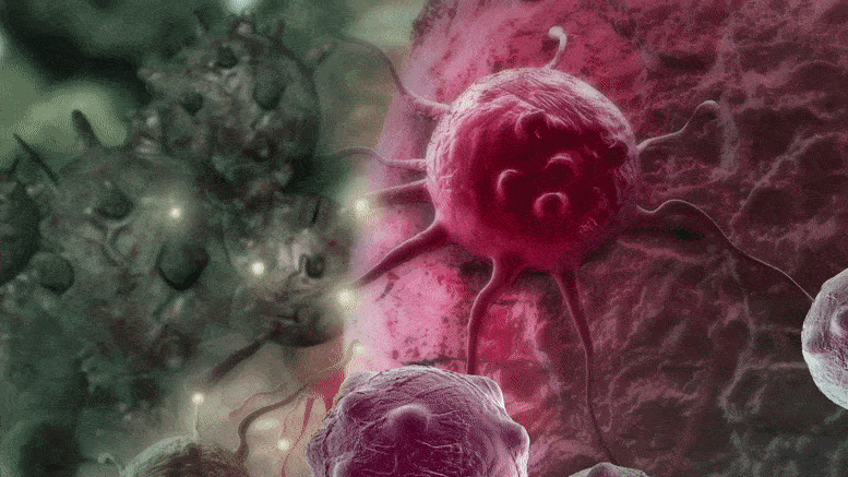
The researchers think that pyrvinium, a cheap drug created in the 1960s could be modified for use in the treatment of cancer.
Researchers from the University of Auckland have discovered that new combinations and formulations of older medicines are promising for the treatment of bowel cancer.
According to a team of University of Auckland scientists, the use of old drugs in new combinations is showing potential in the treatment of bowel cancer.
“While there have been advances in treatments for this disease in recent years, the development of new medicines is expensive and time-consuming,” lead researcher Professor Peter Shepherd says. “As a possible solution to this problem, our group has been investigating whether using old drugs in new ways could provide a faster and cheaper way of treating this disease.”
The researchers looked into several cancer drugs that would soon lose their patent protection. In their lab-based tests, scientists discovered that combining two of these drugs significantly increased the overall efficacy in treating bowel, or colorectal, cancer.
The groundwork for this study, according to Shepherd, has been laid by advancements in our understanding of how cancers function.
“In recent years, research has led to a rapid increase in our understanding of how colorectal cancer develops. In particular, some subtypes of the disease rely on the development of small blood vessels and on proteins called BRAF and beta-catenin. The research group identified existing drugs that target these and investigated the possibility that combining them could have powerful anti-cancer effects.”
Two older drugs have shown considerable promise in studies conducted at the University of Auckland. One is axitinib, an anticancer drug. The other is pyrvinium, a low-cost threadworm drug created in the 1960s that the researchers believe could be modified for use in cancer treatment. In one set of tests, the researchers discovered that combining axitinib with another older BRAF-targeting drug, vemurafenib, significantly increased its effectiveness. Axitinib works by reducing the growth of small blood vessels.
Both these drugs are used in other contexts to treat other types of cancer and will soon be off-patent and so the cost of using them in treatment will drop greatly, Shepherd says.
In a second set of studies, the group found evidence that pyrvinium, which targets beta-catenin, could also increase the efficacy of vemurafenib.
Dr. Khanh Tran who performed most of the experiments says, “This work suggests that existing drugs might be able to be repurposed to treat this type of cancer which could significantly reduce the cost of such therapy.”
Tran continues, “Since the drugs we used are already in use for other purposes, it makes it much easier to develop clinical trials to see how the findings of our studies will actually translate to improved outcomes for patients with this disease.”
Next, the researchers are planning a randomized, controlled clinical trial.
Groundbreaking research reveals that 7-dehydrocholesterol (7-DHC) serves as an antioxidant, protecting cells from ferroptosis. This challenges prior assumptions about 7-DHC and could significantly impact cancer treatment and our understanding of related diseases.
Recent research shows that 7-dehydrocholesterol is an antioxidant that protects cells from ferroptosis, offering new avenues for cancer treatment and disease understanding.
In a groundbreaking study, a team led by Würzburg Professor José Pedro Friedmann Angeli has shown that the cholesterol precursor 7-dehydrocholesterol (7-DHC) plays a crucial role as an antioxidant: it integrates into the cell membranes and protects the cells by preventing a certain type of cell death, known as ferroptosis.
“Until now, the accumulation of 7-DHC was only associated to neurodevelopmental defects, now we show that it actually increases cellular fitness and could promote a more aggressive behavior in cancers such as Burkitt’s lymphoma and neuroblastoma,” says Friedmann Angeli.
The newly discovered protective function of 7-DHC opens up exciting prospects for further improving the treatment of cancer and other diseases associated with ferroptosis: “It gives us new opportunities to test potential inhibitors that target cholesterol biosynthesis and are already established in medical practice.”
Teams From Würzburg, Dresden, Munich and Heidelberg Involved
The researchers report this in the journal Nature. In addition to the Würzburg team from the Rudolf Virchow Zentrum – Center for Integrative and Translational Bioimaging, the following scientists contributed to the study: Dr. Maria Fedorova (Dresden University of Technology), Marcus Conrad (Helmholtz Munich), Derek Pratt (University of Ottawa), and Andreas Trumpp and Hamed Alborzinia (German Cancer Research Center, DKFZ Heidelberg).
Observing Changes in 7-DHC Levels
High cholesterol levels are associated with health problems such as heart disease and diabetes. Most studies focus on how cholesterol contributes directly to these diseases.
In this area, the discovery of the cholesterol precursor 7-DHC as an antioxidant opens up new possibilities: Studies on changes in 7-DHC levels could provide crucial new insights into the diseases. In addition, drugs that specifically block 7-DHC production should be researched in combination with other drugs — this could have a positive effect in the treatment of some cancers.
Possible Effects on Tumour Development
“Our next research goal is to investigate the effects of 7-DHC accumulation during tumor development,” says Würzburg ferroptosis expert José Pedro Friedmann Angeli.
The team responsible for the publication in Nature also calls for further epidemiological studies. Background: There are drugs authorized by the US Food and Drug Administration (FDA) that can inhibit the DHCR7 enzyme. These include trazodone, which is prescribed around 20 million times a year in the USA, sometimes even for off-label use to treat insomnia.
“Studies have shown that people taking this drug have elevated plasma levels of 7-DHC. Epidemiological studies are crucial to better understand possible effects here,” says Friedmann Angeli. These studies would help to find out whether there is a connection between patient groups who regularly take ferroptosis-modulating drugs such as trazodone and cancer incidence, the occurrence of metastases, or other critical aspects of public health.
A new study finds that an artificial intelligence model can predict whether Crohn’s disease will recur after surgery.
A deep learning model trained to analyze histological images of surgical specimens accurately classified patients with and without Crohn’s disease recurrence, investigators report in The American Journal of Pathology.
According to researchers, more than 500,000 individuals in the United States have Crohn’s disease. Crohn’s disease is a chronic inflammatory bowel disease that damages the digestive system lining. It can cause digestive system inflammation, which may result in abdominal pain, severe diarrhea, exhaustion, weight loss, and malnutrition.
Many people end up needing surgery to treat their Crohn’s disease. Even after a successful operation, recurrence is common. Now, researchers are reporting that their AI tool is highly accurate at predicting the postoperative recurrence of Crohn’s disease. It also linked recurrence with the histology of subserosal adipose cells and mast cell infiltration.
Using an artificial intelligence (AI) tool that simulates how humans visualize and is trained to identify and categorize pictures, researchers created a model that predicts the postoperative recurrence of Crohn’s disease with high accuracy by evaluating histological images. The AI tool also identified previously unknown differences in adipose cells and substantial disparities in the degree of mast cell infiltration in the subserosa, or outer lining of the gut, when comparing individuals with and without disease recurrence. Elsevier’s The American Journal of Pathology published the findings.
The 10-year rate of postoperative symptomatic recurrence of Crohn’s disease, a chronic inflammatory gastrointestinal illness, is believed to be 40%. Although there are scoring methods to measure Crohn’s disease activity and the existence of postoperative recurrence, no scoring system has been devised to predict whether Crohn’s disease will return.
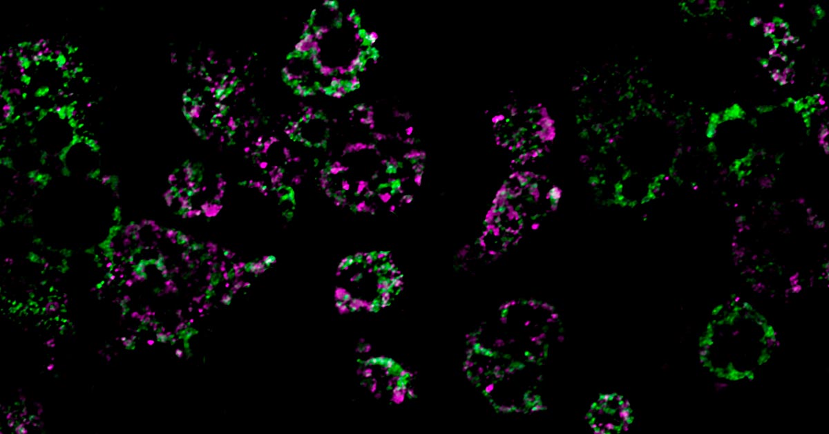
Sixty-eight patients with Crohn’s disease were classified according to the presence or absence of postoperative recurrence within two years. The investigators performed histological analysis of surgical specimens using deep learning EfficientNet-b5, a commercially available AI model designed to perform image classification. They achieved a highly accurate prediction of postoperative recurrence (AUC=0.995) and discovered morphological differences in adipose cells between the two groups. Credit: The American Journal of Pathology
“Most of the analysis of histopathological images using AI in the past have targeted malignant tumors,” explained lead investigators Takahiro Matsui, MD, Ph.D., and Eiichi Morii, MD, Ph.D., Department of Pathology, Osaka University Graduate School of Medicine, Osaka, Japan. “We aimed to obtain clinically useful information for a wider variety of diseases by analyzing histopathology images using AI. We focused on Crohn’s disease, in which postoperative recurrence is a clinical problem.”
The research involved 68 Crohn’s disease patients who underwent bowel resection between January 2007 and July 2018. They were divided into two groups based on whether or not they had postoperative disease recurrence within two years after surgery. Each group was divided into two subgroups, one for training and the other for validation of an AI model. Whole slide pictures of surgical specimens were cropped into tile images for training, labeled for the presence or absence of postsurgical recurrence, and then processed using EfficientNet-b5, a commercially available AI model built to perform image classification. When the model was tested with unlabeled photographs, the findings indicated that the deep learning model accurately classified the unlabeled images according to the presence or absence of disease occurrence.
Following that, prediction heat maps were created to identify areas and histological features from which the machine learning algorithm could accurately predict recurrence. All layers of the intestinal wall were shown in the photos. The heatmaps revealed that the machine learning algorithm correctly predicted the subserosal adipose tissue layer. However, the model was less precise in other regions, such as the mucosal and proper muscular layers. Images with the greatest accurate predictions were taken from the non-recurrence and recurrence test datasets. The photos with the greatest predictive results all had adipose tissue.
Because the machine learning model achieved accurate predictions from images of subserosal tissue, the investigators hypothesized that subserosal adipose cell morphologies differed between the recurrence and the non-recurrence groups. Adipose cells in the recurrence group had a significantly smaller cell size, higher flattening, and smaller center-to-center cell distance values than those in the nonrecurrence group.
“These features, defined as ‘adipocyte shrinkage,’ are important histological characteristics associated with Crohn’s disease recurrence,” said Dr. Matsui and Dr. Morii.
The investigators also hypothesized that the differences in adipocyte morphology between the two groups were associated with some degree or type of inflammatory condition in the tissue. They found that the recurrence group had a significantly higher number of mast cells infiltrating the subserosal adipose tissue, indicating that the cells are associated with the recurrence of Crohn’s disease and the “adipocyte shrinkage” phenomenon.
To the investigators’ knowledge, these findings are the first to link postoperative recurrence of Crohn’s disease with the histology of subserosal adipose cells and mast cell infiltration. Dr. Matsui and Dr. Morii observed, “Our findings enable stratification by the prognosis of postoperative Crohn’s disease patients. Many drugs, including biologicals, are used to prevent Crohn’s disease recurrence, and proper stratification can enable more intensive and successful treatment of high-risk patients.”
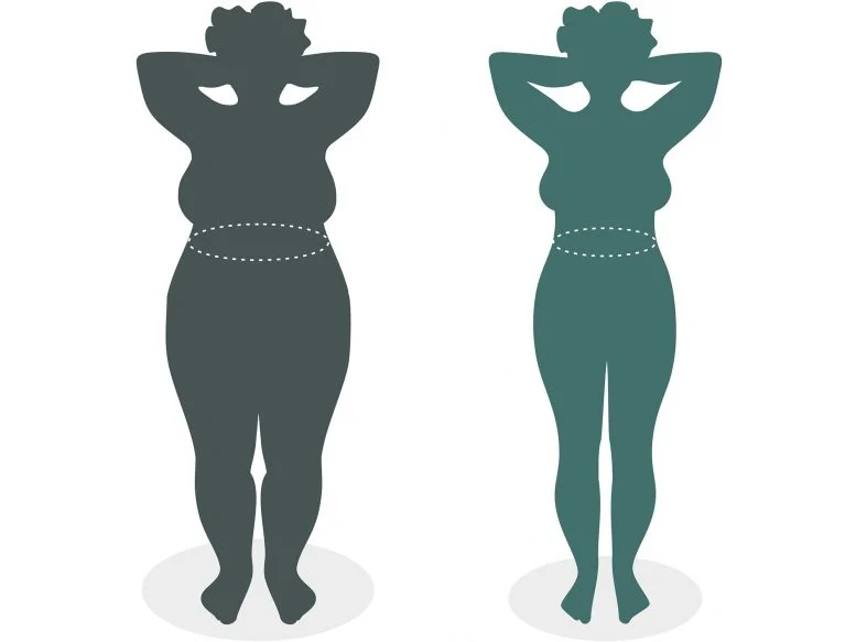
A study from UC San Diego reveals that obesity causes mitochondrial dysfunction in fat cells, driven by a specific gene. Deactivating this gene in mice prevents obesity, suggesting new treatment approaches.
UC San Diego Study reveals key mechanism behind obesity-related metabolic dysfunction.
The number of people with obesity has nearly tripled since 1975, resulting in a worldwide epidemic. While lifestyle factors like diet and exercise play a role in the development and progression of obesity, scientists have come to understand that obesity is also associated with intrinsic metabolic abnormalities. Now, researchers from the University of California San Diego School of Medicine have shed new light on how obesity affects our mitochondria, the all-important energy-producing structures of our cells.
Researchers at the University of California San Diego School of Medicine have discovered that a high-fat diet causes mitochondrial fragmentation in fat cells, reducing their fat-burning capacity.
Mitochondrial Dysfunction in Obesity
In a study published today (January 29, 2024) in the journal Nature Metabolism, the researchers found that when mice were fed a high-fat diet, mitochondria within their fat cells broke apart into smaller mitochondria with reduced capacity for burning fat. Further, they discovered that this process is controlled by a single gene. By deleting this gene from the mice, they were able to protect them from excess weight gain, even when they ate the same high-fat diet as other mice.
“Caloric overload from overeating can lead to weight gain and also triggers a metabolic cascade that reduces energy burning, making obesity even worse,” said Alan Saltiel, PhD, professor in the Department of Medicine at UC San Diego School of Medicine. “The gene we identified is a critical part of that transition from healthy weight to obesity.”
These colored streaks are mitochondrial networks within fat cells. Researchers from UC San Diego discovered that a high-fat diet dismantles mitochondria, resulting in weight gain. Credit: UC San Diego Health Sciences
Adipose Tissue and Obesity
Obesity, which affects more than 40% of adults in the United States, occurs when the body accumulates too much fat, which is primarily stored in adipose tissue. Adipose tissue normally provides important mechanical benefits by cushioning vital organs and providing insulation. It also has important metabolic functions, such as releasing hormones and other cellular signaling molecules that instruct other tissues to burn or store energy.
In the case of caloric imbalances like obesity, the ability of fat cells to burn energy starts to fail, which is one reason why it can be difficult for people with obesity to lose weight. How these metabolic abnormalities start is among the biggest mysteries surrounding obesity.
The study was led by Alan Saltiel, PhD, professor in the Department of Medicine at UC San Diego School of Medicine. Credit: UC San Diego Health Sciences
Research Findings and Potential Therapies
To answer this question, the researchers fed mice a high-fat diet and measured the impact of this diet on their fat cells’ mitochondria, structures within cells that help burn fat. They discovered an unusual phenomenon. After consuming a high-fat diet, mitochondria in parts of the mice’s adipose tissue underwent fragmentation, splitting into many smaller, ineffective mitochondria that burned less fat.
In addition to discovering this metabolic effect, they also discovered that it is driven by the activity of single molecule, called RaIA. RaIA has many functions, including helping break down mitochondria when they malfunction. The new research suggests that when this molecule is overactive, it interferes with the normal functioning of mitochondria, triggering the metabolic issues associated with obesity.
“In essence, chronic activation of RaIA appears to play a critical role in suppressing energy expenditure in obese adipose tissue,” said Saltiel. “By understanding this mechanism, we’re one step closer to developing targeted therapies that could address weight gain and associated metabolic dysfunctions by increasing fat burning.”
By deleting the gene associated with RaIA, the researchers were able to protect the mice against diet-induced weight gain. Delving deeper into the biochemistry at play, the researchers found that some of the proteins affected by RaIA in mice are analogous to human proteins that are associated with obesity and insulin resistance, suggesting that similar mechanisms may be driving human obesity.
“The direct comparison between the fundamental biology we’ve discovered and real clinical outcomes underscores the relevance of the findings to humans and suggests we may be able to help treat or prevent obesity by targeting the RaIA pathway with new therapies,” said Saltiel “We’re only just beginning to understand the complex metabolism of this disease, but the future possibilities are exciting.”
New findings, including the discovery of Eoneophron infernalis, suggest that dinosaurs, particularly caenagnathids, were not declining in diversity before the asteroid impact, contradicting earlier theories of their vulnerability.
Were dinosaurs already on their way out when an asteroid hit Earth 66 million years ago, ending the Cretaceous, the geologic period that started about 145 million years ago? It’s a question that has vexed paleontologistslike us for more than 40 years.
In the late 1970s, debate began about whether dinosaurs were at their peak or in decline before their big extinction. Scientists at that time noted that while dinosaur diversity seemed to have increased in the geologic stage that spanned 83.6 million to 71.2 million years ago, the number of species on the scene seemed to decrease during the last few million years of the Cretaceous. Some researchers have interpreted this pattern to mean that the asteroid that struck the Gulf of Mexico was simply the final blowfor an already vulnerable group of animals.
However, others have argued that what looks like a decrease in the diversity of dinosaurs may be an artifact of how hard it is to accurately count them. Fossil formations might preserve different dinosaurs more or less often based on factors like their favored environment and how easily their bodies fossilized there. The accessibility of various outcrops could influence what kinds of fossils researchers have so far found. These biases are a problem because fossils are what paleontologists must rely on to conclusively answer how healthy dinosaur populations were when the asteroid hit.
At that crucial moment, what was really happening to dinosaur diversity? Discovery, identification and description of new dinosaurs provide vital clues. This is where our work comes in. Close examination of what we’d thought was a juvenile specimen of an already-known species of dinosaur from this time period revealed that it was actually part of an adult from a completely new species.
Our work focusing on the life stage of our specimen demonstrates that dinosaur diversity may not have been declining before the asteroid hit, but rather that there are more species from this time period yet to be discovered – potentially even through reclassification of fossils already in museum collections.
Clues Inside the Bones of a Birdlike Dinosaur
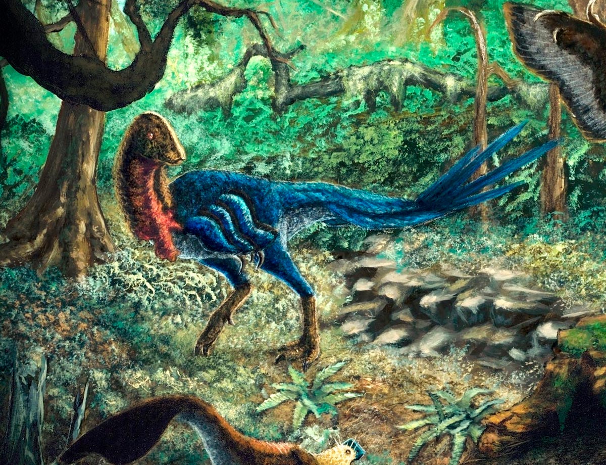
Our new study focused on four hindlimb bones – a femur, a tibia, and two metatarsals. They were unearthed in South Dakota, in rocks of the Hell Creek Formation, and date to the final 2 million years of the Cretaceous.
The only known species of caenagnathid from this time and region was Anzu, sometimes called the “chicken from Hell.” Covered in feathers and sporting wings and a toothless beak, Anzu was between roughly 450 and 750 pounds (200 and 340 kilograms). Despite its fearsome nickname, though, its diet is a matter of debate. It was likely an omnivore, eating both plant material and small animals.
Because our specimen was significantly smaller than Anzu, we simply assumed it was a juvenile. We chalked up the anatomical differences we noticed to its juvenile status and smaller size – and figured the animal would have changed had it continued to grow. Anzu specimens are rare, and no definite juveniles have been published in the scientific literature, so we were excited to potentially learn more about how it grew and changed throughout its lifetime by looking inside its bones.
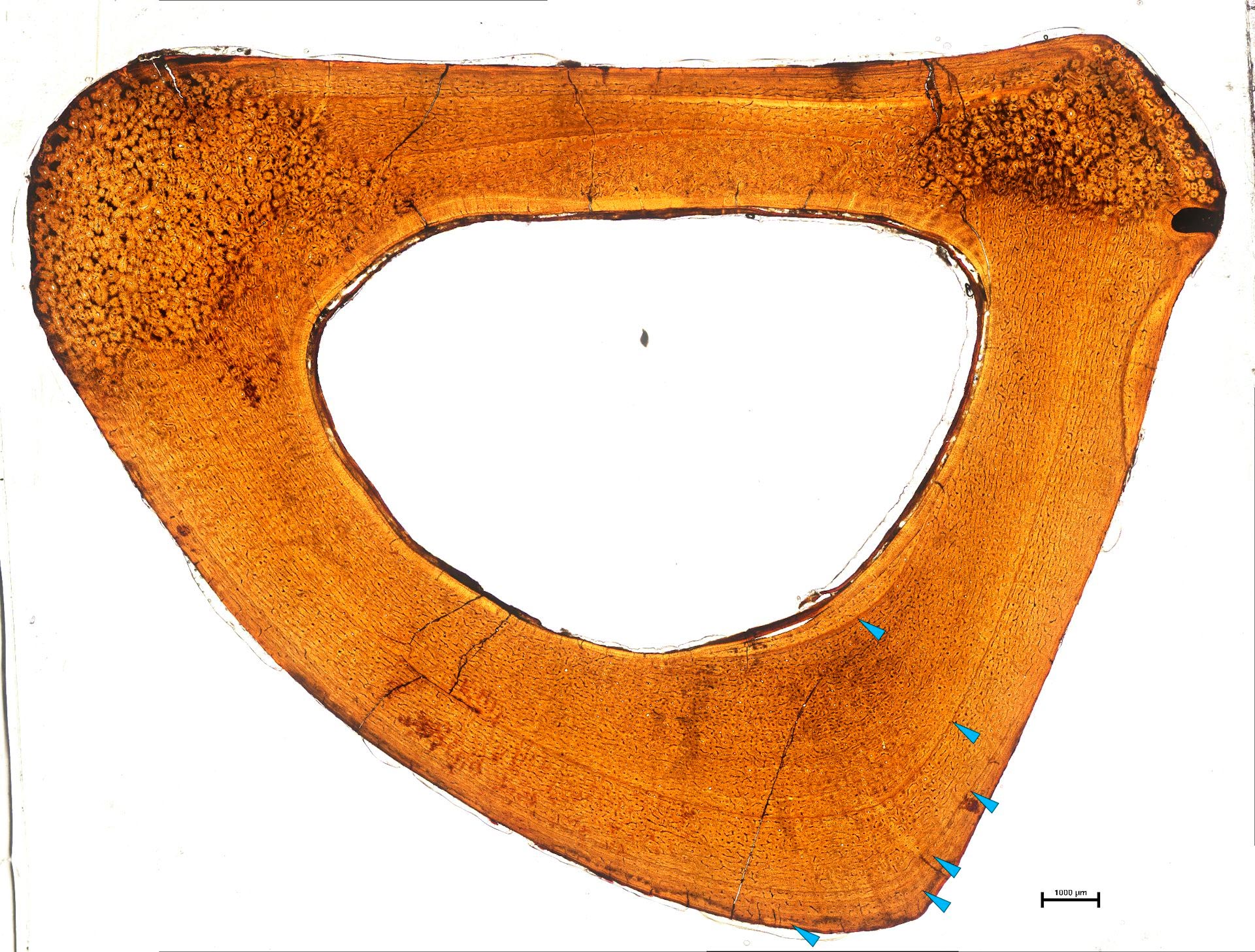
Just like with a tree’s rings, bone records rings called lines of arrested growth. Each annual line represents part of a year when the animal’s growth slowed. They would tell us how old this animal was, and how fast or slow it was growing.
We cut through the middle of three of the bones so that we could microscopically examine the internal anatomy of the cross-sections. What we saw completely uprooted our initial assumptions.
Teal markers point to lines of arrested growth on the cross-section of fossilized bone. Toward the outside of the bone, the lines are much closer together, reflecting less growth per year. Researchers counted exactly six lines, meaning this animal was between 6 and 7 years old when it died. Credit: Holly Woodward
In a juvenile, we would expect lines of arrested growth in the bone to be widely spaced, indicating rapid growth, with even spacing between the lines from the inside to the outside surface of the bone. Here, we saw that the later lines were spaced progressively closer together, indicating that this animal’s growth had slowed and it was nearly at its adult size.
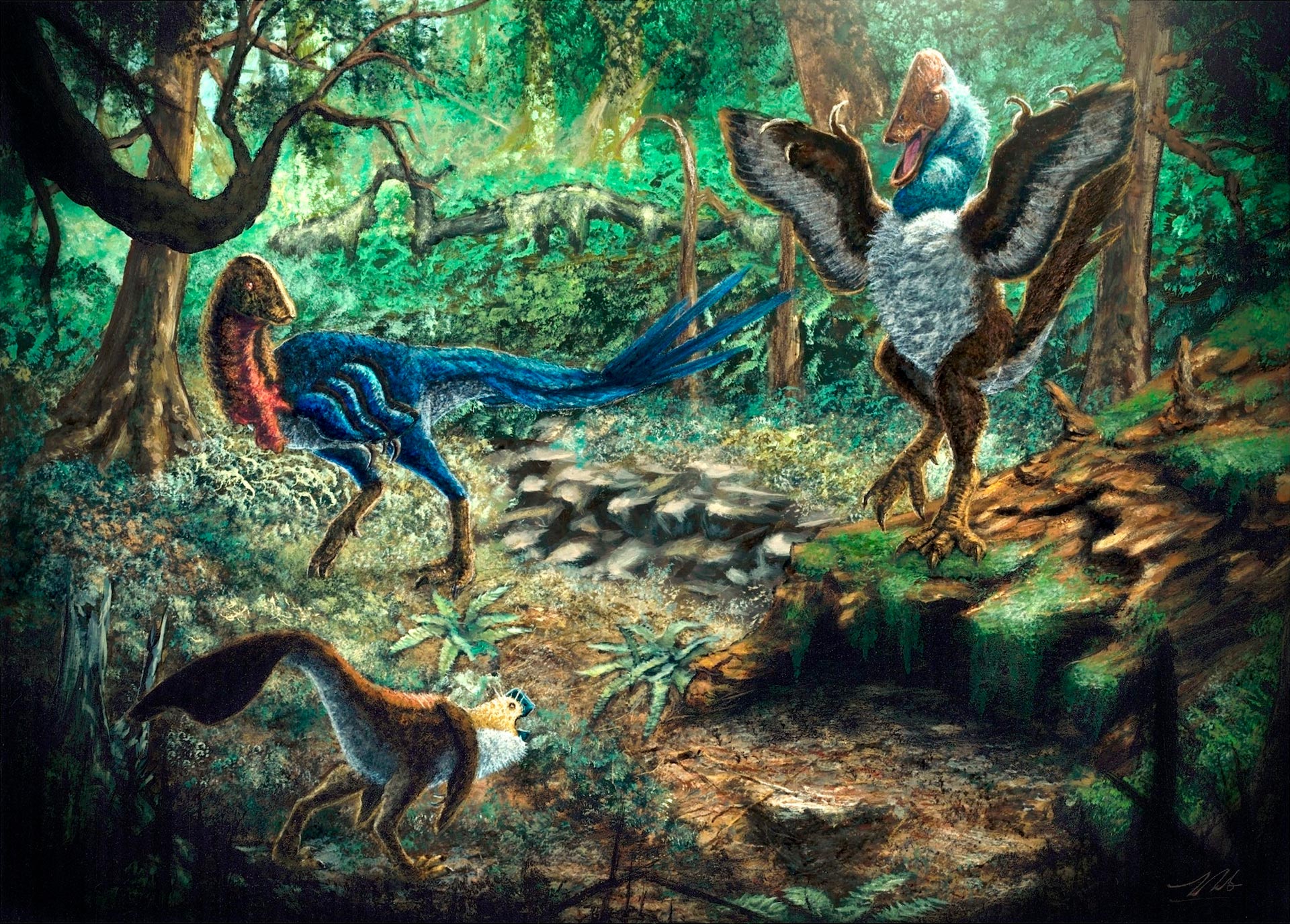
This was no juvenile. Instead, it was an adult of an entirely new species, which we dubbed Eoneophron infernalis. The name means “Pharaoh’s dawn chicken from Hell,” referencing the nickname of its larger cousin Anzu. Traits unique to this species include ankle bones fused to the tibia, and a well-developed ridge on one of its foot bones. These weren’t features a young Anzu would outgrow, but rather unique aspects of the smaller Eoneophron.
Expanding the Caenagnathid Family Tree
With this new evidence, we started making thorough comparisons with other members of the family to determine where Eoneophron infernalis fit within the group.
It also inspired us to reexamine other bones previously believed to be Anzu, as we now knew that more caenagnathid dinosaurs lived in western North America during that time. One specimen, a partial foot bone smaller than our new specimen, appeared distinct from both Anzu and Eoneophron. Where once there was one “chicken from Hell,” now there were two, and evidence for a third: one large (Anzu), weighing as much as a grizzly bear, one medium (Eoneophron), humanlike in weight, and one small and yet unnamed, close in size to a German shepherd.
Comparing Hell Creek with older fossil formations such as the famous Dinosaur Park Formation of Alberta that preserves dinosaurs that lived between 76.5 million and 74.4 million years ago, we find not only the same number of caenagnathid species, but also the same size classes. There, we have Caenagnathus, comparable to Anzu, Chirostenotes, comparable to Eoneophron, and Citipes, comparable to the third species we found evidence for. These parallels in both species count and relative sizes offer compelling evidence that caenagnathids remained stable throughout the last part of the Cretaceous.
Our new discovery suggests that this dinosaur group was not declining in diversity at the very end of the Cretaceous. These fossils show that there are still new species to be discovered, and support the idea that at least part of the pattern of decreasing diversity is the result of sampling and preservation biases.
Did large dinosaurs go extinct the way a Hemingway character quipped he went broke: “gradually, then suddenly”? While there are plenty of questions still outstanding in this extinction debate, Eoneophron adds evidence that caenagnathids were doing quite well for themselves before the asteroid ruined everything.
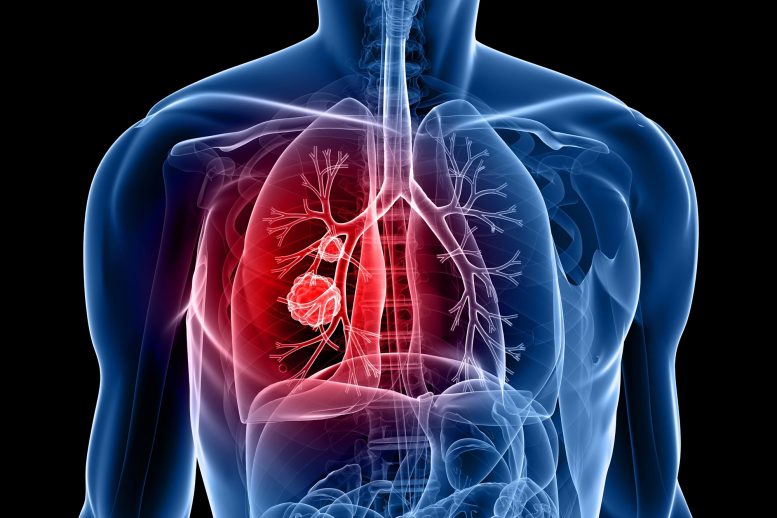
According to a new study in the journal Clinical Cancer Research, patients with non-small cell lung cancer, the most common type of lung cancer, who were treated with neoadjuvant nivolumab had improved five-year recurrence-free and overall survival rates.
Study reports five-year survival outcomes.
Patients with resectable non-small cell lung cancer (NSCLC) who were treated with neoadjuvant nivolumab had improved five-year recurrence-free and overall survival rates compared with historical outcomes.
The research will be published today, February 15, 2023, in Clinical Cancer Research, a journal of the American Association for Cancer Research (AACR), a non-profit organization dedicated to advancing cancer research and improving patient outcomes through education, collaboration, and advocacy..
Patrick Forde, MBBCh, the senior author of the study, is an associate professor of oncology and director of the Thoracic Oncology Clinical Research Program at the Sidney Kimmel Comprehensive Cancer Center at Johns Hopkins.
Samuel Rosner, MD, is co-first author of the study and is a medical oncology fellow at the Sidney Kimmel Comprehensive Cancer Center at Johns Hopkins and a member of Forde’s research group.
NSCLC Background
NSCLC is the most common type of lung cancer and is a leading cause of cancer-related death worldwide. Despite strides in treating metastatic NSCLC, new treatments for earlier-stage disease have only recently emerged, according to Forde.
Rosner added that there is great interest in optimizing neoadjuvant strategies for earlier-stage NSCLCs that are eligible for surgical resection. Rosner is a medical oncology fellow at the Sidney Kimmel Comprehensive Cancer Center at Johns Hopkins and a member of Forde’s research group.
Forde, Rosner, and colleagues previously reported safety and efficacy results from a phase II clinical trial in which patients with stage I-III resectable NSCLC were treated with two doses of neoadjuvant nivolumab. Major pathological responses were observed in 45 percent of patients, independent of tumor PD-L1 expression, and 73 percent of patients whose tumors were surgically resected were recurrence-free 18 months following surgery.
The latest publication reports the final analyses from this trial, including five-year recurrence-free and overall survival rates for the 20 patients who underwent surgical resection.
“To our knowledge, this is the longest follow-up to date for a PD-1/PD-L1 inhibitor in the neoadjuvant setting for any solid tumor,” said Forde.
Study Results
Among the 20 patients who underwent surgical resection, 12 patients (60 percent) remained recurrence-free five years after surgery, and 16 patients (80 percent) were alive, exceeding the 36 to 68 percent five-year survival rate historically observed for patients with stage I-III NSCLC, Rosner noted. Forde added that the observed patient outcomes after neoadjuvant nivolumab were better than those historically observed among patients treated with neoadjuvant chemotherapy.
The authors also identified major pathologic response after neoadjuvant nivolumab as a potential predictive biomarker of recurrence-free and overall survival. Of the nine patients who had a major pathological response after neoadjuvant nivolumab, eight were alive and cancer-free five years after treatment. One patient experienced a recurrence within the first 10 months after treatment but has since been disease-free after definitive chemoradiation. The one death in this subgroup was unrelated to cancer.
In contrast, six of the 11 patients who did not have a major pathological response experienced disease recurrence, and three of these patients died due to their cancer. These results indicate that a major pathological response following neoadjuvant nivolumab may be associated with a lower risk of disease recurrence and death, although the authors caution that these results are preliminary and require further validation in larger studies.
Neoadjuvant nivolumab did not lead to surgical delays, and there was only one late-onset immune-related adverse event, which occurred 16 months after nivolumab treatment and was successfully managed, the authors noted.
“The results from the five-year follow-up analysis indicate that neoadjuvant nivolumab was safe in long-term follow-up and led to encouraging survival in this patient cohort,” said Forde. “The long-term safety and efficacy data from this study provide further support for the use of nivolumab in the neoadjuvant setting.”
Neoadjuvant nivolumab in combination with chemotherapy was approved by the U.S. Food and Drug Administration in March 2022 for the treatment of lung cancer. “Further studies will help us determine whether select patients may benefit from immunotherapy alone,” Forde noted.
“An interesting finding from the analysis was the difference in outcomes between patients with and without a major pathological response,” said Rosner. “Although the sample size was small, the results illustrate the potential power of pathological response as a predictive biomarker.”
Reference: “Five-Year Clinical Outcomes after Neoadjuvant Nivolumab in Resectable Non-Small Cell Lung Cancer” by Samuel Rosner, Joshua E. Reuss, Marianna Zahurak, Jiajia Zhang, Zhen Zeng, Janis Taube, Valsamo Anagnostou, Kellie N. Smith, Joanne Riemer, Peter B. Illei, Stephen R. Broderick, David R. Jones, Suzanne L. Topalian, Drew M. Pardoll, Julie R. Brahmer, Jamie E. Chaft and Patrick M. Forde, 16 February 2023, Clinical Cancer Research. DOI: 10.1158/1078-0432.CCR-22-2994


