In Another Win for CRISPR, FDA Approves Casgevy for Beta Thalassemia


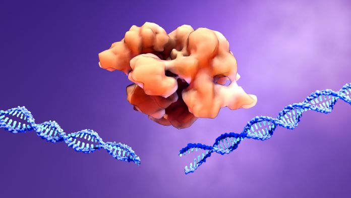
An illustration of a CRISPR-Cas9 gene editing protein cutting a DNA helix
Credit: Design Cells/Getty Images

The U.S. Food and Drug Administration (FDA) has approved Vertex and CRISPR Therapeutics’ Casgevy (exa-cel), a CRISPR/Cas9 gene-edited cell therapy, for the treatment of transfusion-dependent beta thalassemia (TDT) in patients 12 years and older. This is the latest sign the gene and cell therapy market is maturing. This market is already estimated to be worth $15B and expected to grow to more than $80B over the next ten years or so.

Just a few weeks ago the FDA approved Casgevy to treat patients with sickle cell disease. Simultaneously, Bluebird Bio received the FDA OK for Lyfgenia, its gene therapy for sickle cell. In November, the U.K. Medicines and Healthcare products Regulatory Agency (MHRA) granted conditional marketing authorization for Casgevy in both sickle cell disease and TDT. 

With this nod, Casgevy becomes the first treatment for TDT available in the U.S. using CRISPR gene-editing technology. TDT is an inherited blood disease that causes low levels of functioning hemoglobin, which carries oxygen.

“On the heels of the historic FDA approval of Casgevy for sickle cell disease, it is exciting to now secure approval for TDT well ahead of the PDUFA date,” said Reshma Kewalramani, MD, CEO and president of Vertex. “TDT patients deserve new, potentially curative treatment options.”

The cell and gene therapy pipeline is already pretty full, with thousands of trials ongoing. Several are already in Phase II or III, including treatments for cancers and amyloidosis.  

Casgevy administration requires experience in stem cell transplantation. As a result, Vertex is establishing a network of independently operated, authorized treatment centers (ATCs) throughout the U.S. to offer the treatment. All nine current U.S. ATCs can offer Casgevy to eligible patients with either TDT or sickle cell disease. Additional ATCs will be activated in the coming weeks and a complete list of ATCs can be accessed at CASGEVY.com.

TDT requires frequent blood transfusions and iron chelation therapy throughout a person’s life. Due to anemia, patients may experience fatigue and shortness of breath, and infants or develop failure to thrive, jaundice, and feeding problems. Complications of TDT can also include an enlarged spleen, liver and/or heart, misshapen bones and delayed puberty. 

Patients have reduced life expectancy, decreased quality of life, and lowered lifetime earnings and productivity. In the U.S., the median age of death for patients living with TDT is 37 years. Stem cell transplant from a matched donor can cure the condition, but this option, but is only available to a small fraction of people living with TDT because of the lack of available donors.

Vertex and CRISPR Tx charge $2.2 million for a one-time Casgevy treatment. The companies estimate that lifetime healthcare costs to manage TDT in the U.S. exceed $5 million.

Casgevy is a non-viral, ex vivo CRISPR/Cas9 gene-edited cell therapy for certain patients with sickle cell disease or TDT. A patient’s own hematopoietic stem and progenitor cells are extracted and edited. The edits are made at the erythroid specific enhancer region of the BCL11A gene through a precise double-strand break.This change leads to production of high levels of fetal hemoglobin (HbF) in red blood cells. HbF is the form of hemoglobin typically present during fetal development, which then switches to the adult form after birth.
Latest SARS-CoV-2 Variants Evolve Strategies to Overcome Innate Immunity


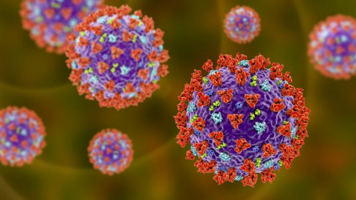
Coronavirus particles, illustration - SARS-CoV-2 virus

Researchers from the University College London (UCL), in the U.K. have unveiled critical insights into the evolving strategies of the latest SARS-CoV-2 variants, such as BA.4 and BA.5, shedding light on their ability to overcome humans’ innate immunity, marking a crucial turning point in the ongoing battle against the COVID-19 pandemic.

Since the beginning of the pandemic, several variants of the SARS-CoV-2 virus have emerged, with Alpha, Delta, and Omicron successively dominating global circulation. Previous research has highlighted the evolutionary tactics of Alpha and Delta, emphasizing their ability to prevent the human innate immune response by disrupting cellular signaling in the airways. According to researchers, this disruption provides the virus with a window of opportunity to establish itself in the body, eventually overwhelming the adaptive immunity developed through prior infection or vaccination.

Unlike its predecessors, the Omicron variant’s evolution was shaped by the pre-existing immunity within the global population resulting from widespread vaccination and prior infections. The adaptive immunity generated produces antibodies targeting the virus’s spike protein, providing protection against infection through a process known as neutralization.

Published in Nature Microbiology, the study focused on examining eight different sub-variants of the Omicron variant to understand how they interact with the human host. Surprisingly, the earliest Omicron variants, BA.1 and BA.2, exhibited reduced abilities to overcome human innate immunity compared to Alpha and Delta. However, more recent variants like BA.5 and XBB have reacquired this ability, employing similar mechanisms observed in Alpha to Delta variants.

Ann-Kathrin Reuschl, first author of the study from the UCL Division of Infection & Immunity, expressed surprise at the initial findings, stating, “This seemed like a backward step in SARS-CoV-2 evolution and may explain reports of reduced disease severity when these variants were dominant.”

Later Omicron variants, echoing the strategies of Alpha to Delta, increased the production of innate immune antagonist proteins, such as nucleocapsid and Orf6, to overcome innate immunity. The researchers emphasized that this evolution demonstrates how the innate immune system acts as an effective gatekeeper for SARS-CoV-2 variant success.

Despite existing immunity from vaccines and prior infection, the study underscores the virus’s persistent ability to infect individuals. To mitigate infection risk, the researchers suggest the use of FFP3 masks or similar protective measures.

“With SARS-CoV-2 we’ve had a unique opportunity to observe how a virus evolves to overcome our defences in real time, giving us an opportunity to predict what a virus with pandemic potential needs to do to be successful. This may help us to assess the risk posed to humans by emerging viruses or new variants of existing viruses. It will be important to continue to monitor the virus as it continues to evolve,” concluded Clare Jolly, a professor at the UCL Division of Infection and Immunity and senior author of the study.


Researchers pinpoint 5 subvariants of Alzheimer’s disease


Could learning more about the different biological types of Alzheimer’s disease help improve treatment in the long run? Image credit: Maskot/Getty Images.

  • About 32 million people globally have Alzheimer’s disease.
  • There is currently no cure for Alzheimer’s, however, some medications are available to treat symptoms and help slow disease progression.
  • Researchers from Amsterdam University Medical Centers and Maastricht University have identified five biological variants directly related to Alzheimer’s disease.
  • Scientists believe these findings may impact how future Alzheimer’s treatments are developed and prescribed.

Approximately 32 million peopleTrusted Source around the world have Alzheimer’s disease, a type of dementia causing memory loss and other types of cognitive issues.

There is currently no cure for Alzheimer’s disease, and researchers are still not completely sure what causesTrusted Source the condition. There are some medicationsTrusted Source available to help treat symptoms and potentially slow the progression of the disease.

Now, researchers from Amsterdam University Medical Centers and Maastricht University in the Netherlands have identified five biological variants directly related to Alzheimer’s disease.

Scientists believe these findings — recently published in the journal Nature AgingTrusted Source — may impact how future Alzheimer’s treatments are developed and prescribed.

Why look for Alzheimer’s biological variants? 

Although researchers do not know the exact cause behind Alzheimer’s disease, most agree the formation of beta-amyloid plaque build-upTrusted Source and tau tanglesTrusted Source characterize it.

Dr. Betty Tijms, associate professor of neurology, brain imaging, and neurodegeneration at Amsterdam University Medical Centers and lead author of this study, told Medical News Today that she and her team decided to look for biological processes other than beta-amyloid and tau that may affect Alzheimer’s disease.

They did that because genetic and tissue proteomic studies previously pointed out that other biological processes beyond amyloid and tau seem to be involved in Alzheimer’s disease.

“But this was difficult to study in patients because the brain is not easily accessible,” Dr. Tijms continued. “New techniques made it possible to measure many proteins in the cerebrospinal fluidTrusted Source, and the levels of those proteins provide a detailed picture of the processes that are ongoing in the brain.“

”So we made use of these innovations to study if certain subgroups of Alzheimer’s disease patients could be identified that share distinct underlying biological processes,” she added.

What are the 5 biological variants of Alzheimer’s disease?

For this study, Dr. Tijms and her team examined a little over 1,000 proteins in the cerebrospinal fluid of 419 people with Alzheimer’s disease. Through this examination, they discovered five biological variants within this group.

According to Dr. Tijms, the first subtype was characterized by abnormal outgrowth of nerve cells.

She explained:

“Outgrowth is a normal process in the brain when connections between nerve cells get damaged, for example, because of the clumping of amyloid-beta. But here we see that this process goes into overdrive, and does not seem to efficiently repair the connections. The immune system of this variant was not activated properly, which may [interfere] with the clearance of protein clumps.”

– Dr. Betty Tijms

“The second subtype had an overactive immune system, which aggravated the disease progression,” Dr. Tijms continued. “The third subtype had problems with the synthesis of proteins — RNA metabolism dysfunctionTrusted Source.”

”The fourth subtype had damage in the choroid plexusTrusted Source, which is the organ in the brain that produces cerebrospinal fluid,” she said. ”[And] the fifth subtype showed leakage of the blood-brain barrier.”

Surprising results may impact drug development

Both researchers found some of the discovered Alzheimer’s disease biological variants surprising.

“I was surprised by the subtype with dysregulated RNA metabolism because such processes have not been highlighted as a key factor in Alzheimer’s disease,” Dr. Pieter Jelle Visser, professor of molecular epidemiology of Alzheimer’s disease at Maastricht University in the Netherlands and senior author of this study, told MNT.

“It was surprising to see the new subtype with choroid plexus dysfunction,” Dr. Tijms also noted. “They had similar effects as [the] blood-brain barrier with low cerebrospinal fluid tau levels, for example, but no indication at all of leakage of blood proteins in the cerebrospinal fluid.“

“To me that indicates that these brain interfaces are really two different entities, with their own roles in Alzheimer’s disease,” she told us.

Both researchers believe these findings may change how Alzheimer’s medications are developed and prescribed in the future.

“The existence of these subtypes suggests that each subtype may need a different treatment,” Dr. Visser explained. “Future trials should take this into account and test their treatment in the subtypes that matched with the working mechanism of the drug.”

“Alternatively, each future Alzheimer’s disease trial should stratify on subtypes such that the subtypes that best respond to treatment can be identified,” he added. “Future trials may also take into account that side effects may differ between subtypes as well.”

Potential to accelerate intervention research

After reviewing this study, Dr. Karen D. Sullivan, a board-certified neuropsychologist, owner of I CARE FOR YOUR BRAIN, and Reid Healthcare Transformation Fellow at FirstHealth of the Carolinas in Pinehurst, NC, told MNT she found this research to be extremely hopeful, as it has been known for a long time that Alzheimer’s disease is an extremely heterogeneous subtype of neurodegenerative diseaseTrusted Source.

“Some patients have a slow and steady decline while others progress quickly,” she continued. “Some have predominantly memory symptoms while others experience primarily visual and spatial impairments. Identifying these five specific disease processes in Alzheimer’s disease is a necessary starting point [for] personalizing brain healthcare interventions.”

Dr. Jennifer Bramen, a senior research scientist at the Pacific Neuroscience Institute in Santa Monica, CA, also not involved in the current study agreed, saying: “There are many known risk factors for Alzheimer’s disease, and each patient has a unique risk profile. Patients exhibit diverse symptoms, progression timelines, and neurodegeneration patterns.”

“If the authors are correct in their hypothesis that different Alzheimer’s disease variants may respond differently to treatments, there’s an opportunity to reassess drugs that showed promise in earlier research but were not effective overall. If true, this has the potential to accelerate intervention research.”

– Dr. Jennifer Bramen

For the next steps in this research, Dr. Sullivan said she would like to see researchers find out if these five biological variants result in distinct clinical syndromes in people living with Alzheimer’s disease.

“What is the specific set of cognitive and behavior symptoms and the prognostic significance of these five variants — is there a cause-and-effect relationship?” she continued. “[And] the ultimate question — and here is where the hope lies — do these five variants respond preferentially to different experimental drugs or from different types of preventative or early interventions that keep the disease process from spreading throughout the brain?”

Healthy microbiome plays a role in skin aging, researchers say


A new study suggests a healthy skin microbiome could help improve signs of skin aging, such as transepidermal water loss. Rob and Julia Campbell/Stocksy

  • A new study revealed a potential link between the skin microbiome and signs of skin aging.
  • Researchers found the balance of good and bad bacteria can change transepidermal water loss, the moisture that evaporates through the skin.
  • To optimize your skin microbiome health, limit the use of harsh ingredients such as salicylic acid.

Numerous factors contribute to skin aging, including environmental, genetics and lifestyle.

Researchers at the Center for Microbiome Innovation (CMI) at the University of California San Diego (UC San Diego) and L’Oréal Research and Innovation discovered the skin microbiome could hold the key to understanding how the skin ages.

Their findings were recently published in Frontiers in Aging.

Skin microbiome diversity shows fewer signs of aging

For the study, the research team analyzed 13 previous L’Oréal studies, which included skin clinical data of more than 1,000 female participants ages 18–70.

Researchers found a link between skin microbiome diversity and , generally viewed as a key sign of skin aging. They also found a negative association between microbiome diversity and transepidermal water loss, which is the moisture that evaporates through the skin.

“The most compelling aspect of the study was that there were indeed taxa that appeared to be linked to the grade of crow’s feet wrinkles, separate from age, despite the strong relationship between wrinkles and age,” Dr. Se Jin Song, the director of research at the UC San Diego Center for Microbiome Innovation, told Medical News Today.

“This is an exciting starting point for further research that can help us definitively determine whether they actually play a role in the development of wrinkles and what that role might be,” she added.

While this study revealed specific microbes of interest, further research will focus on elucidating what role they might play in skin aging.

Dr. Jin Song posed the following questions: “Are they interacting with our skin cells in a particular way? Are they affecting other members of the skin microbiome? Are they producing specific chemicals or compounds that are beneficial for skin appearance?”

“Answering these questions will help pave the path to applications that leverage the relationship between the microbiome and skin health,” she stated.

How does the microbiome affect skin health?

“What researchers are finding is the balance of the good and bad bacteria can change transepidermal water loss,” said Dr. Shilpi Khetarpal, a dermatologist at the Cleveland Clinic, not involved in the current research. “Think of a grape turning into a raisin. With our skin, it works the same way. The microbiome can influence the hydration.”

It can also change how our skin reacts to environmental factors — the sun, pollution, etc. Taking all of this into consideration, it’s evident our skin microbiome can directly influence how we age, Dr. Khetarpal added.

In reference to the study, “this work opens doors to new opportunities for discovering technologies that leverage the microbiome for maintaining healthy-looking skin,” said Dr. Qian Zheng, Head of Advanced Research, North America at L’Oréal.

“The microbial markers and aging signs identified in this study will steer future research, enabling the generation of larger, more harmonized data to accelerate our understanding of the microbiome in the context of healthy skin and skin aging,” Dr. Zheng further noted.

A significant challenge ahead is to validate or refute the causal role of these markers through in vitro methods and in vivo interventional studies, Zheng added. Additionally, identifying specific microbiome markers for each aging sign presents an exciting avenue for personalized skin care solutions.

Gut-skin axis: What’s the connection?

A growing body of research suggests it may be beneficial to focus on skin health and gut health.

StudiesTrusted Source show the gut and skin microbiota fight against pathogens, alleviating inflammation and regulating the immune system.

“I think what’s interesting is in the last several years, there’s more buzz surrounding the skin microbiome when the gut microbiome was previously focused on,” Dr. Khetarpal said.

“As time goes on, we’re learning the skin microbiome affects acne, rosacea, aging, and other skin conditions. It’s fascinating. We’re seeing this whole new trend in how we care for our skin and new conditions,” Dr. Khetarpal added.

Researchers continue to explore the connection between the gut and skin and whether a healthy gut could lead to healthier, more youthful skin.

“I believe there is an indirect link between the gut microbiome and skin microbiome. For instance, whether you’re taking oral antibiotics or topical antibiotics, it is possible to influence the two microbiomes. As a result, optimizing both the skin microbiome and gut microbiome will provide [the] best results and [improve] the overall appearance of the skin.”

— Dr. Shilpi Khetarpal, dermatologist

How to restore your skin microbiome

While everyone has different skin types, there are some general guidelines for healthier skin.

“Minimize the use of harsh ingredients that will disrupt the microbiome,” Dr. Khetarpal said. “For example, with normal skin use a gentle cleanser and moisturizer. And avoid salicylic acid because it can cause irritation and dryness.”

“The microbiome plays a crucial role in maintaining skin health and function,” Dr. Zheng said.

“Our focus on understanding the microbiome profile of healthy skin and the specific biomarkers associated with various conditions, including aging, is a testament to our commitment to pioneering in this field.”

“This knowledge allows us to identify differentiating and efficacious technologies inspired by the microbiome. We leverage this understanding to develop a variety of actives, including prebiotics and postbiotics, that promote beneficial microbes or target harmful ones. We also explore bio-derived materials like endolysins and even probiotics to reshape the skin microbiome towards a healthier state.”
— Dr. Qian Zheng, head of advanced research, North America at L’Oréal

Do cold water training, cinnamon supplements aid blood sugar control in diabetes?


Could swimming in cold water and taking cinnamon supplements improve blood sugar control? Image credit: Diego Martin/Stocksy.

  • Diabetes is a condition affecting blood glucose (sugar) levels.
  • Type 1 diabetes occurs when a person’s body stops producing insulin, the hormone that controls blood glucose. In type 2, the body stops responding to insulin.
  • For many people with diabetes, a healthy diet and appropriate exercise can help control blood glucose and minimize the risk of complications, such as heart disease, chronic kidney disease and vision problems.
  • A new study in rats with diabetes has found that a combination of cold-water swimming and cinnamon supplements lowered blood glucose levels.
  • The researchers suggest that this method may help people with diabetes control their blood sugar levels.

Diabetes is a chronic health condition where blood glucose control is impaired. It is major cause of mortality and ill health globally.

In 2021, some 529 million peopleTrusted Source (more than 6% of the global population) were living with diabetes. Of these, 96% had type 2 diabetes, the risk of which is greatly increased by poor diet and high body mass indexTrusted Source.

The prevalence of diabetes is increasing rapidly. Within 20 years, studies predict that there will be more than 700 million people with diabetes worldwide, and by 2050, that number could be as high as 1.3 billionTrusted Source.

Can lifestyle interventions help treat diabetes?

People with type 1 diabetes do not produce insulin, so need to control their blood sugar levels with regular insulin injections or an insulin pump.

However, only some people with type 2 diabetes need additional insulin. For most, changes in diet and exercise, together with medication, are key to managing the condition.

Now, a study in rats with diabetes has found that cold-water swimming, combined with cinnamon supplements, helped regulate blood glucose levels and enhanced insulin sensitivity.

The study appears in Nature Nutrition & DiabetesTrusted Source.

Dr. Ishita Prakash Patel, a board-certified endocrinologist with Texas Diabetes and Endocrinology in Austin, not involved in this study, commented on its findings for Medical News Today, cautioning that: “This was a relatively small study, short duration, using rats. […] Further studies in humans would be useful.”

Do cold swimming and cinnamon aid blood sugar control?

Exercise is known to help with blood glucose control, and studies have suggested that, as well as having other potential health benefits, cold exposure may help with glucose metabolism by increasing peripheral insulin sensitivity, as well as thermogenesisTrusted Source and glucose uptake in muscles.

One small studyTrusted Source found that 10 days of exposure to cold — 14–15 degrees Celsius (°C), or 57–59 degrees Fahrenheit (°F) — increased peripheral insulin sensitivity by around 43% in eight people with type 2 diabetes.

The researchers in this study suggest that this may be because cold exposure increases the mass and activity of brown adipose tissueTrusted Source (brown fat), which is specialized for energy expenditure.

Studies investigating the effects of cinnamon supplementation on blood glucose have shown differing effects. One studyTrusted Source found that 3–6 grams (g) of cinnamon a day had a positive effect on blood glucose levels in healthy adults, keeping them within the normal range.

However, another, in 70 people with type 2 diabetesTrusted Source, found no difference in blood glucose effect between those taking 1g cinnamon supplements daily for 30 or 60 days and those taking placebo.

Dr. Patel explained to Medical News Today:

“Cold adaptation has been shown to improve glucose tolerance and insulin sensitivity by stimulating brown fat. Cinnamon supplementation has been noted to increase insulin sensitivity and decrease post-prandial blood sugars through enhanced signaling pathways.”

Rat study investigates use of cinnamon supplements, cold water swimming

In the current study, researchers investigated the effect of cold-water swimming with and without cinnamon supplementation, and cinnamon supplementation alone, on blood glucose in rats with diabetes.

Of the 91 rats, 78 had symptoms consistent with diabetes and 13 did not. The researchers divided them into seven groups of 13, as follows:

  1. healthy controls
  2. controls with diabetes
  3. cold water (5°C/ 41°F) swim training
  4. cold water swim training plus cinnamon supplement (200 milligrams per kilogram [mg/kg] body weight)
  5. warm water (35–36°C/95–97°F) swim training
  6. warm water swim training plus cinnamon supplement (200mg/kg)
  7. cinnamon supplement (200mg/kg) only.

The rats in the swimming groups swam for up to 4 minutes per day in a specially designed swimming tank measuring 100 centimeters (cm) in length, 50 cm in width, and 50 cm in depth.

The researchers took blood and plasma samples from the rats (which were anesthetized) 48 hours after the last training session.

They then analyzed these samples for levels of TBC1D4 and TBC1D1Trusted Source — two proteins that help control skeletal muscle glucose transport. In addition, they assessed HbA1cTrusted Source, to determine average blood glucose levels.

Cinnamon plus cold water swimming may lower blood sugar

In rats that exercised in warm water and in controls with diabetes, all measures increased, showing that there was no improvement in blood glucose control.

In rats that underwent cold water swim training and received cinnamon supplements, blood glucose levels reduced significantly compared with the other groups.

The researchers propose that this might be “a beneficial alternative exercise method in comparison with traditional approaches for improving glucose indices.”

However, they advise that further research is needed to fully investigate the mechanisms involved.

Could people use these methods?

Dr. Patel noted that, although the current research was conducted in rat models of diabetes, its findings ”could certainly be applicable to humans, as these are benign interventions that can be implemented for patients, even if they have only a marginal effect.”

“Some studies have shown higher levels of cinnamon supplement to have a small effect on blood sugar, and this can be added to the daily diet. In addition, cold water swimming or a cold shower can also be added to the daily routine,” she added.

Although this research suggests cold-water swimming could help people with type 2 diabetes control their blood glucose levels, cold water swimmingTrusted Source does have some risks, as well as potential benefits, so should not be undertaken without medical advice.

Cinnamon supplementation is generally safeTrusted Source, although excessive amounts of coumarin, which is found in cinnamon, can cause dangerous side effects, and it should not be used by people at risk of liver disease.

As with any change to a person’s diet and exercise regime, it is advisable to consult a healthcare professional before starting.

A Diet Lacking in Fiber Wreaks System-Wide Havoc, Scientists Find


It’s more than just weight gain—it’s chronic inflammation and weak immunity.


The first two episodes of the Netflix documentary series Rotten touch upon important issues in our relationship to food. The first focuses on the dangers of colony collapse in bee populations as well as international companies filling bottles with ingredients that definitely are not honey. The second deals with food allergies, particularly focused on the largest: peanuts.

Top Stories

While these are distinct issues, two themes weave these stories together. First, the impact of our environment on health. Humans have gone to great lengths to separate from nature. Yet we interact with whatever environment we live within. Effects of sedentary existences lived apart from the planet’s rhythms include the slow destruction of our bodies and pretty much every species we come into contact with.

The booming almond industry needs pollinators, which stressed beekeepers (and bees) travel hundreds or thousands of miles to accomplish in California’s central valley each season—adding to the stress. Colony collapse is rampant given the diseases these nomadic bees are now sharing. This is but one example of interdependence that we often overlook. No pollination, no honey, no almonds, no—a lot.

The rapid onset of food allergies over the course of only one generation provides another example of our exile from nature’s rhythms. We would never eat foods apart from the environment they were grown or captured within until recently. Industrial monocultures are likely, at least in part, to blame for this stunning increase in any or all of the eight allergens, which leads us to the second theme in these episodes: our microbiome.

These 8 foods make up 90% of all food allergies in the U.S.

The bacteria that live inside of our guts is arguably the most important feature of our entire body. While the brain receives the bulk of praise, scientists have more recently raised an inquisitive eyebrow regarding all the data emerging on the microbiome. The relationship between our nervous system and gut (which has its own nervous system) is exceptionally influential on health.

Our microbiome also directly interacts with our environment. While Purell has proven beneficial for soldiers in foreign territories, constantly sanitizing your hands weakens your immune system when in home territory. Synopsis: let your kids play in dirt. You play in dirt too. Those bacteria are strengthening.

Yet we have many weird relationships with our environment and the foods we eat, often in the invented cause of “purity.” One example is juicing, heralded as the perfect (and profitable) “cleansing” mechanism. Drink juice for five or ten days and your body “resets.” But juice is no different than soda, as you’ve removed the most beneficial part of the fruit: fiber.

We’ve long known fiber is essential to our diet, in order to “get things moving.” Otherwise known as roughage, dietary fiber is comprised of soluble and insoluble fiber. Both play critical roles in defecation. While too much fiber can cause intestinal gas and bloating, too little, a hallmark of a highly processed diet heavy on sugar, means we’ll turn to laxatives instead of eating the fruits, plants, and grains that offer an abundance of it.

Fiber also reduces the risk of heart disease, arthritis, and diabetes, and has been shown to lower mortality rates. But its role in digestion is particularly important. The food we consume is broken down by enzymes, its nutrients absorbed by our intestines. The molecules we cannot absorb, fiber, either pass through or, as it turns out, become food for gut microbes.

A recent study published in Cell Host and Microbe investigates mice on a low-fiber, high-fat diet. The gut bacterial population crashed, triggering immune reactions. A similar experiment, published in the same journal, discovers that the effects of a low-fiber diet are wide-ranging:

Along with changes to the microbiome, both teams also observed rapid changes to the mice themselves. Their intestines got smaller, and its mucus layer thinner. As a result, bacteria wound up much closer to the intestinal wall, and that encroachment triggered an immune reaction.

Continuation of this diet causes chronic inflammation; the mice also got fatter and developed high blood sugar. In both cases, the inclusion of a fiber called inulin dramatically improved their health and gut bacteria population. The researchers, which include Georgia State University’s Andrew T. Gewirtz, realized that fiber serves as an essential food for an entire population of bacteria.

“One way that fiber benefits health is by giving us, indirectly, another source of food, Dr. Gewirtz said. Once bacteria are done harvesting the energy in dietary fiber, they cast off the fragments as waste. That waste — in the form of short-chain fatty acids — is absorbed by intestinal cells, which use it as fuel,” writes Carl Zimmer for The New York Times.

The “peaceful coexistence” of bacteria in the microbial system is disturbed on a low-fiber diet. Famine breaks out. Bacteria dependent upon fiber starve, followed by the bacteria that depend upon them for sustenance. A colony collapse. What follows isn’t a disappearance, but an aggravation.

“Inflammation can help fight infections, but if it becomes chronic, it can harm our bodies. Among other things, chronic inflammation may interfere with how the body uses the calories in food, storing more of it as fat rather than burning it for energy,” writes Zimmer.

Obesity isn’t the only thing fiber fights. It is also believed to help combat or prevent immune disorders. A fiber supplement probably won’t cut it, however, since what our microbiome truly craves is a variety of fiber sources, which, fortunately, can be found in the produce aisle.

Smarter faster: the Big Think newsletter

Subscribe for counterintuitive, surprising, and impactful stories delivered to your inbox every Thursday

Fields marked with an * are required

We begin life with a disadvantage regarding fiber. In his book, Catching Fire, British primatologist Richard Wrangham writes that our relatively small colon means we cannot utilize plant fiber nearly as effectively as great apes. Cooked food provides an important means for intaking more fiber (and other nutrients) than raw plants, but thing is, we have to eat those plants.

A diet filled with processed foods and fiber supplements is not going to cut it. Our microbiome craves what it has evolved to need in order to survive. Without those requirements those bacteria perish, initiating system-wide havoc in our bodies. Sans fiber we’re not honoring the environment that gave birth to us, and that environment is certainly speaking back.

Anti-inflammatory drugs like ibuprofen are used to treat chronic pain. What if they cause it?


Drugs that stifle acute inflammation may prevent the body from healing properly.

chronic pain

Key Takeaways

  • Chronic pain affects as many as 50 million Americans, and it is commonly thought to be exacerbated by inflammation.
  • However, a new study study suggests that excessively fighting inflammation can actually hinder bodily healing, causing pain to stick around longer.
  • If confirmed in a randomized clinical trial, the finding implicates non-steroidal anti-inflammatory (NSAID) medications like ibuprofen and aspirin in causing chronic pain.

In treating a sprained ankle, a strained muscle, or various other injuries, doctors regularly prescribe, and patients often reach for, anti-inflammatory painkillers like ibuprofen, aspirin, or dexamethasone. But a provocative new paper published to Science Translational Medicine suggests that reducing inflammation in the short-term could actually stifle healing, resulting in chronic pain over the long-term.

Top Stories

“What we’re saying here is pretty radical,” Jeffrey Mogil, a neuroscientist at McGill University and a senior author on the paper, told Stat. Mogil and his nineteen co-authors, hailing from institutions around the world, backed their assertion with three fairly convincing lines of evidence.

The case against anti-inflammatory drugs

They first collected blood samples from 98 patients in Italy seeking treatment for acute lower back pain, tracking any changes in gene expression over three months. Some of the patients went on to develop chronic lower back pain, while others did not. The researchers found that patients whose pain resolved showed increased activation in genes involved in inflammation. Their immune cells ratcheted up the fundamental immune process then quickly pared it down. Those same inflammatory genes remained relatively inert in patients who went on to develop chronic pain. To the researchers, this suggested that a vigorous, short-term inflammatory response speeds healing and resolves pain.

Mogil and his co-authors subsequently tested this hypothesis in injured mice, giving one group an over-the-counter anti-inflammatory drug, one group the anti-inflammatory steroid dexamethasone, and another group saline (salt water) as a placebo over the course of a few days. While the mice that received the anti-inflammatory and the steroid experienced greater pain relief in the short term, their pain took far longer to resolve overall compared to the mice that received saline — on the order of months instead of weeks.

Lastly, the researchers pored through the UK Biobank, a large-scale biomedical database containing in-depth genetic and health information from half a million UK participants, searching for records of patients with acute lower back pain who treated their symptoms with various painkillers. They found that patients who used non-steroidal anti-inflammatory drugs like ibuprofen or aspirin were 76% more likely to develop chronic back pain compared to patients who used other painkillers that didn’t reduce inflammation.

Featured Videos

Taken together, these lines of evidence present a strong case against fighting early inflammation.

However, the researchers only looked at lower back pain. Moreover, findings in mice studies regularly fail to replicate in humans. And finally, the UK Biobank study is subject to confounding variables. Perhaps patients who took NSAIDs had far worse back pain and inflammation than patients who didn’t take NSAIDs, and it was because their back injuries were more troublesome that they went on to develop chronic pain.

A clinical trial is coming

While the researchers’ finding would be paradigm-changing if confirmed — suggesting that clinicians should be more willing to allow early inflammation to run its course, and that pain sufferers at home might want to consider reaching for acetaminophen rather than ibuprofen — the study did not come out of nowhere. In the past few years, scientists have been starting to realize that acute inflammation (perhaps from an injury) and chronic inflammation (say, from obesity) are quite different. The former is good and the latter bad.

The redness, swelling, and pain from acute inflammation are signs that blood is flowing to the area, bringing along rampaging immune cells (that clear the area of contaminants and damaged cells) as well as chemicals that stimulate healing. You don’t want the immune cells to stick around too long, risking “friendly fire,” but you also don’t want to force the healing compounds out too early. Right now, medicine may be doing the latter when it comes to treating pain and inflammation. It’s only when inflammation is too debilitating that clinicians might want to prescribe anti-inflammatory medications.

We’ll know more in the near future, as the researchers are planning to conduct a “straightforward” randomized clinical trial in humans, comparing rates of chronic pain in people given either anti-inflammatory pain medicines or pain medicines that don’t tamp down inflammation.

How does Alzheimer’s disease erode memory? New findings on risk gene offer insights


A recent study reveals how nerve insulation becomes impaired in the brains of Alzheimer’s patients.

A silhouette of a person holding a cell phone in front of a tree.

The E4 variant of the apolipoprotein E gene (APOE4) is the strongest genetic risk factor for Alzheimer’s disease: One copy increases the risk of developing Alzheimer’s three- to four-fold, while two copies increase it 15-fold. The variant is present in up to half of individuals with the disease. 

APOE4 was known to play a role in transporting cholesterol in brain cells, but its precise function was unclear. Now, research published in the journal Nature shows that it impairs the fatty tissue that insulates nerve fibers. The findings provide an insight into the mechanisms underlying memory loss in Alzheimer’s, and they offer new treatment strategies. 

New insights into Alzheimer’s risk factors

Joel Blanchard of MIT’s Picower Institute for Learning and Memory and his colleagues examined post-mortem tissue from the prefrontal cortex of eight people who carried two copies of APOE4 and had been diagnosed with Alzheimer’s, twelve who carried one copy of the variant, and twelve non-carriers, who had two copies of APOE3.   

The researchers used single-cell transcriptomics to measure gene expression levels in various cell types, including different types of neurons and glial cells. Comparing the scores from the three groups of samples, they identified 484 molecular processes or pathways that were perturbed by APOE4

These included up-regulation of inflammatory and immune-related pathways; down-regulation of pathways involved in synaptic processes; and increased cellular stress and altered metabolism. These changes were seen not only in various types of neurons, but also in the different types of glial cells.

Further analysis identified 17 lipid-related processes altered by APOE4, including metabolism of steroids, fatty acids and triglycerides. Crucially, the variant also increased synthesis of cholesterol in glial cells oligodendrocytes, which form the fatty myelin tissue that insulates nerve fibers in the brain. 

The observed changes occurred in a dose-dependent manner, with the tissue samples from Alzheimer’s patients exhibiting larger changes than those from people who carried one copy of APOE4

The researchers also cultured human oligodendrocytes containing different forms of the APOE gene. Normally, cholesterol is transported to the plasma membrane, but in cells with APOE4, it accumulated in the organelles, causing an exaggerated stress response.  

These cells also formed less myelin than cells without APOE4, likely because of reduced transport of cholesterol to their membranes. Myelination was, however, restored by cyclodextrin, a small molecule drug that promotes cholesterol transport. 

Cyclodextrin also improved learning and memory in aged mice carrying two copies of APOE4, which exhibit accelerated neurodegeneration and Alzheimer’s-like symptoms. 

Taken together, the results establish a clear link between APOE4, cholesterol, and myelin formation in the brain. They show how these factors combine to produce the memory loss characteristic of Alzheimer’s, and point to cholesterol synthesis and transport pathways as a promising target for new drugs to treat the disease.

Brain stimulation could boost learning


People who had their brains zapped while learning a new task in VR were better at it in the real world.

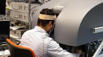
A man using a machine to control a surgical robot. Wires are connected to the back of his head.

Applying a painless electric current to the back of a person’s head while they learn a new task in VR can improve their performance in the real world, according to a small study out of Johns Hopkins University.

The challenge: Simulating surgery in VR can help doctors practice the operation without putting patients’ lives at risk. However, going from VR to the OR isn’t simple.

“Training in virtual reality is not the same as training in a real setting, and we’ve shown with previous research that it can be difficult to transfer a skill learned in a simulation into the real world,” said Jeremy D. Brown, a roboticist at Johns Hopkins University (JHU).

“The group that didn’t receive stimulation struggled a bit more to apply the skills they learned.”Guido Caccianiga

What’s new? In a new study, Brown and his colleagues demonstrate how electrically stimulating a person’s brain while they learn to control a surgical robot in VR simulations can improve their performance when they’re given control of the robot in the real world. 

“The group that didn’t receive stimulation struggled a bit more to apply the skills they learned in virtual reality to the actual robot, especially the most complex moves involving quick motions,” said researcher Guido Caccianiga. “The groups that received brain stimulation were better at those tasks.”

The details: During their study, the JHU team placed small electrodes on the backs of 18 volunteers’ heads. This allowed them to noninvasively stimulate the cerebellum, a part of the brain that plays a key role in learning new movements.

The volunteers, who didn’t have any experience in surgery or robotics, were then trained to use a surgical robot to lace a needle through three tiny holes, an exercise that mimics movements a surgeon might make during an operation.

This training took place in VR, and half of the participants received brain stimulation for the duration of it, while the others’ brains were stimulated only briefly at the beginning. 

The volunteers were then challenged to perform the task using an actual surgical robot, and according to the researchers, those who’d received the extended simulation were better at it.

“It’s very hard to claim statistical exactness, but we concluded people in the study were able to transfer skills from virtual reality to the real world much more easily when they had this stimulation,” said Brown.

Looking ahead: This isn’t the first study to claim a link between brain stimulation and enhanced learning, and the researchers acknowledge the need for larger studies to validate their finding that brain stimulation can improve VR training.

However, if the results hold up, they believe brain stimulation could one day be used to speed up training for doctors and others learning new skills in VR.

“What if we could show that with brain stimulation you can learn new skills in half the time?” said Caccianiga. “That’s a huge margin on the costs because you’d be training people faster.”