Brain stimulation could boost learning


People who had their brains zapped while learning a new task in VR were better at it in the real world.

A man using a machine to control a surgical robot. Wires are connected to the back of his head.

Applying a painless electric current to the back of a person’s head while they learn a new task in VR can improve their performance in the real world, according to a small study out of Johns Hopkins University.

The challenge: Simulating surgery in VR can help doctors practice the operation without putting patients’ lives at risk. However, going from VR to the OR isn’t simple.

“Training in virtual reality is not the same as training in a real setting, and we’ve shown with previous research that it can be difficult to transfer a skill learned in a simulation into the real world,” said Jeremy D. Brown, a roboticist at Johns Hopkins University (JHU).

“The group that didn’t receive stimulation struggled a bit more to apply the skills they learned.”Guido Caccianiga

What’s new? In a new study, Brown and his colleagues demonstrate how electrically stimulating a person’s brain while they learn to control a surgical robot in VR simulations can improve their performance when they’re given control of the robot in the real world. 

“The group that didn’t receive stimulation struggled a bit more to apply the skills they learned in virtual reality to the actual robot, especially the most complex moves involving quick motions,” said researcher Guido Caccianiga. “The groups that received brain stimulation were better at those tasks.”

The details: During their study, the JHU team placed small electrodes on the backs of 18 volunteers’ heads. This allowed them to noninvasively stimulate the cerebellum, a part of the brain that plays a key role in learning new movements.

The volunteers, who didn’t have any experience in surgery or robotics, were then trained to use a surgical robot to lace a needle through three tiny holes, an exercise that mimics movements a surgeon might make during an operation.

This training took place in VR, and half of the participants received brain stimulation for the duration of it, while the others’ brains were stimulated only briefly at the beginning. 

The volunteers were then challenged to perform the task using an actual surgical robot, and according to the researchers, those who’d received the extended simulation were better at it.

“It’s very hard to claim statistical exactness, but we concluded people in the study were able to transfer skills from virtual reality to the real world much more easily when they had this stimulation,” said Brown.

Looking ahead: This isn’t the first study to claim a link between brain stimulation and enhanced learning, and the researchers acknowledge the need for larger studies to validate their finding that brain stimulation can improve VR training.

However, if the results hold up, they believe brain stimulation could one day be used to speed up training for doctors and others learning new skills in VR.

“What if we could show that with brain stimulation you can learn new skills in half the time?” said Caccianiga. “That’s a huge margin on the costs because you’d be training people faster.”

New Technology May Help Inform Brain Stimulation


Summary: A new technique that uses ultrafast fMRI is able to capture brain activity at sub-second levels. The technique allows for real-time monitoring of the brain under stimulation conditions.

Source: University of Queensland

Brain stimulation, such as Deep brain stimulation (DBS), is a powerful way to treat neurological and psychiatric disorders. While it has provided therapeutic benefit for sufferers of Parkinson’s, Alzheimer’s, and addiction for more than a decade, its underlying neural mechanism is not yet fully understood.

Researchers at the Queensland Brain Institute (QBI) are now one step closer to unravelling the mystery of brain activity to better understand this mechanism and potentially predict DBS outcomes.

The brain is a highly complex network of circuits organised hierarchically with wide-ranging connections. Connections go in different directions, forwards and backwards, and between neurons that are either excitatory – the accelerators of a response – or inhibitory – the brakes modifying a response.

“Say you want to move your hand – once that signal is initiated, we expect that the activity that follows depends on the brain’s neural networks,” Associate Professor Kai-Hsiang Chuang said.

“What we don’t fully understand is how or when these structural and functional components of the brain interact to eventually lead to the outcome of moving your hand.”

Functional MRI (fMRI) is the most popular technique used to study brain networks. fMRI tracks blood flow and oxygenation changes following neural activity, thereby indirectly measuring the functional connections being formed, and giving us an indication of where brain activity is propagating.

Brain activity, however, isn’t as simple as a signal travelling from area to area.

The team at the Chuang laboratory have developed a new ultrafast fMRI technique with a vastly increased temporal resolution, enabling them to capture the dynamics of brain activity at a sub-second level.

Associate Professor Chuang said the new technique had led to more comprehensive understanding of how and when the brain’s structural and functional connections interact.

“The first new discovery we made is that brain activity not only propagates through structural wiring but follows certain preferential circuits depending on their excitatory and inhibitory neuronal distribution,” he said.

“Communication between brain regions of similar cell types becomes more fluent, and the brain activity stronger.”

This shows a brain map with the stimulated areas highlighted orange and blue
Mouse brain activity under optogenetic stimulation detected by ultrafast fMRI technique. Red shows a positive response (reflecting excitatory activity) and blue shows a negative response (reflecting inhibitory activity).

The Chuang group tracked the brain activity of mice both while stimulated and at rest using their ultrafast fMRI technique. When the brain was stimulated, activity followed the structural wiring in the forward direction — from A to B and then B to C. When the brain was at rest, activity was more dependent on cell type organisation and less on structural wiring, propagating between C and B but not with A, if that’s where the preferential circuit was.

This means that how information is processed is actually dependent on your state, where it was previously thought that brain activity functioned in the same way whether at rest or busy doing a task.

“The second discovery we made was that the blood signal detected by fMRI could reflect the network organisation and cell type distribution,” Associate Professor Chuang said.

“These findings have significant implications for how brain structure shapes function, and how to predict activity based on the knowledge of this structure. More practically, what we now know will impact the design of DBS and other brain stimulation techniques.

“The next steps are to work with clinicians versed in brain stimulation to determine how we can utilise this knowledge combined with human data to help improve our understanding of DBS.”

This more comprehensive understanding could enable us to better predict DBS results and potentially improve its design for better therapeutic outcomes.

Abstract

Hemodynamic transient and functional connectivity follow structural connectivity and cell type over the brain hierarchy

The neural circuit of the brain is organized as a hierarchy of functional units with wide-ranging connections that support information flow and functional connectivity. Studies using MRI indicate a moderate coupling between structural and functional connectivity at the system level.

However, how do connections of different directions (feedforward and feedback) and regions with different excitatory and inhibitory (E/I) neurons shape the hemodynamic activity and functional connectivity over the hierarchy are unknown.

Here, we used functional MRI to detect optogenetic-evoked and resting-state activities over a somatosensory pathway in the mouse brain in relation to axonal projection and E/I distribution.

Using a highly sensitive ultrafast imaging, we identified extensive activation in regions up to the third order of axonal projections following optogenetic excitation of the ventral posteriomedial nucleus of the thalamus.

The evoked response and functional connectivity correlated with feedforward projections more than feedback projections and weakened with the hierarchy.

The hemodynamic response exhibited regional and hierarchical differences, with slower and more variable responses in high-order areas and bipolar response predominantly in the contralateral cortex.

Electrophysiological recordings suggest that these reflect differences in neural activity rather than neurovascular coupling. Importantly, the positive and negative parts of the hemodynamic response correlated with E/I neuronal densities, respectively. Furthermore, resting-state functional connectivity was more associated with E/I distribution, whereas stimulus-evoked effective connectivity followed structural wiring.

These findings indicate that the structure–function relationship is projection-, cell-type- and hierarchy-dependent. Hemodynamic transients could reflect E/I activity and the increased complexity of hierarchical processing.

Brain Stimulation Leads to Long-Lasting Memory Improvement


Novel intervention selectively boosts working and long-term memory

A photo of a senior man struggling to remember something.

Noninvasive electrical brain stimulation led to selective improvements in working and long-term memory in older adults that lasted for at least a month, a randomized double-blind study showed.

Among 150 people ages 65 to 88, investigational transcranial alternating current stimulation for 20 minutes over 4 consecutive days produced selective boosts in auditory-verbal working memory and long-term memory, reported Robert Reinhart, PhD, of Boston University, and colleagues in Nature Neuroscience.

Low-frequency modulation in the parietal cortex improved working memory on days 3 and 4, and at 1 month after the intervention. High-frequency activity in the prefrontal cortex bettered long-term memory on days 2 to 4 and 1 month later.

“We found that by applying extremely weak electrical current safely and noninvasively to the prefrontal cortex at a high frequency, we could selectively improve long-term memory in older people aged 65 to 88 years old without changing short-term memory,” Reinhart said in a press briefing.

“And conversely, we found that we can apply the same kind of specialized alternating current, but this time now to the parietal cortex farther back in the brain at a low frequency and by doing this, we could selectively improve short-term memory in older people without changing long-term memory,” he added.

“That is, based on the spatial location and the frequency of the electrical stimulation, we can improve either short-term memory or long-term memory separately,” he noted.

The rate of memory improvement over 4 days predicted the size of memory benefits 1 month later, the researchers found. Moreover, people with lower baseline cognitive function experienced larger, more enduring memory improvements.

“Older people with poor general cognitive functioning at baseline coming into the experiment were the individuals who showed the largest improvements during both the intervention and the 1-month time point, which we think bodes well for transferring this intervention over to a proper clinical study of people with Alzheimer’s disease who are suffering from more severe memory impairments,” Reinhart observed.

The study involved 150 older adults who received electrical currents through a cap embedded with electrodes. Mean baseline scores of participants ranged from 25.45 to 27.4 on the 30-point Montreal Cognitive Assessment (MoCA) test. Scores of 18-25 on the MoCA suggest mild cognitive impairment, while scores of 26 or higher are considered normal.

Participants heard and were asked to recall five lists of 20 words. Targeting the inferior parietal lobule at 4 Hz boosted participants’ ability to recall words from the end of the list, indicating working memory. Targeting the dorsolateral prefrontal cortex at 60 Hz improved recall from the beginning of the list, reflecting long-term memory storage.

“Statistically, the effects were moderate to large” and most participants experienced memory benefits, Reinhart said. “Specifically, 85% to 90% experienced the memory improvements during the intervention that were then predictive of the benefits we saw at the 1-month timepoint after the intervention.”

The findings build on research from Reinhart’s lab in 2019 that showed stimulating temporal and prefrontal brain areas simultaneously improved memory for at least 50 minutes.

That work reported short-lasting improvements in visuospatial working memory only, Reinhardt noted. In the new study, the researchers aimed to target additional memory systems and induce more long-lasting memory benefits.

“The advance of this work over and above that of our 2019 research was that we constructed a repetitive intervention, one where we’re stimulating people 20 minutes each day for 4 consecutive days, unlike in 2019 where it was just a one-shot stimulation,” he said.

While promising, the intervention still is in early stages, the researchers acknowledged. What’s not clear is whether it has benefits for people with neurodegenerative diseases, especially those at risk for dementia.

But as people age, noninvasive brain stimulation may help improve daily activities and could be personalized based on functional or anatomical characteristics, possibly leading to more lasting effects, they suggested.

“There’s been remarkable progress in the neurosciences over the decades on characterizing the brain circuits and networks that underpin our memory capacity, both short-term and long-term,” Reinhart pointed out.

“There’s also evidence showing that rhythmic or oscillatory brain activity patterns in certain regions are important for organizing and retrieving memory,” he added. “What we need now are really innovative technologies that allow us to act on these large-scale oscillatory brain networks and determine whether it’s possible to protect or even enhance memory for older people.”

Brain Stimulation of Novel Target Improves Depression


Direct electrical stimulation of the lateral orbitofrontal cortex (OFC) acutely improves mood in patients with depressive symptoms, new research shows.

OFC stimulation “broadly modulates mood-related circuitry, revealing a new brain stimulation target with strong therapeutic potential,” the investigators, led by Edward F. Chang, MD, professor of neurosurgery, University of California San Francisco, write.

The OFC, a small region on the lower surface of the brain just above the eyes, is “one of the least understood regions in the brain, but it is richly connected to various brain structures linked to mood, depression and decision making, making it very well positioned to coordinate activity between emotion and cognition,” Chang said in news release.

The study was published online November 29 in Current Biology.

Shedding New Light

The investigators studied 25 patients with epilepsy who were implanted with intracranial electrodes for seizure localization. Baseline depression traits ranged from mild to severe based on the Beck Depression Inventory.

Unilateral stimulation of the lateral OFC “consistently produced acute, dose-dependent mood-state improvement across subjects with baseline depression traits.”

This “stimulation induced neural features associated with positive mood states,” the researchers report. Sham stimulation of the OFC produced no discernible change in mood.

“Our findings shed light on how brain activity relates to mood symptoms and how symptom-relieving stimulation effects that activity,” co-study leader Kristin Sellers, PhD, postdoctoral researcher in Chang’s lab, told Medscape Medical News.

“These observations advance our understanding of the brain networks that underlie mood disorders and suggest ways in which we might use that understanding to develop therapies that are better-informed and more effective,” said Sellers.

Questions Remain

Heather Dawes, PhD, co-director of the Defense Advanced Research Projects Agency (DARPA) Systems-Based Neurotechnology for Emerging Therapies (SUBNETS) program at UCSF, cautioned that there is still “much work to be done to see whether this stimulation approach can help patients with depression actually recover from their illness; so far, we’ve only been able to look at short-term effects.”

“Some of this continuing work will be conducted in the context of clinical trials, using implanted recording and stimulation devices to enable longer tracking of potential therapeutic effects. In addition, we are working to optimize the effect, and to make sure that stimulation of this part of the brain is safe and effective as a possible longer-term treatment,” Dawes, who worked on the study, told Medscape Medical News.

Helen Mayberg, MD, behavioral neurologist and founding director of the Center for Advanced Circuit Therapeutics, Icahn School of Medicine at Mount Sinai in New York City, agrees that more study is needed and urged caution in interpreting the study data.

“There are a lot of areas in the brain that you can stimulate and get an acute effect, so this is another interesting piece of the puzzle,” but it’s unclear if the effect is long-lasting, she told Medscape Medical News.

The OFC, said Mayberg, is “an area of the brain that connects to areas that we already do stimulate, so the results are certainly not contradictory, but it’s also not a paradigm shift.”

Mayberg said it’s also important to note that depression was not the primary diagnosis in these patients. “These were epilepsy patients and some of them had high Beck Depression Inventory scores, which basically is a trait depression rating. It’s not a diagnosis of depression. This needs to be replicated in patients with major depression.”

New Device Can Ease Chronic Pain Without Drugs, Thanks to Brain Stimulation


IN BRIEF

This new method of pain treatment can prevent risky side-effects such as addiction, dependence, and overdose-related deaths – and it does so using electricity.

OPIOID MEDICINES

Abuse of prescription pain killers or opioid medicines is common. But then again, how else can you treat chronic pain? Unfortunately, addiction is a terrible side-effect that can lead to overdose-related deaths.

But now a research team from the University of Arlington seems to have found a better and more efficient solution: Electrical stimulation.

By delivering electrical currents—which can block pain signals at the spinal cord level—into a deep, middle brain structure, it might be possible to treat chronic pain without the intervention of drugs. At the same time, the technique can spur the release of dopamine, which helps with the emotional distress typically associated with long-term pain.

A SHOCKING STUDY

“This is the first study to use a wireless electrical device to alleviate pain by directly stimulating the ventral tegmental area of the brain,” said Yuan Bo Peng, UTA psychology professor. “While still under laboratory testing, this new method does provide hope that in the future we will be able to alleviate chronic pain without the side effects of medications.”

Yuan Bo Peng, UTA Psychology Professor.

The team experimented with a custom-built wireless implant, which through electrical stimulation of the ventral tegmental area effectively reduced the sensation of pain, even blocking pain signals in the spinal cord.

This could greatly benefit the almost two million Americans who are addicted or dependent on opioid medicines. The Centers for Disease Control that 165,000 Americans died of opioid-related overdoses from 1999 to 2014.

“Until this study, the ventral tegmental area of the brain was studied more for its key role in positive reinforcement, reward and drug abuse,” said Peng. “We have now confirmed that stimulation of this area of the brain can also be an analgesic tool.”

Source:futurism.com

New Device Can Ease Chronic Pain Without Drugs, Thanks to Brain Stimulation


IN BRIEF

This new method of pain treatment can prevent risky side-effects such as addiction, dependence, and overdose-related deaths – and it does so using electricity.

OPIOID MEDICINES

Abuse of prescription pain killers or opioid medicines is common. But then again, how else can you treat chronic pain? Unfortunately, addiction is a terrible side-effect that can lead to overdose-related deaths.

But now a research team from the University of Arlington seems to have found a better and more efficient solution: Electrical stimulation.

By delivering electrical currents—which can block pain signals at the spinal cord level—into a deep, middle brain structure, it might be possible to treat chronic pain without the intervention of drugs. At the same time, the technique can spur the release of dopamine, which helps with the emotional distress typically associated with long-term pain.

A SHOCKING STUDY

“This is the first study to use a wireless electrical device to alleviate pain by directly stimulating the ventral tegmental area of the brain,” said Yuan Bo Peng, UTA psychology professor. “While still under laboratory testing, this new method does provide hope that in the future we will be able to alleviate chronic pain without the side effects of medications.”

Yuan Bo Peng, UTA Psychology Professor. 

The team experimented with a custom-built wireless implant, which through electrical stimulation of the ventral tegmental area effectively reduced the sensation of pain, even blocking pain signals in the spinal cord.

This could greatly benefit the almost two million Americans who are addicted or dependent on opioid medicines. The Centers for Disease Control that 165,000 Americans died of opioid-related overdoses from 1999 to 2014.

“Until this study, the ventral tegmental area of the brain was studied more for its key role in positive reinforcement, reward and drug abuse,” said Peng. “We have now confirmed that stimulation of this area of the brain can also be an analgesic tool.”

POPULAR BRAIN STIMULATION DOESN’T BOOST IQ


Despite its popularity, using a weak electric current to boost brainpower doesn’t live up to the hype. A new study shows that the most common form of the treatment actually has a statistically significant detrimental effect on IQ scores.

Published in the journal Behavioural Brain Research, the study adds to the increasing amount of literature showing that transcranial direct current stimulation—tDCS—has mixed results when it comes to cognitive enhancement.

“It would be wonderful if we could use tDCS to enhance cognition because then we could potentially use it to treat cognitive impairment in psychiatric illnesses,” says Flavio Frohlich, study senior author and assistant professor of psychiatry, cell biology and physiology, biomedical engineering, and neurology at the UNC School of Medicine.

“So, this study is bad news. Yet, the finding makes sense. It means that some of the most sophisticated things the brain can do, in terms of cognition, can’t necessarily be altered with just a constant electric current.”

TWO TYPES OF BRAIN STIMULATION

Frohlich, though, says that using less common alternating current stimulation—so-called tACS—could be a better approach, one that he has been investigating. Earlier this year, Frohlich’s lab found that tACS significantly boosted creativity, likely because he used it to target the brain’s natural electrical alpha oscillations, which have been implicated in creative thought.

With tDCS, scientists don’t target these brain waves, which represent neuronal patterns of communication throughout regions of the brain. Instead, they use tDCS to target brain structures, such particular regions of the cortex.

“All of our brain structures look more or less the same, but the reason why we’re all so different is that the electrical brain activities in our brains are very different,” Frohlich says. “We have to better understand this and target specific brain activity patterns.”

‘EXPLOSION’ OF STUDIES

Using a weak electrical current to boost the brain’s natural abilities has been around for decades, but the current boom within the science community started in 2000, when German scientists published a paper showing that tDCS could change the excitability of neurons in the motor cortex—the brain region that controls voluntary body movement.

Since then, there’s been an explosion of tDCS studies to try to make neurons more active or less active and therefore change outcomes for a variety of brain functions, such as working memory and cognitive acuity, and for illnesses, such as depression and schizophrenia.

But Frohlich says that scientists still don’t know exactly what the direct current does to neural activity. He also says some of the studies that have made waves were poorly designed. Some studies were not properly double-blinded or properly placebo controlled. Other studies were very small—fewer than 10 people.

A recent meta-analysis of a large number of tDCS papers showed that tDCS is far from a magic pill for cognitive enhancement or brain-related health conditions.

“Aside from stimulating the motor cortex, which has very exciting implications for stroke rehabilitation, I think the jury is still out on tDCS,” says Frohlich, who is a member of the UNC Neuroscience Center.

PUTTING ON THE ELECTRODES

In the new, Frohlich’s team—including graduate student Kristin Sellers, the paper’s first author—recruited 40 healthy adults, each of whom took the standard WAIS-IV intelligence test—the most common and well-validated test of IQ, which includes tests for verbal comprehension, perceptional reasoning, working memory, and processing speed.

A week later, Frohlich’s team divided the participants into two groups. Electrodes were placed on each side of each participant’s scalp, under which sat the frontal cortex. A third electrode, which sent electricity back to the device that that produced the electric current, was placed on top of the scalp.

Duke University collaborator and coauthor Angel Peterchev created imaging simulations to ensure Frohlich’s team targeted the same parts of the cortex that previous tDCS studies had targeted.

Then the placebo group received sham stimulation—a brief electrical current, which led participants to think they had been receiving the full tDCS. The other participants received the standard tDCS for twenty minutes—a weak electrical current of 2 millioamperes.

All participants then retook the IQ tests. Frohlich expected that most, if not all, IQ scores would improve, but that the participants who received tDCS would not improve their scores any more than would the people who did not undergo tDCS

SCORES THAT SANK

Frohlich’s team did find that all scores improved—most likely because of the “practice effect” of previously taking the test. Surprisingly, the participants who did not receive tDCS saw their IQ scores increase by ten points, whereas participants who received tDCS saw their IQ scores increase by just shy of six points, on average.

When Frohlich and colleagues analyzed the test scores, they saw that the scores for three of the four kinds of cognitive tests were very similar between the two groups of participants. But the scores for perceptual reasoning were much lower among people who underwent tDCS.

Perceptual reasoning tests fluid intelligence, which is defined as the ability to think logically and apply innovative problem solving to new problems.

Within the category of perceptual reasoning, the researchers saw the biggest differences in the subcategory of matrix reasoning—when participants viewed two groups of symbols and had to find the one symbol missing from the other group.

“This is one of the classical assays of fluid intelligence,” Frohlich says, “where you have to identify hidden rules and apply logic to find a missing element.”

NO ‘MAGIC PILL’

Frohlich emphasizes, “Our findings do not preclude the possibility that other tDCS paradigms may be less harmful or even beneficial. However it is time to make sure that everybody uses gold standard, placebo-controlled, double-blind study designs. Also, our study demonstrates the importance of more research on how stimulation interacts with brain activity.”

Frohlich stresses that the scientific community should be careful not to create simplistic storylines about tDCS being a “magic pill” for many brain-related conditions.

“There could be dangerous consequences, especially if tDCS is used daily,” he says. “Ours was an acute study. We don’t know what the long-term effects are. There is so much more we need to understand before tDCS is ready for home use without medical supervision”

Frohlich adds, “I think our study demonstrates that we need to think of smarter ways to engage the brain to really target the specific brain dynamics involved in what we want to improve, such as cognition for people with depression or schizophrenia. I think tACS is an option, as well as more sophisticated modalities we’ve yet to develop.”

A New Psychiatry Subspecialty?


While onsite at the 2013 Annual Meeting of the American Psychiatric Association, Medscape spoke with Drs. Edward M. Kantor and Nolan R. Williams about the emerging new field of interventionalist psychiatry and their initiative to develop a training program.[1]

Background

Medscape: What is interventional psychiatry?

Dr. Williams: Interventional psychiatry is an emerging subspecialty that uses brain stimulation techniques to modulate the dysfunctional circuitry underlying medically resistant psychiatric diseases. Physicians who deliver procedures in the spectrum between standard care and surgery are sometimes referred to as “interventionalists” in other areas of medicine (eg, cardiology, radiology, and neurology). Currently, the field of psychiatry does not recognize interventionalists or offer formal training and certification. Our group is proposing the concept of “interventional psychiatrist” in place of procedure-specific terms such as “somatic therapist” or “ECT (electroconvulsive therapy) practitioner,” which fail to encompass the scope of brain stimulation strategies. It is not meant to replace current psychiatric therapies (medication and psychotherapy) but rather to enhance the practice of psychiatry with an additional set of tools. This can be viewed much in the same way that interventional cardiologists do not replace general cardiologists.

Dr. Kantor: I also see this bringing great opportunity for collaboration across neurology, neurosurgery, and psychiatry, which rarely occurs in other settings. This alone may advance our liaison activities and communication and, more than anything else, will facilitate better care between the disciplines and really help focus us on the whole person — mind, body, and brain — as opposed to one at a time, in isolation.

Medscape: How do you recommend that interventional psychiatry be incorporated into clinical training?

Dr. Kantor: It’s an emerging area of our field, where older techniques like ECT are being adapted for better efficacy with fewer side effects, and new techniques are coming of age. It’s not currently accredited on the training side, but my guess is that as it formalizes over the next 2-3 years, that process will naturally evolve. As a residency director, I support an educational plan that outlines minimum competencies, experience, and oversight. We already have begun exploring the paradigm with the American Association of Directors of Psychiatric Residency Training (AADPRT) and relevant specialty societies. I think the education has to begin with programs like ours at Medical University of South Carolina (MUSC), where the resources are more established, there are enough cases and mentors, and we can train practitioners and research scientists within psychiatry, in a way best suited to work within the existing graduate medical education framework.

A component of basic understanding and clinical exposure in residency would likely be the minimum expectation. On top of that, an optional, more formal track, using senior elective time, would be fairly easy to plan for within the existing training structure. Currently, not all programs have the expertise, but I believe that there is a desire among those that do to collaborate with needed tools like shared guidelines, online and remote learning, and course-based experiences. That said, I imagine that there will never be a one-size-fits-all plan that works in every institution.

Dr. Williams: Psychiatry is rapidly changing. New methods for noninvasively and invasively stimulating the brain have powerful therapeutic potential, but they require background knowledge (eg, circuits, physics of electricity) that is foreign to most psychiatrists. Interventional psychiatry is an emerging subspecialty that needs to be formally recognized and developed at various levels of psychiatric training. Academic centers will have to adapt to ensure adequate training to those who will be providing these neuromodulatory interventions, in order to avoid mistakes of an earlier era and to make sure that psychiatrists are the ones to perform the procedures, rather than other specialists who are clinically unfamiliar with the psychiatric disease management. Establishing formal training programs will ensure that psychiatry is ready to meet the challenges of treatment-resistant psychiatric illness with a properly trained cohort of interventional psychiatrists. We have an interventional psychiatry fellowship program at MUSC, and there are a few others starting around the country.

Approaches to Neuromodulation

Medscape: Can you walk our readers through the primary types of neuromodulation techniques used in psychiatry currently, and also in which conditions they are used, both on- and off-label?

Dr. Williams: Yes.

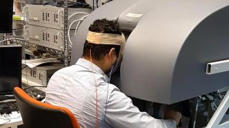
Transcranial magnetic stimulation (TMS): There are currently 2 different TMS coils that have been approved by the US Food and Drug Administration (FDA) for the acute treatment of depressed patients who have failed to respond to at least 1 antidepressant medication. Interventional psychiatrists need to understand the fundamental principles behind TMS and demonstrate competency in the delivery and programming of TMS paradigms. A recent multisite, naturalistic, observational study of acute treatment outcomes in clinical practice[2] demonstrated greater than 50% efficacy in sicker populations using TMS.

ECT: ECT is an effective acute treatment for a wide array of neuropsychiatric diseases (eg, depression, mania, psychosis) and remains the single most effective therapy for treatment-resistant depression (TRD). Although ECT has been used for nearly a century, advances in the way that it is delivered have greatly reduced side effects. Shorter pulse widths and unilateral electrode configurations have been shown to diminish cognitive side effects. Interventional psychiatrists should receive comprehensive ECT training that addresses indications and contraindications, length and timing of treatment, pulse programming, and maintenance therapy.

Focal electrically administered seizure therapy (FEAST): A new type of ECT has been developed, called FEAST. This unidirectional electrical stimulation with a novel electrode placement and geometry has been proposed as a means to initiate seizures in the prefrontal cortex prior to secondary generalization, but it is still in the research phase.[3]

Vagus nerve stimulation (VNS): VNS was FDA approved in 1997 to treat epilepsy, and in 2005 it became the first invasive neuromodulation device approved by the FDA as a treatment for a psychiatric disorder (chronic TRD). Unfortunately, VNS was FDA approved prior to any Class 1 evidence of efficacy; thus, insurance companies have been reluctant to reimburse for the implant. Nevertheless, the effects of VNS appear to be remarkably durable. Interventional psychiatrists should be the lead contacts on VNS consults and programming for TRD.

Deep brain stimulation (DBS): DBS is typically used to manage movement disorders but is now being investigated as a therapy for a variety of neuropsychiatric conditions such as obsessive-compulsive disorder (OCD), Tourette syndrome [still in research phase], addiction [still in research phase], and TRD [still in research phase]. In 2009, the FDA granted a somewhat controversial humanitarian device exemption for use of DBS for treatment-resistant OCD. Interventional psychiatrists will play a critical role in developing the field of functional neurosurgery for psychiatric disorders. Pertinent skills include patient consultation, intraoperative assessment, postoperative programming, troubleshooting, and integrating device settings with medications (psychopharmacology). There has also been an explosion in psychiatric side effects of DBS used for neurologic conditions like Parkinson disease. The interventional psychiatrist should be adequately trained to troubleshoot these issues.

Transcranial direct-current stimulation (TDCS): This is a therapy that involves an energy source that delivers a constant weak (typically ≤ 1 mA) electrical current through scalp electrodes. This therapy is not grandfathered in by the FDA as a device currently in practice, although other, similar devices are. A recent study[4] from Brazil demonstrated that when combined with sertraline, there is a synergistic effect in treating depression. There are limited data currently, but it seems to have great promise and low cost.

Putting Neuromodulation Into Practice

Medscape: Can you expand on how these techniques might be incorporated into care in conjunction with psycho- and pharmacotherapy?

TMS: In the pivotal trials, the patients were not on any medications. In the real world, TMS is typically combined with medications and therapy. There are now studies looking at combining therapy with TMS for a synergistic effect.

ECT: There are medications that, when used alongside ECT (venlafaxine/nortriptyline) or after ECT (lithium), increase the chances of improvement and better cognitive outcomes.

DBS: Typically this intervention can eventually replace medications; many of the studies reduced/removed medications once the device was working. In many instances, DBS (particularly in depression) will allow for patients to better participate in therapies that they would not have been able to participate in before.

TDCS: The most efficacy that has been shown to date is in combination with sertraline. This will potentially be a role for TDCS in enhancing therapeutic efficacy.

Medscape: Tell us about the interventional psychiatry training program at MUSC and how you envision the program evolving and affecting care.

Dr. Williams: Drs. Mark George and Baron Short have developed a 1-year interventional psychiatry fellowship with the first fellow, Dr. Jon Snipes, finishing June 30, 2013. A second fellow, Dr. Suzanne Kerns, will begin in July 2013.

We feel that interventional psychiatry should be present at 3 levels: (1) a core curriculum of introductory knowledge and experience during psychiatry residency training for all psychiatrists; (2) a neuromodulation elective track during residency at some locations; and eventually (3) a formal interventional psychiatry fellowship that leads to an approved subspecialty certification process under the American Board of Medical Specialties (ABMS).

Base resident education: Psychiatry residents should have an introductory-level understanding of the brain circuits underlying behavior and how they can be modulated using invasive and noninvasive brain stimulation. This fundamental knowledge should improve the quality of patient management by ensuring that patients are aware of the full complement of available therapeutic interventions. Ideally, all psychiatric residents would have a core curriculum that includes brain stimulation consultation and observation of ECT and TMS.

Interventional psychiatry track: Psychiatry residents who have a specific interest in brain stimulation should have the option of pursuing a dedicated training track within their residency program. Under this proposal, interested residents would be required to manage ECT and TMS treatment cases, from initial consultation to acute therapy and maintenance treatments. Psychiatrists who are currently performing these duties could be grandfathered into this arrangement.

Interventional psychiatry fellowship: Psychiatrists who wish to pursue the most rigorous training program should have the option of pursuing a 1-year fellowship that includes focused training in all of the aforementioned techniques. This training would occur at institutions with robust neuromodulation programs in collaboration with neurology, neurosurgery, and neuroradiology. Fellowship trainees should receive hands-on exposure in established (ECT, TMS, DBS, VNS) and emerging (eg, TDCS) neuromodulatory technologies. Additionally, fellowship trainees should have experience with the tools used to measure the effects of neuromodulation, such as functional MRI and EEG.

Previous Page


Stimulating The Brain’s Immune Response May Provide Treatment For Alzheimer’s Disease


A new target for the prevention of adverse immune responses identified as factors in the development of Alzheimer’s disease (AD) has been discovered by researchers at the University of South Florida’s Department of Psychiatry and the Center of Excellence for Aging and Brain Repair.

Their findings are published online in the Journal of Neuroscience.

The CD45 molecule is a receptor on the surface of the brain’s microglia cells, cells that support the brain’s neurons and also participate in brain immune responses.

Previous studies by the USF researchers showed that triggering CD45 was beneficial because it blocked a very early step in the development of Alzheimer’s disease. In the present study, the researchers demonstrated in Alzheimer’s mouse models that a loss of CD45 led to dramatically increased microglial inflammation.

Although the brain’s immune response is involved in Alzheimer’s disease pathology, “this finding suggests that CD45 on brain immune cells appears critically involved in dampening harmful inflammation,” said study senior author Jun Tan, M.D., Ph.D., a professor of psychiatry and Robert A. Silver chair at the Rashid Laboratory for Developmental Neurobiology, USF Silver Child Development Center and research biologist for Research and Development Service at the James A. Haley Veteran’s Hospital.

The investigators also found an increase in harmful neurotoxins, such as A beta peptides, as well as neuron loss in the brains of the test mice.

“In short, CD45 deficiency leads to increased accumulation of neurotoxic A beta in the brains of old Alzheimer’s mice, demonstrating the involvement of CD45 in clearing those toxins and protecting neurons,” Dr. Tan said. “These findings are quite significant, because many in the field have long considered CD45 to be an indicator of harmful inflammation. So, researchers assumed that CD45 was part of the problem, not a potential protective factor.”

The next step is to apply these findings to develop new Alzheimer’s disease treatments, said Paula Bickford, Ph.D., a professor in the USF Department of Neurosurgery and senior career research scientist at the James A. Haley Veteran’s Hospital.

“We are already working with Natura Therapeutics, Inc. to screen for natural compounds that will target CD45 activation in the brain’s immune cells,” Dr. Bickford said.