Beating-Heart Septal Myectomy: An Innovative Approach for Hypertrophic Obstructive Cardiomyopathy.


Introduction

Hypertrophic cardiomyopathy (HCM) accounts for a significant portion of inherited cardiovascular diseases. The incidence of HCM is estimated to be approximately 2 to 5 per 1,000 people in the general adult population.1-3 HCM often leads to chest pain, dyspnea, and heart failure and is among the important causes of sudden cardiac death. At present, there is a consensus that left ventricular outflow tract (LVOT) obstruction is an important cause of clinical symptoms in most patients with hypertrophic obstructive cardiomyopathy (HOCM). For patients who receive drug therapy but still have significant symptoms or are drug intolerant, septal-reduction therapy (SRT) should be strongly considered. Surgical myectomy (SM) has been the gold standard of SRT because of its efficacy and proven safety. In experienced high-volume centers, postoperative patients of hemodynamic obstruction have a remission rate of more than 95% and have the potential to return to normal life expectancy.4-6 Although the risk of mortality and complications associated with surgery and cardiopulmonary bypass (CPB) is generally low in these patients, morbidity and mortality remain important concerns.

With the advancement in medical technology and the increase in patient desires, there is a strong impetus to evaluate other HOCM treatment options beyond surgery. Alcohol septal ablation (ASA) for treatment of HOCM has been accepted as a less invasive and more effective alternative in suitable patients. In recent years, the emergence of a new interventional therapy, percutaneous intramyocardial septal radiofrequency ablation (PIMSRA [Liwen procedure]), adopts a transapical approach under the guidance of transthoracic echocardiography (TTE) to insert a radiofrequency electrode directly into the hypertrophied ventricular septum. Liwen procedure can reliably achieve the therapeutic goal of the resting left LVOT gradient <30 mm Hg.7-9

Recently, transapical beating-heart septal myectomy (TA-BSM), inspired by strategies such as transapical aortic valve implantation, has shown us a trend that navigates the development of myectomy.10 The pericardium is exposed through a small thoracic incision, and a newly designed device is used to go through the apex of the heart and into the left ventricle, and then advance to the outflow tract under real-time echocardiographic guidance. Hypertrophic septal myocardial tissue, which is captured by the beating-heart myectomy device with negative pressure and a puncture needle, is then excised and removed. In this first-in-human clinical trial, 47 patients received TA-BSM and were followed up for 3 months. Forty-two patients were considered to achieve successful outcome (maximal LVOT gradient were decreasing from 86 [range: 67-114] mm Hg to 19 [range: 14-28] mm Hg). In terms of safety, 1 patient died in hospital (2%). One patient had iatrogenic ventricular septal perforation (2%). One patient had apical tear and was converted to sternotomy (2%). One patient required a permanent pacemaker implantation (2%).

Similarities and Distinctions of PIMSRA and TA-BSM

TA-BSM appears to be feasible as an SRT and represents an important technologic innovation with impressive results. As a minimal invasive, beating-heart treatment without CPB, TA-BSM is of great worldwide significance in the innovation of treatment for HOCM. Although short-term data from the preliminary series cannot confirm the efficacy and safety of this technique, the results of further large-scale, long-term studies are fully expected. As original Asian/Chinese procedures, TA-BSM and PIMSRA share many similarities: 1) both provides SRT for drug-resistant HOCM without CPB or sternotomy; 2) both avoid the difficulties of relying on vascular anatomy for catheter-based coronary approach and minimize complications such as myocardial infarction during ASA; and 3) both have relatively small trauma, concise inserting route, and rapid post-procedure recovery. Specific data are shown in Table 1.

ApproachFirst Author,
Year
CountryPeriodNMean Age, ySCDI, %Follow-Up, monthsResting LVOTG, mm HgNovel Device Success, %Peri-Mortality, %New-Onset LBBB, %New-Onset RBBB, %Pacemaker,
%
VT,
%
VSP,
%
LVAT,
%
PIMSRALiu L, 20187China2016-20171540.76.1688/11a0000000
PIMSRAZhou M, 20228China2016-202020046.919 (6-50)79/14a115.500.500
PIMSRAWang Z 20229China2019-20206847.74.31275/12a10001.58.80000
TA-BSMFang J, 202310China20224749.14.0366/11a97.92.138.302.102.12.1

LBBB = left bundle branch block; LVAT = left ventricular apical tear; LVOTG = left ventricular outflow tract gradient; Peri = peri-procedural (<30 days); PIMSRA = percutaneous intramyocardial septal radiofrequency ablation; RBBB = right bundle branch block; TA-BSM = transapical beating-heart septal myectomy; VSP = ventricular septal perforation; VT = ventricular tachycardia.

a P < 0.05.

The principles and mechanisms of the TA-BSM and PIMSRA are different. TA-BSM is essentially a myectomy, but PIMSRA is accomplished by inserting a radiofrequency needle and ablating the myocardium supplied by the left anterior descending (LAD) coronary artery, including the septal perforators. The localized “therapeutic infarction” is induced by ablation energy delivered in the myocardium supplied by LAD artery distribution with minimal injury to the surrounding tissues. These effects eliminate or reduce the LVOT gradient immediately after the procedure, which might result from hypokinesia of the left ventricular (LV) wall and the uncoordinated movement caused by thermal myocardial necrosis. The continuous reduction in the LVOT gradient and septal thickness, as well as the broadened LVOT, possibly result from a local remodeling process caused by ongoing fibrosis and shrinkage of the ablation-induced septal lesion. TA-BSM is an open surgical procedure that requires a small incision—minimally invasiveness—and a pouch suture after instrument removal. PIMSRA is an extremely minimal invasive approach with no surgical wound and therefore less trauma. TA-BSM needs to excise the myocardium from the endocardial surface, which can potentially affect the conduction system and cardiac hemodynamics. The puncture needle of PIMSRA enters the interventricular septum directly from the apex of the heart, without direct contact with the endocardium. Effective management of ablation energy during the procedure can effectively reduce the impact on the conduction system and hemodynamics. TA-BSM is guided by transesophageal echocardiography (TEE), which provides excellent images yet more complex peri-operative set-up. PIMSRA uses transthoracic echo imaging, which is convenient for the comparison of pre- and postprocedure data. In addition, the safety of early clinical trials is different. As a new procedure, TA-BSM has major adverse events (MAEs) including ventricular septal perforation, requirement for permanent pacemaker implantation, left ventricular apical tear, and sternotomy conversion, whereas the 30-day MAEs of PIMSRA are intraprocedural pericardial effusion, hypotension, ventricular tachycardia, and 1 case of late aneurysm of the septal branch.

Significance of Innovation in Asia

The fruitful achievements of innovation in the field of cardiovascular medicine in China and Asia in recent years have helped to advance new therapies for patients with structural heart diseases. PIMSRA and TA-BSM are fueled by patient demand for minimally invasive treatment for HOCM, especially in Asia.

Pioneering the Future Path

Given the rapid advancements in interventional techniques, several trends may be expected in the development of the SRT field in HOCM for the next few years. As the TA-BSM and PIMSRA have different indications and contraindications, reducing the respective complications will be the next priority. Hierarchical management will be more precise, especially for perioperative management of critical patients. Standardized training will be developed to truly achieve individual treatment. Device evolution based on the integration of medicine and engineering will contribute to higher intelligence and less trauma in treatment.

As a first-in-human study, TA-BSM showed promising feasibility and encouraging initial results. We applaud the TA-BSM team for their innovation of transapical beating-heart myectomy to treat patients with severe HOCM and are cautiously optimistic about optimizing PIMSRA (Liwen procedure) and TA-BSM for more eligible HOCM patients in the future.

Exercise’s Dopamine-Driven Cognitive Boost


Summary: Recent research has revealed a significant link between exercise and improved cognitive performance, attributing this enhancement to increased dopamine levels. This discovery, involving sophisticated PET scans to monitor dopamine release in the brain during exercise, indicates that dopamine plays a vital role in boosting reaction times and overall brain function.

The study’s implications are far-reaching, suggesting potential therapeutic applications for conditions influenced by dopamine, like Parkinson’s disease and ADHD. The research underscores the importance of voluntary exercise for cognitive health, differentiating it from involuntary muscle stimulation.

Key Facts:

  1. The study used positron emission tomography (PET) scans to track dopamine release in the brain during exercise, linking it to improved cognitive performance.
  2. Findings suggest that voluntary exercise, not just forced muscle movement, is essential for this dopamine-related enhancement of brain function.
  3. The research has potential therapeutic implications for various dopamine-influenced conditions, including Parkinson’s disease and ADHD.

Source: University of Portsmouth

A study exploring the mechanisms behind why cognitive performance improves in response to exercise, has found that dopamine plays a key role.

The neurotransmitter and hormone – which is tied to pleasure, satisfaction and motivation – is known to increase when you work out. New findings suggest it is also linked to faster reaction time during exercise.

The researchers behind the discovery say it could lead to a new therapeutic pathway for cognitive health, because of dopamine’s significant role in several conditions including Parkinson’s disease, schizophrenia, ADHD, addiction, and depression.

This shows a woman exercising.
The results revealed that when a participant cycled lying down in the machine, their brain increased the amount of dopamine release, and that this process was linked with improved reaction time.

They measured the release of dopamine in the brain using a sophisticated scanning device, known as a positron emission tomography (PET). It tracks the metabolic and biochemical activity of the cells in the body.

The results revealed that when a participant cycled lying down in the machine, their brain increased the amount of dopamine release, and that this process was linked with improved reaction time. 

Dr Joe Costello, from the University’s School of Sport, Health & Exercise Science (SHES), said: “We know cardiovascular exercise improves cognitive performance, but the exact mechanisms behind this process have not been rigorously investigated in humans until now.

“Using novel brain imaging techniques, we were able to examine the role dopamine plays in boosting brain function during exercise, and the results are really promising. Our current study suggests the hormone is an important neuromodulator for improved reaction time.

“These findings support growing evidence that exercise prescription is a viable therapy for a host of health conditions across the lifespan.”

As part of the study, three experiments were carried out with 52 male participants overall. In the first, individuals were asked to carry out cognitive tasks at rest and while cycling in the PET scanner, so the team could monitor the movement of dopamine in their brain. 

The second used electrical muscle stimulation to test whether forced muscle movement to stimulate exercise would also improve cognitive performance. The final experiment combined both voluntary and involuntary exercise.

In the experiments where voluntary exercise was carried out, cognitive performance improved. This was not the case when only forced electrical stimulation was used.

Soichi Ando, Associate Professor in the Health & Sports Science Laboratory at the University of Electro-Communications in Japan, said: “We wanted to remove voluntary muscle movement for part of the study, to see if the process in which acute exercise improves cognitive performance is present during manufactured exercise. But our results indicate that the exercise has to be from the central signals of the brain, and not just the muscle itself.

“This suggests that when we tell our central command to move our body during a workout, that’s the process which helps the dopamine release in the brain.”

The team’s previous study examined the relationship between oxygen levels, cognitive performance and exercise, to test the theory that the more oxygen we breathe during a workout, the more awake our brain is. They found no change to an individual’s reaction time when cycling both inside and outside of an environment with low levels of oxygen (hypoxia). 

“These latest findings support our previous theory that cognitive performance during exercise is affected by changes to brain regulating hormones, including dopamine”, added Dr Costello.

“There could also be a number of other psychophysiological factors including cerebral blood flow, arousal and motivation that play a part.”

The paper, published in The Journal of Physiology, says further studies are urgently needed to fully understand how dopamine release is linked to cognitive performance following exercise. 

The authors also recognise limitations to the sample size being relatively small, and recommend more participants are needed in future experiments, from a range of populations including women and older individuals, over a longer period of time. 

The study was a collaboration between the University of Portsmouth and University of Chichester in England; the University of Electro-Communications, Tohoku University, Meiji Yasuda Life Foundation of Health and Welfare, and Setsunan University in Japan; University Sultan Zainal Abidin in Malaysia; and Da-Yeh University in Taiwan.

The neuromodulatory role of dopamine in improved reaction time by acute cardiovascular exercise

Acute cardiovascular physical exercise improves cognitive performance, as evidenced by a reduction in reaction time (RT). However, the mechanistic understanding of how this occurs is elusive and has not been rigorously investigated in humans.

Here, using positron emission tomography (PET) with [11C]raclopride, in a multi-experiment study we investigated whether acute exercise releases endogenous dopamine (DA) in the brain.

We hypothesized that acute exercise augments the brain DA system, and that RT improvement is correlated with this endogenous DA release. The PET study (Experiment 1: n = 16) demonstrated that acute physical exercise released endogenous DA, and that endogenous DA release was correlated with improvements in RT of the Go/No-Go task.

Thereafter, using two electrical muscle stimulation (EMS) studies (Experiments 2 and 3: n = 18 and 22 respectively), we investigated what triggers RT improvement. The EMS studies indicated that EMS with moderate arm cranking improved RT, but RT was not improved following EMS alone or EMS combined with no load arm cranking.

The novel mechanistic findings from these experiments are: (1) endogenous DA appears to be an important neuromodulator for RT improvement and (2) RT is only altered when exercise is associated with central signals from higher brain centres.

Our findings explain how humans rapidly alter their behaviour using neuromodulatory systems and have significant implications for promotion of cognitive health.

Breast cancer cells and adipocytes in hypoxia: metabolism regulation


Abstract

Adipocytes play a significant role in breast cancer due to the unique histological structure of the breast. These have not only been detected adjacent to breast cancer cells but they have also been implicated in cancer development. Adipocytes in obese individuals and tumor microenvironment (TME) have a common feature, that is, hypoxia. The increased expression of hypoxia-inducible factor (HIF)-1α is known to alter the metabolism and functions of adipocytes. In this study, we described the mechanism linking the hypoxia-sensing pathway manifested by HIF to adipocytes and breast cancer and discussed the mechanism underlying the role of hypoxic adipocytes in breast cancer development from the perspective of metabolic remodeling. The processes and pathways in hypoxic adipocytes could be a promising target in breast cancer therapy.

1 Introduction

Breast cancer is the most commonly diagnosed cancer in women and has also surpassed lung cancer. It is one of the five most deadly cancers worldwide, accounting for an estimated 1 in 4 cancer cases and 1/6 of cancer deaths [1]. Although the intrinsic characteristics of cancer cells contribute to the development of breast cancer, the tumor microenvironment (TME) and individual differences play a crucial role as well. Adipose tissue is a highly active endocrine organ that releases adipokines to regulate several physiological processes such as energy production, neuroendocrine functions, immune regulation, and reproduction [2]. In addition, it contributes to the growth and development of the breasts [3] and is involved in the changes of breasts at different ages [4]. Studies have reported an association between adipocytes and breast cancer that leads to increased tumor invasion, migration, and drug resistance [5]. Hypoxia-inducible factor (HIF)-1α is a key mediator of hypoxia, whose activation causes adipocyte dysfunction and metabolic abnormalities [6,7,8], consequently affecting cancer progression. In this review, we described the effects of adipose tissue on breast development and explored the effects of adipocytes on breast cancer under hypoxic conditions from the perspective of metabolic reprogramming.

2 Adipose tissue in the breast

As an endocrine organ, adipose tissue releases different adipokines such as leptin and adiponectin to regulate several physiological processes [9], such as energy production, neuroendocrine functions, immune regulation, and reproduction. In the breast, the percentage of adipose tissue volume to total breast volume varies from 7 to 56%, and adipose tissue weight accounts for 3.7–37% [10]. Adipokines released by the adipose tissue in the breasts induce the differentiation of the mammary epithelium and participate in the development of the breasts. Furthermore, adipocytes in the breasts undergo differentiation during pregnancy and lactation. During pregnancy, white adipocytes in the breast differentiate into milk-producing glands with abundant lipids through the integrin-secreted phosphoprotein 1 (SPP1) signaling pathway and are termed pink adipose tissue [11]. During pregnancy, the activated peroxisome proliferator-activated receptor γ (PPARγ) promotes the reverse conversion of pink adipose into white adipose [12]. Thus, mammary adipose tissue not only contributes to the development of the whole mammary gland development but also participates in the whole process of mammary tissue during pregnancy, lactation, and degeneration.

3 Adipose tissue and breast cancer

The interaction between adipocytes and breast cancer is a significant factor driving breast cancer invasion and metastasis; this interaction occurs during all stages of breast cancer progression [13]. Pathological conditions are characterized by adipose tissue hypoxia that can promote the pro-tumor environment in the breast.

The hypoxic state of adipose tissue in obese individuals is associated with inadequate perfusion due to reduced capillary density in the tissue, reduced oxygen diffusion owing to increased cell size, and inadequate oxygen supply because of elevated adipocyte oxygen consumption. Adipocyte hypertrophy causes capillary obstruction and reduces blood flow, causing inflammation, which further reduces blood flow by damaging the capillary endothelium and making it dysfunctional [14]. In addition, the excessive diameter of hypertrophic adipocytes does not allow oxygen to diffuse properly, thereby reducing the availability of oxygen to adipocytes. Saturated fatty acids could stimulate adenine nucleotide translocase, which increases non-coupled mitochondrial respiration, thereby increasing the consumption of adipocyte oxygen [15]. Thus, adipocytes do not have access to adequate blood flow and oxygen, and hypoxia and ischemia can cause a further elevation in oxidative stress in adipocytes, producing a range of carcinogenic effects.

4 Hypoxia-inducible factor-1α expression in breast cancer

HIF-1α is a transcription factor activated by obese adipose tissue under hypoxic conditions. Under normoxic conditions, prolyl hydroxylase-2 (PHD-2) hydroxylates proline residues on the N-TAD of HIF-1α, which triggers the interaction of HIF-1α with von Hippel-Lindau tumor suppressor protein (VHL). Subsequently, the binding of ubiquitination ligase leads to the ubiquitination and degradation of HIF-1α under the action of 26s protease [16].

During hypoxia, HIF hydroxylation and ubiquitination are inhibited, causing its accumulation in cells and inducing the transcription of multiple hypoxia-responsive genes, which, consequently, regulate the progression of breast cancer.

HIF-1α affects glucose metabolism in tumor cells by inducing the genes encoding glucose transporter 1 (GLUT1) and glycolytic enzymes [17]. Among the factors affecting tumor angiogenesis, the most critical one is vascular endothelial growth factor (VEGF), and HIF-1α affects tumor cell glucose metabolism by regulating VEGF, hepatocyte growth factor (HGF), and vascular cell adhesion molecule 1 (VCAM1) [18], whereas HIF-1α increases the expression of matrix metalloproteinase-2 (MMP-2) and MMP-9. The expression of MMP-9 disrupts the extracellular matrix and transcribes integrins to promote the targeted movement of breast tumor cells [19], eventually inducing tumor invasion and metastasis. Hypoxia in the TME significantly contributes to tumor immune escape; HIF-1α causes tumor immune escape by promoting macrophage polarization [20], inhibiting the recruitment of regulatory T cells [21], and suppressing the anti-tumor activity and immune cell infiltration of CD8+ T cells [22]. Recent studies have shown that the NF-κB, RAS-RAF-MEK-ERK, PI3K/Akt/mTOR, and JAK-STAT signaling pathways regulate the expression of HIF-1α, which in turn drives the biological processes of breast cancer cell proliferation, angiogenesis, and BCSCs enrichment [23,24,25,26].

HIF-1α is regulated by a complex network and drives the development of breast cancer through glycolysis, metastasis, angiogenesis, breast cancer stem cells enrichment and activation, and immune escape, indicating that targeting HIF-1α is of great significance for the treatment of breast cancer.

5 Adipose tissue affects breast cancer by promoting multiple metabolic pathways

5.1 Glycolysis

The majority of tumor cells obtain ATP under aerobic conditions primarily through the glycolytic pathway—known as the Warburg effect. HIF-1α not only induces the expression of GLUT to uptake extracellular glucose [17] but also increases glycogen synthesis and catabolism, allowing cancer cells to produce sufficient ATP and metabolic intermediates for the synthesis of nucleotides, amino acids, and fatty acids that can provide energy and biosynthetic substrates to neighboring cells [27].

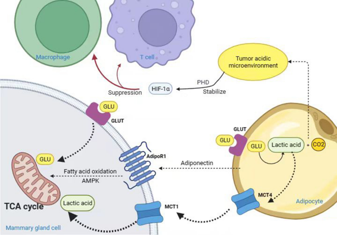
A “reverse Warburg effect” exists between breast cancer cells and adipocytes. During this effect, byproducts of adipocyte glycolysis, such as lactate, are excreted from adipocytes by monocarboxylate transporter 4 (MCT4), which is taken up by tumor cells in response to MCT1 and used for mitochondrial oxidative phosphorylation in breast cancer cells [28]. Furthermore, differential expression of MCT between breast cancer cells and adipocytes enhances the invasiveness of breast cancer. The highly proliferative estrogen receptor-negative breast cancer subtype expresses high levels of MCT1 and is related to poor outcomes in patients [29].

Hypoxia is the primary driver of glycolysis and lactate production. Hypoxia facilitates lactate production in adipocytes, and approximately 50 to 70% of glucose is converted to lactate in adipose tissue. Large amounts of lactate, CO2, and other metabolites accumulate in the TME to create an acidic environment, which suppresses immune cell functions and allows tumor cells to escape the immune system.

Studies have demonstrated that adipocyte-derived lactate contributes to developing an adipose pro-inflammatory microenvironment. This could be because first, an increase in the levels of intracellular lactate initiates apoptosis of adipocytes, thereby initiating an inflammatory response [30]. Second, lactate binds to the catalytic structural domain containing PHD2, thereby stabilizing HIF-1α and reducing its degradation [31]. Third, lactate induces M1 macrophages to polarize to the M2 type, causing immunosuppression and tissue remodeling in the tumor zone (as shown in Fig. 1) [32].

figure 1
Fig. 1

In summary, in breast cancer, HIF-1α mainly promotes glycolysis by inducing genes encoding glucose transporters such as GLUT1 and glycolytic enzymes.

5.2 Lipid metabolism

Under hypoxia, both breast and ovarian cancer cells enhance the rate of adipocyte lipolysis [33]. Cohen’s team found through RNA-seq that the para-cancerous adipose metabolism pathway changed significantly, high levels of creatine are released from adipocytes and taken up by adjacent cancer cells, mainly through the Slc6a8 and Smad2/3 pathways, thereby promoting the progression of breast cancer [34].

A study demonstrated that following co-culturing of breast cancer cells with adipocytes, adipocyte-derived free fatty acids were transferred into breast cancer cells. Through the AMPK pathway, elevating the levels of carnitine palmitoyl transferase 1 (CPT1A) and electron transfer proteins [35], consequently driving fatty acid metabolism to maintain a high level of ATP and enhance the ability of breast cancer tumor cells to grow, survive, and multiply.

Both adipocyte-released lipids and lipids entering the cancer cells are reduced in HIF-deficient adipocytes, such that the lipid availability of such adipocytes is low. Therefore, the release of fatty acids by adipocytes and subsequently reuptake by cancer cells depends on the hypoxic HIF-1α pathway.

The above indicated that fatty acid secreted by obese fat plays an important role in breast cancer proliferation. Inhibiting the production of FASN may be one of the directions for the treatment of breast cancer in the future.

6 Combined application of targeted HIF-1α for diet control, drugs, chemotherapy and immunotherapy

During cancer treatment, dysregulation of cell metabolism in tumor areas is associated with drug resistance. HIF-associated metabolic pathways participate in the development of adipocyte-associated cancers.

Several therapeutic targets can be proposed for HIF-dependent metabolic reprogramming, such as reducing adipose tissue, altering adipocyte metabolic mode, and targeting HIF to prevent or treat breast cancer. Multiple therapeutic approaches are available to target HIF-1α, such as inhibiting the regulation of upstream target genes of HIF-1α, modulating protein modifications to promote HIF-1α protein hydroxylation and degradation, inhibiting HIF-1α protein synthesis, and inhibiting the regulation of downstream target genes of HIF-1α.

Diet control is the simplest method to lose fat. In addition, reducing caloric intake regulates the levels of relevant hormones in the body to protect postmenopausal women with breast cancer. In the WHI DM trial published in 2020, a dietary intervention in 48,835 postmenopausal women without previous breast cancer, a significant reduction in breast cancer deaths persisted after a long-term cumulative 19.6 years of follow-up (359 [0.12%] v 652 [0.14%] deaths) [36]. Low-fat diets prevent and treat breast cancer by reducing the production and secretion of adipokines and insulin, enhancing immunity, and diminishing drug resistance [37]. Certain substances extracted from broccoli, soybean, and ginger can hinder tumor progression by promoting the self-renewal of mesenchymal stem cells, preventing the differentiation of preadipocytes into mature adipocytes, and inhibiting the transcription levels of HIF.

Cardamonin is a flavonoid that increases the sensitivity of tumors to chemotherapeutic drugs [38]. In addition, it reduces glucose uptake and lactate production by inhibiting the transcriptional levels of HIF-1α, thereby enhancing mitochondrial oxidative phosphorylation [39]. Curcumin is a compound extracted from the rhizomes of turmeric [40], and its nanosuspension is effective in alleviating the hypoxic and inflammatory states in the TME of triple-negative breast cancer, reducing the stability of HIF-1α and increasing its susceptibility to degradation [41].

The glucose analog 2-DG is phosphorylated by the hexokinase and competes with glucose-6-phosphate to inhibit glycolysis, causing apoptosis due to energy deficiency in cancer cells [42]. Combining 2-DG with radiotherapy or chemotherapy to inhibit glycolysis could enhance the therapeutic effect of radiotherapy and chemotherapy. Therefore, a combination of cell metabolism inhibitors and chemotherapeutic agents can be used to treat breast cancer by targeting HIF-associated adipocyte metabolic pathways.

Regulation of immune metabolism has been known to control cancer progression. CD8+ T cells have a very high rate of glycolysis, which is further enhanced by the overexpression of HIF-1α, thus increasing the levels of CD8+ T cell immune infiltration and activity. However, increased glycolysis enhances the competition for glucose between adipocytes and cancer cells within the tumor area and immune cells [43], causing the exhaustion or death of immune cells. Studies on programmed cell death protein 1 (PD-1) and T lymphocytes have demonstrated that PD-1 activates T cell apoptosis and suppresses immune responses [44]. The PD-1 blocking therapy improves the competition of T cells for glucose and restores their glycolytic ability, thereby manifesting their immune efficacy [43]. Future therapies could consider combining treatments inhibiting tumor metabolism with agents enhancing the glycolytic ability of T cells to promote optimal anti-tumor immunity.

7 Conclusions and future directions

Adipocytes are intricately related to the development of breast tumors, with hypoxia acting as the key crosstalk between adipocytes and their neighboring breast cancer cells. Under hypoxia, metabolic reprogramming of adipocytes allows breast cancer cells to maintain hypoxia.

Cancer cells are highly plastic and their morphology can change dynamically in response to external signals in the microenvironment. In addition, epithelial–mesenchymal transformation increases their resistance to chemotherapy and metastatic capacity through plasticity [45, 46]. Hypoxia, induced either by adipocytes or within the tumor microenvironment, has been identified as a critical factor in promoting cancer stem cell populations and activating pathways associated with increased migration and metastasis. Under hypoxic conditions, HIF-1α regulators increase Snail1 levels and induce EMT by activating the COX-2/PGE2 pathway [47]. Reverse differentiation of cancer cells into epithelial cells is now being used to treat cancer, which greatly reduces the proliferation of cancer cells and increases their sensitivity to chemotherapy [48]. This strategy has led to the development of adipogenesis therapy, which inhibits cancer cell metastasis by transdifferentiating breast cancer cells into adipocytes. Factors affecting breast cancer cell differentiation into adipocytes and intra- and extracellular influences on differentiation are not well understood. Therefore, more research is warranted to discover mechanisms that can be used to selectively induce the differentiation of cancer cells into adipocytes.

The immune impact of HIF-dependent metabolism of adipocytes is highly complex, involving several signaling pathways and multiple cell interaction mechanisms. Therefore, it could be used as a target for cancer immunotherapy. In the future, it is necessary to explore different crosstalk mechanisms of adipocytes and breast cancer cells under hypoxia and strategies related to immunotherapy.

The role of cardiac pericytes in health and disease: therapeutic targets for myocardial infarction


Abstract

Millions of cardiomyocytes die immediately after myocardial infarction, regardless of whether the culprit coronary artery undergoes prompt revascularization. Residual ischaemia in the peri-infarct border zone causes further cardiomyocyte damage, resulting in a progressive decline in contractile function. To date, no treatment has succeeded in increasing the vascularization of the infarcted heart. In the past decade, new approaches that can target the heart’s highly plastic perivascular niche have been proposed. The perivascular environment is populated by mesenchymal progenitor cells, fibroblasts, myofibroblasts and pericytes, which can together mount a healing response to the ischaemic damage. In the infarcted heart, pericytes have crucial roles in angiogenesis, scar formation and stabilization, and control of the inflammatory response. Persistent ischaemia and accrual of age-related risk factors can lead to pericyte depletion and dysfunction. In this Review, we describe the phenotypic changes that characterize the response of cardiac pericytes to ischaemia and the potential of pericyte-based therapy for restoring the perivascular niche after myocardial infarction. Pericyte-related therapies that can salvage the area at risk of an ischaemic injury include exogenously administered pericytes, pericyte-derived exosomes, pericyte-engineered biomaterials, and pharmacological approaches that can stimulate the differentiation of constitutively resident pericytes towards an arteriogenic phenotype. Promising preclinical results from in vitro and in vivo studies indicate that pericytes have crucial roles in the treatment of coronary artery disease and the prevention of post-ischaemic heart failure.

Key points

  • Cardiac pericytes interact with endothelial cells through physical and paracrine mechanisms to maintain normal vascular homeostasis.
  • In the infarcted heart, pericytes have crucial roles in angiogenesis, scar formation, and stabilization and control of the inflammatory response.
  • Persistent ischaemia and accrual of age-related risk factors can lead to pericyte depletion and dysfunction; nevertheless, some age-related cardiac defects might be treatable using pharmacotherapeutic approaches or by supplying the heart with exogenous pericytes alone or in combination with other cell types.
  • A greater understanding of the molecular mechanisms underlying the numerous functions of cardiac pericytes could uncover novel therapeutic solutions for coronary artery disease.

Animal models to study cardiac regeneration


Permanent fibrosis and chronic deterioration of heart function in patients after myocardial infarction present a major health-care burden worldwide. In contrast to the restricted potential for cellular and functional regeneration of the adult mammalian heart, a robust capacity for cardiac regeneration is seen during the neonatal period in mammals as well as in the adults of many fish and amphibian species. However, we lack a complete understanding as to why cardiac regeneration takes place more efficiently in some species than in others. The capacity of the heart to regenerate after injury is controlled by a complex network of cellular and molecular mechanisms that form a regulatory landscape, either permitting or restricting regeneration. In this Review, we provide an overview of the diverse array of vertebrates that have been studied for their cardiac regenerative potential and discuss differential heart regeneration outcomes in closely related species. Additionally, we summarize current knowledge about the core mechanisms that regulate cardiac regeneration across vertebrate species.

Key points

  • Cardiac regeneration potential tends to be robust in fish, amphibians and neonatal mammals, but is restricted in adult mammals; however, cardiac regeneration potential in several model organisms defies this trend.
  • Cardiac regeneration potential is determined by multiple highly interconnected processes, including cardiomyocyte proliferation, cardiac fibrosis, neovascularization, immune response and energy metabolism.
  • Mammalian cardiomyocytes exit the cell cycle postnatally due to changes in structure and energy metabolism; partial in vivo reprogramming of adult mammalian cardiomyocytes can increase their proliferation capacity.
  • Fibrosis in the injured heart is both beneficial and detrimental; altering fibrotic tissue composition and mechanical properties might improve adult mammalian heart regeneration.
  • Rapid neovascularization of the wound is a hallmark of heart regeneration and is absent in the adult mammalian heart; lymphatic coronary vessels modify the immune response after myocardial infarction via immune cell clearance.
  • The immune response to cardiac injury consists of multiple phases; restricting the initial inflammatory phase and promoting the subsequent reparative phase represents a strategy to improve heart regeneration.

Modified Radical Mastectomy vs Oncoplastic Breast-Conserving Surgery: Quest for Recurrence Risk Model


Yuan et al, of the First People’s Hospital of Changde City, Hunan, China, and colleagues aimed to evaluate the clinical efficacy of modified radical mastectomy vs oncoplastic breast-conserving surgery for early-stage breast cancer treatment. Published in the American Journal of Cancer Research, this retrospective analysis revealed that despite longer operation times for oncoplastic breast-conserving surgery, patients experienced significantly less intraoperative bleeding, postoperative drainage, and hospitalization compared with the modified radical mastectomy group. Additionally, patients undergoing oncoplastic breast-conserving surgery exhibited higher subjective satisfaction, better quality-of-life scores, and comparable objective outcomes.

Postoperative complications and recurrence rates did not significantly differ between the two groups. However, multivariate Cox regression analysis identified lymph node metastasis and molecular type as independent prognostic factors for disease-free survival. A risk model based on these variables predicted recurrence effectively, according to the investigators, with an AUC of 0.852. Lower risk scores correlated with significantly higher disease-free survival rates.

The investigators focused on the medical data of 149 patients with early-stage breast cancer treated at their institution from between January 2018 and January 2022. A total of 104 patients were treated with modified radical mastectomy (the control group), and 45 patients were treated with oncoplastic breast-conserving surgery (the observation group). The groups were comparable in terms of age, body mass index, tumor diameter, tumor stage, lymph node metastasis, and molecular type, the authors noted.

The investigators’ key finding suggests that compared with modified radical mastectomy, oncoplastic breast-conserving surgery may reduce the surgical incision and enhance patient satisfaction, without increasing complication or recurrence risks. The risk model, developed through Cox regression, has potential clinical value in predicting breast cancer recurrence and facilitating personalized patient management and treatment planning. It may contribute to the ongoing quest for effective early-stage breast cancer treatments, emphasizing the importance of tailoring interventions based on individual risk factors identified through a risk model.

Physical Activity is Medicine


The frequent lack of appropriate statistical evidence regarding physical activity benefits in patients with cancer has meant that either epidemiologic or retrospective observational studies have provided the basis for our knowledge. The level of evidence for results has been weak and the basis of a controversial and not well-established issue. Scientific and clinical evidence is much more limited for less frequent and low survival cancers such as pancreatic cancer. Conducting randomized controlled trials on physical activity implementation in individuals with cancer is challenging due to the low observed adherence to trial recommendations and completion of quality-of-life questionnaires; the interference of environmental, social, and structural barriers; comorbidities as well as symptoms derived from cancer and its treatment; and the high number of patients needed, which is difficult to achieve in uncommon and deadly cancers. In the article by Neuzillet et al1 elsewhere in this issue, we can learn from a well-designed randomized trial on the positive effects of increasing physical activity in quality-of-life parameters for patients with advanced pancreatic cancer who are receiving treatment.

We have known that healthy habits contribute to a healthier life. Patients with heart disease, type 2 diabetes, hypertension, and cancer, which are quite frequent and major causes of morbidity and mortality in our Western world, do benefit from a systematic increase in physical activity. Patients desire to be more active and would like help and guidance from their doctor. The American Cancer Society recommends that individuals maintain healthy behaviors regarding diet and physical activity in order to reduce cancer risk.2 Indeed, in a recent analysis, the combination of risk factors (excess body weight, physical inactivity, poor diet, and alcohol consumption) accounted for at least 18.2% of cancer cases and 15.8% of cancer deaths in the United States.3 Feeling fit could help patients physically and psychologically cope with the development of cancer, as well as with the therapeutic strategies needed to cure or control this disease. The beneficial effect of physical activity implementation in cancer prevention and during cancer treatment has been demonstrated.4 This finding is potentially the result of a dose–response association5 and independent of critical confounders such as smoking or increased body mass index.6 Even individuals who were sedentary throughout most of their adult life (age 19–60 years) but routinely increased physical activity in their 50s or 60s showed a lower risk for all-cause mortality (hazard ratio [HR], 0.65; 95% CI, 0.62–0.68), cardiovascular disease–related mortality (HR, 0.57; 95% CI, 0.53–0.61), and cancer-related mortality (HR, 0.84; 95% CI, 0.77–0.92).7

A systematic review and meta-analysis of hundreds of epidemiologic studies with several million participants showed that there was a 10% to 20% reduction in bladder, breast, endometrial, renal, colon, and gastroesophageal cancers in people with a dynamic lifestyle. A 40% to 50% reduction in mortality was also seen in people with prostate, breast, and colorectal cancer who were physically active.8

On the other side, cancer treatments damage patient’s tissues and can contribute to accelerated biological aging. Psychological stress from cancer and its treatments favors prolonged toxicities, impairs essential repair mechanisms, and promotes a lack of will to do what has been recommended. Therapeutic objectives are to control disease growth and dissemination, reduce stress, improve sleep, increase physical activity according to patient’s performance status, improve self-esteem, preserve mental well-being, sustain a healthy diet, and maintain a healthy body weight.

Benefits of regular physical activity include stress reduction, sleep quality improvement, mood enhancement, muscle strength and tone enhancement, and cardiovascular and lung fitness augmentation for improving quality of life and aging outcomes. This is especially relevant for patients with sedentary lifestyles. More recently, the use of smart devices has increased adherence to physical exercise programs.

At the molecular level, the enhancement of immune function is a strong candidate to explain these effects, at least partly. Evidence has shown a stimulating effect of vigorous physical activity on immune effectors, mainly natural killer (NK) cells and CD8+ T cells, against tumors, as well as a decrease in inflammatory markers such as prostaglandin E2 and an increase in gut microbiota diversity, while also potentially reducing the levels of tumor-promoting bacteria.9

During physical exercise, skeletal muscle secretes myokines into the bloodstream, such as IL-6, IL-7, and IL-15, that can circulate free or are packaged into exosomes. IL-6 causes an anti-inflammatory effect by inducing the release of IL-1 receptor antagonist (IL-1RA), and it can also bind to NK cells to accelerate their mobilization into the bloodstream. IL-7 helps to preserve thymic mass and enhances the output of naive T cells. IL-7, together with IL-15, plays a relevant role in T-cell survival and CD8+ T-cell homoeostasis. As recently reported in pancreatic cancer,9,10 regular physical activity may act on inflammation and immunity through IL-15–mediated activation of CD8+ T cells, decreasing tumor growth and enhancing sensitivity to chemotherapy and anti–PD-1 antibodies. These molecular findings underline the potential benefit of constant physical exercise and point to IL-15 as a useful resource for pancreatic cancer treatment.9 The diminution in myokine secretion owing to the aging-related decline in skeletal muscle mass is a fact that triggers immune senescence, a condition linked to cancer immune evasion.11

The cognitive benefits of systematic physical activity are tied to an increased plasticity and reduced inflammation within the hippocampus, and to a decelerated neurodegeneration. One of the underlying mechanisms is an increase in clusterin, a complement cascade inhibitor, that reduces neuroinflammatory gene expression.12

A highly disease-specific microbiota-based classification model confined to pancreatic cancer–enriched species has been identified and could be useful as a control group for future randomized trials and for the early detection of pancreatic cancer.13 In contrast to dysbiosis, changes toward increased microbial abundance, diversity, and composition observed with physical activity training significantly change the function of the gut microbiota in favor of the patient, including improvements in immune function and inflammatory profiles.14 Many other physical activity mechanisms of action remain to be discovered.

Physical activity programs must be properly tailored to a patient’s characteristics by an expert team. In this way, a positive impact can be made on the health status of patients with cancer, even those with advanced-stage cancer, as well as persons living beyond cancer. All should have a lifestyle as active as possible to avoid complications that could occur when physical activity is not adequately prescribed and closely followed. In clinical practice, rehabilitation professionals must ask patients about their daily or weekly physical activity, and must record the information. Patients should then be given a written prescription specifying the type of exercise, frequency, and duration appropriate for them. Patients can start small and low, and increase duration and frequency over time. It is crucial for patients to measure the physical activity performed. Health monitoring wearables are helpful and support adherence to the recommendations. Clinicians should follow up on their patients’ physical activity program, asking about it at every contact. Reinforcement of this advice is relevant, and pointing out that physical activity can help preserve functional independence can be particularly motivating for patients. If rehabilitative services are not available, patients should be referred to another institution that provides them.

And last but not least, there is room for improvement, given that exercise is the most cost-effective medical intervention for patients with cancer. No financial toxicity is associated with it. Our health teams will not be properly organized until physical exercise and the professionals involved in it are part of the pancreatic cancer treatment circuit within the multidisciplinary team.

Exogenous Ochronosis from Skin-Lightening Cream


A 55-year-old woman presented to the dermatology clinic with a 1-year history of skin darkening on her face. Two years before presentation, she had started applying a skin-lightening cream containing hydroquinone to her face daily to treat melasma. On physical examination, bluish-brown patches with background erythema and telangiectasias were observed on the cheeks, nasal bridge, and perioral region, with lesser involvement on the forehead (Panel A). Dermoscopy of the affected areas revealed hyperchromic, pinpoint macules (Panel B). A skin-biopsy sample from the left cheek showed extracellular deposition of yellow-brown, banana-shaped bodies in the dermis (Panel C, hematoxylin and eosin stain). A diagnosis of exogenous ochronosis from skin-lightening cream was made. Ochronosis is a hyperpigmentation disorder that results from the accumulation of ochre-colored deposits in tissue. It is deemed endogenous when related to alkaptonuria and exogenous when related to the use of skin-lightening agents. Exogenous ochronosis is challenging to treat and may not be reversible. Counseling regarding the importance of photoprotection and cessation of the use of the skin cream was given to the patient. A topical emollient and retinoid gel were also prescribed. At 2 months of follow-up, the patient had minimal abatement of the hyperpigmentation.

Testosterone Treatment and Fractures in Men with Hypogonadism


Abstract

Background

Testosterone treatment in men with hypogonadism improves bone density and quality, but trials with a sufficiently large sample and a sufficiently long duration to determine the effect of testosterone on the incidence of fractures are needed.

Methods

In a subtrial of a double-blind, randomized, placebo-controlled trial that assessed the cardiovascular safety of testosterone treatment in middle-aged and older men with hypogonadism, we examined the risk of clinical fracture in a time-to-event analysis. Eligible men were 45 to 80 years of age with preexisting, or high risk of, cardiovascular disease; one or more symptoms of hypogonadism; and two morning testosterone concentrations of less than 300 ng per deciliter (10.4 nmol per liter), in fasting plasma samples obtained at least 48 hours apart. Participants were randomly assigned to apply a testosterone or placebo gel daily. At every visit, participants were asked if they had had a fracture since the previous visit. If they had, medical records were obtained and adjudicated.

Results

The full-analysis population included 5204 participants (2601 in the testosterone group and 2603 in the placebo group). After a median follow-up of 3.19 years, a clinical fracture had occurred in 91 participants (3.50%) in the testosterone group and 64 participants (2.46%) in the placebo group (hazard ratio, 1.43; 95% confidence interval, 1.04 to 1.97). The fracture incidence also appeared to be higher in the testosterone group for all other fracture end points.

Conclusions

Among middle-aged and older men with hypogonadism, testosterone treatment did not result in a lower incidence of clinical fracture than placebo. The fracture incidence was numerically higher among men who received testosterone than among those who received placebo

Class 4 Laser Therapy for a Rotator-Cuff Injury: Case Study


WHAT YOU NEED TO KNOW

  • Rotator-cuff injuries, ranging from tendinopathies to tears, affect a significant portion of the population.
  • Conventional treatments range from conservative approaches, such as physical therapy and non-steroidal anti-inflammatory drugs (NSAIDs), to surgical interventions.
  • However, recent advances in therapeutic modalities have introduced class 4 laser therapy as a potential alternative.

Rotator-cuff injuries are a common cause of shoulder pain and functional impairment.1 Various treatment modalities2 are available to manage these injuries, with class 4 laser therapy emerging as a promising non-invasive option. This case study explores the application of class 4 laser therapy in the rehabilitation of a patient with a rotator-cuff injury, analyzing its effectiveness, safety and implications for clinical practice.

Background

Rotator-cuff injuries, ranging from tendinopathies to tears, affect a significant portion of the population, particularly individuals engaged in overhead activities or repetitive motions.3 These injuries can lead to pain, reduced range of motion and diminished quality of life.

Conventional treatments range from conservative approaches, such as physical therapy and non-steroidal anti-inflammatory drugs (NSAIDs), to surgical interventions. However, recent advances in therapeutic modalities have introduced class 4 laser therapy as a potential alternative for managing rotator-cuff injuries.4-5

The Case

“Jack,” a 42-year-old male, presented with complaints of persistent shoulder pain, limited range of motion, and difficulty performing daily activities following a traumatic fall. Magnetic resonance imaging (MRI) revealed a partial-thickness tear in the supraspinatus tendon of his right shoulder. Physical examination indicated pain and weakness during abduction and external rotation.

Jack’s treatment plan comprised an individualized combination of chiropractic adjustments to the upper thoracic and cervical spine, class 4 laser therapy, and instruction on proper home exercises. Laser was integrated into the plan due to its reported benefits in reducing pain and promoting tissue healing.6

Class 4 laser therapy involves the application of red and infrared laser wavelengths to stimulate tissue healing and modulate inflammation. It works through the process of photobiomodulation,7 in which photons are absorbed by cells, leading to enhanced cellular metabolism, increased circulation, and reduced pain proprioception.8

Over the course of six weeks, Jack underwent 14 class 4 laser therapy sessions, each lasting around 10 minutes. A laser probe emitting laser wavelengths of 650, 810, 915 and 980 nanometers (nm) was applied to the cervical spine and cervical nerves extending into the brachial plexus, and targeted the supraspinatus muscle and supporting structures of the affected shoulder.

Treatment was delivered from all angles to target all affected tissues. The laser was set at a power output of 9.2 watts, delivering continuous-wave and various pulse frequencies.9 Outcome measures consisted of the following:

  • Visual Analog Scale (VAS): Jack’s pain levels were assessed using the VAS, with scores recorded before and after each laser therapy session.
  • Range of Motion (ROM): Shoulder abduction and external rotation were measured using a goniometer to evaluate improvements in range of motion.
  • Strength Testing: Isometric strength of the supraspinatus and deltoid muscles was assessed using a handheld dynamometer.
  • Patient-Reported Outcome Measures: Functional assessments10 were used to evaluate improvements in daily functioning and quality of life.

Results and Clinical Takeaway

After the six-week treatment period, Jack exhibited significant improvements in pain reduction, range of motion, and strength. The VAS scores consistently decreased after each laser therapy session, indicating a reduction in pain levels. His ROM increased by approximately 30% in both abduction and external rotation. Isometric strength testing showed a 20% improvement in supraspinatus and deltoid muscle strength. Moreover, the DASH11 score decreased from 52 to 18, suggesting enhanced functional ability and quality of life.

The positive outcomes observed in this case study align with previous research on class 4 laser therapy for rotator-cuff injuries.12-13 The mechanism of photobiomodulation has been shown to accelerate tissue repair by promoting cellular metabolism, collagen synthesis and angiogenesis.14 The analgesic effect of laser therapy may contribute to improved range of motion and muscle activation, as observed in Jack’s case.

The non-invasive nature of class 4 laser therapy offers several advantages, including minimal side effects and the potential to reduce reliance on NSAIDs or invasive procedures. However, the optimal dosage parameters, such as wavelength, power and treatment duration, remain areas of ongoing research and discussion.

This case study highlights the potential benefits of integrating class 4 laser therapy into the chiropractic treatment plan for rotator-cuff injuries. The observed improvements in pain reduction, range of motion and muscle strength suggest laser therapy could serve as an effective adjunct.

As the field of laser therapy continues to evolve, it has the potential to revolutionize the management of musculoskeletal injuries, offering patients non-invasive options for pain relief and functional improvement.