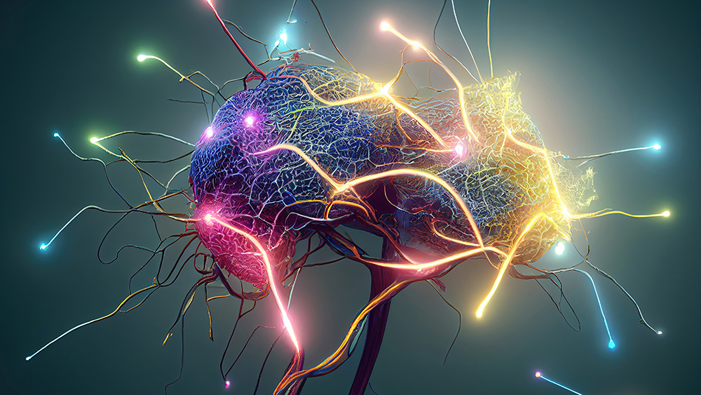
Summary: A recent study showcases a significant leap in the study of brain oscillations, particularly ripples, which are crucial for memory organization and are affected in disorders like epilepsy and Alzheimer’s. Researchers have developed a toolbox of AI models trained on rodent EEG data to automate and enhance the detection of these oscillations, proving their efficacy on data from non-human primates.
This breakthrough, stemming from a collaborative hackathon, offers over a hundred optimized machine learning models, including support vector machines and convolutional neural networks, freely available to the scientific community. This development opens new avenues in neurotechnology applications, especially in diagnosing and understanding neurological disorders.
Key Facts:
AI-Driven Innovation: The study introduces a toolbox of AI models capable of detecting brain ripples, key in memory organization and neurological diseases.
Cross-Species Application: Initially trained on rodent data, these models have been successfully tested on non-human primate EEG data, indicating potential for human application.
Open-Source Contribution: Over a hundred machine learning models from the project are now openly available for research use and further development, demonstrating the collaborative spirit of the scientific community.
Source: CSIC
The study of brain oscillations has advanced our understanding of brain function. Ripples are a type of fast oscillations underlying the organization of memories. They are affected in neurological disorders such as epilepsy and Alzheimer’s.
For this reason, they are considered an electroencephalographic (EEG) biomarker. However, ripples exhibit various waveforms and properties that can be missed by standard spectral methods.
The model toolbox emerged as a result of a hackathon, which resulted in a short list for the best detection models.
Recently, the neuroscience community called for the need to better automate, harmonize, and improve the detection of ripples across a range of tasks and species. In the study, the authors used recordings obtained in laboratory mice to train a toolbox of machine learning models.
“We have tested the ability of these models using data from non-human primates that were collected at Vanderbilt University (Nashville, USA) by Saman Abbaspoor and lab leader Kari Hoffman as part of the Brain Initiative.
“We found that it is possible to use rodent EEG data to train AI algorithms that can be applied to data from primates and possibly human, provided the same type of recording techniques are used.”, De la Prida explains.
The model toolbox emerged as a result of a hackathon, which resulted in a short list for the best detection models. These architectures were then harmonized and optimized by the authors who now provide all codes and data openly to the research community.
Models include some of the best-known supervised learning architectures, such as support vector machines, decision trees, and convolutional neural networks.
“We have identified more than one hundred possible models from the different architectures that are now available for application or retraining by other researchers.”, commented Andrea Navas Olivé and Adrián Rubio, who are first authors of the work.
“This bank of AI models will provide new applications in the field of neurotechnologies and can be useful for detection and analysis of high-frequency oscillations in pathologies such as epilepsy, where they are considered clinical markers” concludes De la Prida, who is part of the CSIC’s AI-HUB connection aimed at advancing the use of AI and its applications.
Abstract
A machine learning toolbox for the analysis of sharp-wave ripples reveals common waveform features across species
The study of sharp-wave ripples has advanced our understanding of memory function, and their alteration in neurological conditions such as epilepsy is considered a biomarker of dysfunction.
Sharp-wave ripples exhibit diverse waveforms and properties that cannot be fully characterized by spectral methods alone.
Here, we describe a toolbox of machine-learning models for automatic detection and analysis of these events.
The machine-learning architectures, which resulted from a crowdsourced hackathon, are able to capture a wealth of ripple features recorded in the dorsal hippocampus of mice across awake and sleep conditions. When applied to data from the macaque hippocampus, these models are able to generalize detection and reveal shared properties across species.
We hereby provide a user-friendly open-source toolbox for model use and extension, which can help to accelerate and standardize analysis of sharp-wave ripples, lowering the threshold for its adoption in biomedical applications.
A new study shows an association between sleep apnea symptoms and an increased risk of memory and thinking problems.
A new study analyzes the impact of sleep apnea symptoms on memory and thinking.
Sleep apnea is a sleep disorder that sometimes causes people to stop breathing.
The study subjects participated in a survey where they reported any symptoms of sleep apnea and difficulty with remembering things.
The study shows an association between sleep apnea symptoms and a higher rate of memory and thinking problems.
Getting a good night’s sleep is important for many reasons, from having the energy to go about one’s daily tasks to optimal brain performance.
Sleep apnea can interfere with this, and according to the National Council on Aging, it may impact around 39 million adults in the United States.
While experts know sleep apnea can impact quality of life and even contribute to mood disordersTrusted Source, there is still more to learn.
A researcher based in Boston recently conducted a cross-sectional study to determine whether a correlation between sleep apnea and thinking and memory problems exists.
The findings showed that having sleep apnea symptoms correlated with a 50% increase in memory and thinking problems.
Most people with sleep apnea have obstructive sleep apnea (OSA), but some experts have said that OSA is underdiagnosed.
For instance, researchersTrusted Source note, “it is believed that more than 85% of patients with clinically significant OSA have never been diagnosed.”
With the notion that sleep apnea could be underdiagnosed in mind, researcher Dr. Dominique Low wanted to learn more about a possible connection between sleep apnea and cognition. Dr. Low works at Boston Medical Center in Massachusetts and is a member of the American Academy of Neurology.
Dr. Low pulled data from a government-funded survey called the 2017–18 National Health and Nutrition Examination Survey (NHANES) to establish a potential link between sleep apnea and thinking and memory.
The study participants included 4,257 adults ages 20 and older. Of the questionnaires they completed for the NHANES, they answered questions about sleep quality, memory, and thinking.
Dr. Low used the data from these questionnaires to determine how people who reported sleep apnea symptoms compared to people without these symptoms.
The participants also answered questions on their memory quality, whether they had any periods of confusion, and if the participants thought they had trouble making decisions.
A total of 1,079 participants reported sleep apnea symptoms, including snoring and gasping for breath while asleep.
Of people who indicated they had sleep apnea symptoms, 33% also reported symptoms of memory and thinking problems. This is significantly higher than the number of people without sleep apnea symptoms who reported such problems, which was only 20% of that group.
After adjusting for other factors like age, race, and gender, Dr. Low observed that people with sleep apnea symptoms had a 50% higher chance of having thinking and memory issues compared to participants who didn’t report sleep apnea symptoms.
“Our study found participants who had sleep apnea symptoms had greater odds of having memory or thinking problems,” Dr. Low said in a news release. “These findings highlight the importance of early screening for sleep apnea.”
Despite the implications of these findings, it’s important to note that a correlation does not indicate causation. Scientists must conduct further research that does not rely solely on self-reported symptoms to establish the effects of sleep apnea symptoms on memory and thinking.
Dr. Joey R. Gee, a neurologist at Providence Mission Hospital in Mission Viejo, California, spoke with Medical News Today about how sleep apnea may impact memory. Dr. Gee was not involved with the study.
“Apnea may have an impact due to poor oxygenation through the night or also impairing appropriate sleep cycles with frequent arousals,” Dr. Gee noted. “Impaired executive functions, such as working memory and attention through the day, are greatly impacted.”
Dr. Gee said that while untreated sleep apnea may impact cognitive function, the risk could be reduced with appropriate treatment.
“Just as untreated sleep apnea increases the risk of impairment in executive function and attention, treatment can substantially reduce the risk of progressing cognitive decline,” Dr. Gee said.
Dr. Thomas Kilkenny, the director of the Institute of Sleep Medicine at Staten Island University Hospital in New York, not involved in the study, emphasized the importance of treating sleep apnea as soon as it is detected.
“If the patient can be treated early in OSA, these brain damages will not occur,” Dr. Kilkenny told MNT. “There will be a decrease in the amount of cognitive decline in OSA patients.”
Dr. David Merrill, a geriatric psychiatrist and director of the Pacific Neuroscience Institute’s Pacific Brain Health Center at Providence Saint John’s Health Center in Santa Monica, California, not involved with the study, shared his thoughts on the findings with MNT:
“With high quality, restorative sleep, the brain’s function is enhanced and protected as we age,” explained Dr. Merrill. “If sleep is chronically disrupted, this can lead to a number of health issues, including headaches, fatigue, and memory loss that worsens over time. The disrupted, poor-quality sleep seen in sleep disorders leads to both acute and chronically worsening changes in the brain. Normally, a good night’s sleep literally allows for repair and restoration of brain function to the levels seen at the beginning of the prior day.”
Dr. Merrill also spoke about the importance of treating sleep apnea and noted that it is a risk factor for developing dementia. While that may sound scary, he said that using a CPAP machine can help reduce risk.
“Research studiesTrusted Source have shown that even 4 hours per night using a CPAP device results in significantly less worsening of cognitive decline over time,” said Dr. Merrill.
Sleep apnea, including obstructive sleep apnea and central sleep apnea, can affect people of all ages but, as the National Council on Aging notes (link above), it is most prevalent in middle-aged and older adults.
A person’s partner may notice additional symptoms such as snoring or gasping for breath while asleep.
“Signs of obstructive sleep apnea are usually readily apparent,” Dr. Kilkenny said.
“Loud snoring, restlessness, and daytime fatigue are the hallmarks of OSA,” he noted. “If someone snores even to a minor degree, they should bring this to the attention of their physician so they can get tested for OSA before damage occurs.”
People with sleep apnea can treat it using a continuous positive airway pressure (CPAP) machine.
They may also try to improve symptoms by making lifestyle changes such as losing weight. They may also have surgery or use an oral appliance.
Cognitive symptoms after coronavirus disease 2019 (Covid-19), the disease caused by severe acute respiratory syndrome coronavirus 2 (SARS-CoV-2), are well-recognized. Whether objectively measurable cognitive deficits exist and how long they persist are unclear.
METHODS
We invited 800,000 adults in a study in England to complete an online assessment of cognitive function. We estimated a global cognitive score across eight tasks. We hypothesized that participants with persistent symptoms (lasting ≥12 weeks) after infection onset would have objectively measurable global cognitive deficits and that impairments in executive functioning and memory would be observed in such participants, especially in those who reported recent poor memory or difficulty thinking or concentrating (“brain fog”).
RESULTS
Of the 141,583 participants who started the online cognitive assessment, 112,964 completed it. In a multiple regression analysis, participants who had recovered from Covid-19 in whom symptoms had resolved in less than 4 weeks or at least 12 weeks had similar small deficits in global cognition as compared with those in the no–Covid-19 group, who had not been infected with SARS-CoV-2 or had unconfirmed infection (−0.23 SD [95% confidence interval {CI}, −0.33 to −0.13] and −0.24 SD [95% CI, −0.36 to −0.12], respectively); larger deficits as compared with the no–Covid-19 group were seen in participants with unresolved persistent symptoms (−0.42 SD; 95% CI, −0.53 to −0.31). Larger deficits were seen in participants who had SARS-CoV-2 infection during periods in which the original virus or the B.1.1.7 variant was predominant than in those infected with later variants (e.g., −0.17 SD for the B.1.1.7 variant vs. the B.1.1.529 variant; 95% CI, −0.20 to −0.13) and in participants who had been hospitalized than in those who had not been hospitalized (e.g., intensive care unit admission, −0.35 SD; 95% CI, −0.49 to −0.20). Results of the analyses were similar to those of propensity-score–matching analyses. In a comparison of the group that had unresolved persistent symptoms with the no–Covid-19 group, memory, reasoning, and executive function tasks were associated with the largest deficits (−0.33 to −0.20 SD); these tasks correlated weakly with recent symptoms, including poor memory and brain fog. No adverse events were reported.
CONCLUSIONS
Participants with resolved persistent symptoms after Covid-19 had objectively measured cognitive function similar to that in participants with shorter-duration symptoms, although short-duration Covid-19 was still associated with small cognitive deficits after recovery. Longer-term persistence of cognitive deficits and any clinical implications remain uncertain.
Poor memory and difficulty thinking or concentrating (commonly referred to as “brain fog”) have been implicated in syndromes occurring after coronavirus disease 2019 (Covid-19) — a situation that has led to suggestions that Covid-19 may have lasting cognitive consequences.1-7 However, objective data on cognitive performance are largely lacking, and how long such deficits may persist and which cognitive functions are most vulnerable are unclear.
In this observational study, our primary hypothesis was that there would be measurable cognitive deficits after Covid-19 that would scale with covariates of illness duration and severity. We secondarily speculated that objective impairments in executive and memory functions would be observable in persons with prolonged symptoms, especially poor memory or brain fog.8-10 We addressed these hypotheses by analyzing cognitive-task performance data9,11 that were obtained in the Real-Time Assessment of Community Transmission (REACT) cohort in England
Discussion
In this large community-based study, we found that Covid-19 was associated with longer-term objectively measurable cognitive deficits. The difference of approximately −0.2 SD in the global cognitive score in the groups of participants who had symptoms that had resolved, as compared with the no–Covid-19 group, is classified as “small” according to Cohen’s effect sizes24; this deficit would equate to a difference of −3 points on a typical IQ scale, in which 1 SD equals 15 points. Participants with unresolved persistent symptoms had a greater mean difference of approximately −0.4 SD. This downward shift was most evident at the distribution extreme,25 with a probability of task performance below the cutoff point for moderate impairment (−2 SD) that was 2.4 times as high among these participants as that in the no–Covid-19 group. ICU admission was associated with larger cognitive differences relative to the no–Covid-19 group (−0.63 SD, equivalent to a difference of −9 IQ points), with the probability of a score that was below −2 SD being 3.6 times as high as that in the no–Covid-19 group; this finding aligns with previous findings of medium-to-large-scale cognitive deficits in patients hospitalized in a critical care unit.2,26,27
Multiple findings indicated that the association between Covid-19 and cognitive deficits attenuated as the pandemic progressed. We found smaller cognitive deficits among participants who had been infected during recent variant periods than among those who had been infected with the original virus or the alpha variant. We also found a small cognitive advantage among participants who had received two or more vaccinations and a minimal effect of repeat episodes of Covid-19. Furthermore, the cognitive deficits that were observed in participants who had been infected during the first wave of the pandemic, when the original virus was predominant, coincided with peak strain on health services and a lack of proven effective treatments at that time, and the probability of hospitalization due to Covid-19 has progressively decreased over time.28 The finding that participants with resolved persistent symptoms had global cognitive deficits that were similar to those with shorter-duration symptoms suggests that persons with unresolved persistent symptoms may have some cognitive improvement once symptoms resolve.20
Our assessment comprised tasks that were designed to measure distinct aspects of cognitive performance that are associated with different brain systems.17 The memory, reasoning, and executive function (i.e., planning) tasks were among the most sensitive to Covid-19–related cognitive differences.9,10,26 We found that performance on these tasks differed according to illness duration and hospitalization. Scores on these tasks also correlated (albeit weakly) with recent poor memory or brain fog among participants with resolved symptoms and those with unresolved symptoms but not in the no–Covid-19 group — a finding that highlights the fact that although such symptoms are imprecise, they can reflect objectively measurable deficits. Poorer memory performance was characterized by equivalent reduced accuracy in immediate and delayed recognition rather than by accelerated forgetting — an observation that points to mechanisms of the medial temporal lobe, such as hippocampal neurogenesis,29,30 and functional interactions with frontoparietal attentional systems.31 Increased inflammation in the medial temporal lobe,32,33 accelerated atrophy of functionally associated regions of the brain,30,34 and disturbed functional dynamics have been reported after Covid-19.35,36
Although previous, often underpowered, studies have offered contradictory evidence for associations between mental health and cognitive deficits after Covid-19,5,37,38 our study was powered to detect small associations with high confidence. Our results confirmed associations of cognitive deficits with mood swings and fatigue but also with a variety of other symptoms. Therefore, it is likely that multiple underlying factors contribute to cognitive deficits after Covid-19. This heterogeneity is exemplified by the distinct cross-task profile of impairments in participants who had been admitted to the ICU, who also had cognitive consequences that have been associated with critical care.39
SARS-CoV-2 infection during the period when the delta variant was predominant was associated with better cognitive performance than infection during periods in which the original virus or alpha variant was predominant, a finding that is contrary to some previous findings (e.g., from clinics caring for persons with “long Covid-19” [also called “long Covid” or “post-Covid syndrome,” involving various constellations of symptoms after the acute phase of Covid-19]).40 Of note, the delta variant occurred in a highly vaccinated population. In addition, participants in our study were recruited by means of community-based random sampling, which resulted in the inclusion of persons with more asymptomatic and milder cases than would occur in hospital- or clinic-based studies but which also excluded persons with the most severe cases (e.g., those who died).
This study has certain limitations, including reliance on subjective reporting to identify persons with persistent symptoms. The relationship of our results to the literature about long Covid is complicated owing to a lack of established, defining criteria for post–Covid-19 syndromes. Consequently, we focused on symptoms that had persisted for at least 12 weeks, and we did not depend on a diagnosis of long Covid, which may require clinical assessment. In the absence of baseline cognitive data before infection, we could not assess cognitive change, and the observational nature of the data means that we could not infer causality.
Our calculation of the global cognitive score included the adjustment of raw performance scores for demographic characteristics and specific preexisting health conditions (as separate variables). Given the observational nature of the data, it is possible that some residual confounding remained. Consequently, in addition to standard regression analyses, we applied propensity-score matching23 as an alternative approach to address confounding. In analyses that closely matched selected participants on the basis of potentially confounding variables, we found a highly consistent pattern of results.
Any study that requires active participant engagement has a degree of participant self-selection bias. With regard to our study, persons with the most severe impairment may not have been able or willing to undertake a cognitive assessment. In addition, certain groups, including women and White persons, were slightly overrepresented in our study sample as compared with the base population, whereas younger persons and those from areas with greater levels of multiple deprivation were underrepresented. However, the sample size in our study meant that all sectors of society were represented and contributed meaningful data to the findings.
In this observational study, we found objectively measurable cognitive deficits that may persist for a year or more after Covid-19. We also found that participants with resolved persistent symptoms had small deficits in cognitive scores, as compared with the no–Covid-19 group, that were similar to those in participants with shorter-duration illness. Early periods of the pandemic, longer illness duration, and hospitalization had the strongest associations with global cognitive deficits. The implications of longer-term persistence of cognitive deficits and their clinical relevance remain unclear and warrant ongoing surveillance.
Engaging the fine motor system to produce letters by hand has positive effects on learning and memory
Handwriting notes in class might seem like an anachronism as smartphones and other digital technology subsume every aspect of learning across schools and universities. But a steady stream of research continues to suggest that taking notes the traditional way—with pen and paper or even stylus and tablet—is still the best way to learn, especially for young children. And now scientists are finally zeroing in on why.
A recent study in Frontiers in Psychology monitored brain activity in students taking notes and found that those writing by hand had higher levels of electrical activity across a wide range of interconnected brain regions responsible for movement, vision, sensory processing and memory. The findings add to a growing body of evidence that has many experts speaking up about the importance of teaching children to handwrite words and draw pictures.
Differences in Brain Activity
The new research, by Audrey van der Meer and Ruud van der Weel at the Norwegian University of Science and Technology (NTNU), builds on a foundational 2014 study. That work suggested that people taking notes by computer were typing without thinking, says van der Meer, a professor of neuropsychology at NTNU. “It’s very tempting to type down everything that the lecturer is saying,” she says. “It kind of goes in through your ears and comes out through your fingertips, but you don’t process the incoming information.” But when taking notes by hand, it’s often impossible to write everything down; students have to actively pay attention to the incoming information and process it—prioritize it, consolidate it and try to relate it to things they’ve learned before. This conscious action of building onto existing knowledge can make it easier to stay engaged and grasp new concepts.
To understand specific brain activity differences during the two note-taking approaches, the NTNU researchers tweaked the 2014 study’s basic setup. They sewed electrodes into a hairnet with 256 sensors that recorded the brain activity of 36 students as they wrote or typed 15 words from the game Pictionary that were displayed on a screen.
When students wrote the words by hand, the sensors picked up widespread connectivity across many brain regions. Typing, however, led to minimal activity, if any, in the same areas. Handwriting activated connection patterns spanning visual regions, regions that receive and process sensory information and the motor cortex. The latter handles body movement and sensorimotor integration, which helps the brain use environmental inputs to inform a person’s next action.
“When you are typing, the same simple movement of your fingers is involved in producing every letter, whereas when you’re writing by hand, you immediately feel that the bodily feeling of producing A is entirely different from producing a B,” van der Meer says. She notes that children who have learned to read and write by tapping on a digital tablet “often have difficulty distinguishing letters that look a lot like each other or that are mirror images of each other, like the b and the d.
Reinforcing Memory and Learning Pathways
Sophia Vinci-Booher, an assistant professor of educational neuroscience at Vanderbilt University who was not involved in the new study, says its findings are exciting and consistent with past research. “You can see that in tasks that really lock the motor and sensory systems together, such as in handwriting, there’s this really clear tie between this motor action being accomplished and the visual and conceptual recognition being created,” she says. “As you’re drawing a letter or writing a word, you’re taking this perceptual understanding of something and using your motor system to create it.” That creation is then fed back into the visual system, where it’s processed again—strengthening the connection between an action and the images or words associated with it. It’s similar to imagining something and then creating it: when you materialize something from your imagination (by writing it, drawing it or building it), this reinforces the imagined concept and helps it stick in your memory.
The phenomenon of boosting memory by producing something tangible has been well studied. Previous research has found that when people are asked to write, draw or act out a word that they’re reading, they have to focus more on what they’re doing with the received information. Transferring verbal information to a different form, such as a written format, also involves activating motor programs in the brain to create a specific sequence of hand motions, explains Yadurshana Sivashankar, a cognitive neuroscience graduate student at the University of Waterloo in Ontario who studies movement and memory. But handwriting requires more of the brain’s motor programs than typing. “When you’re writing the word ‘the,’ the actual movements of the hand relate to the structures of the word to some extent,” says Sivashankar, who was not involved in the new study.
For example, participants in a 2021 study by Sivashankar memorized a list of action verbs more accurately if they performed the corresponding action than if they performed an unrelated action or none at all. “Drawing information and enacting information is helpful because you have to think about information and you have to produce something that’s meaningful,” she says. And by transforming the information, you pave and deepen these interconnections across the brain’s vast neural networks, making it “much easier to access that information.”
The Importance of Handwriting Lessons for Kids
Across many contexts, studies have shown that kids appear to learn better when they’re asked to produce letters or other visual items using their fingers and hands in a coordinated way—one that can’t be replicated by clicking a mouse or tapping buttons on a screen or keyboard. Vinci-Booher’s research has also found that the action of handwriting appears to engage different brain regions at different levels than other standard learning experiences, such as reading or observing. Her work has also shown that handwriting improves letter recognition in preschool children, and the effects of learning through writing “last longer than other learning experiences that might engage attention at a similar level,” Vinci-Booher says. Additionally, she thinks it’s possible that engaging the motor system is how children learn how to break “mirror invariance” (registering mirror images as identical) and begin to decipher things such as the difference between the lowercase b and p.
Vinci-Booher says the new study opens up bigger questions about the way we learn, such as how brain region connections change over time and when these connections are most important in learning. She and other experts say, however, that the new findings don’t mean technology is a disadvantage in the classroom. Laptops, smartphones and other such devices can be more efficient for writing essays or conducting research and can offer more equitable access to educational resources. Problems occur when people rely on technology too much, Sivashankar says. People are increasingly delegating thought processes to digital devices, an act called “cognitive offloading”—using smartphones to remember tasks, taking a photo instead of memorizing information or depending on a GPS to navigate. “It’s helpful, but we think the constant offloading means it’s less work for the brain,” Sivashankar says. “If we’re not actively using these areas, then they are going to deteriorate over time, whether it’s memory or motor skills.”
Van der Meer says some officials in Norway are inching toward implementing completely digital schools. She claims first grade teachers there have told her their incoming students barely know how to hold a pencil now—which suggests they weren’t coloring pictures or assembling puzzles in nursery school. Van der Meer says they’re missing out on opportunities that can help stimulate their growing brains.
“I think there’s a very strong case for engaging children in drawing and handwriting activities, especially in preschool and kindergarten when they’re first learning about letters,” Vinci-Booher says. “There’s something about engaging the fine motor system and production activities that really impacts learning.”
Summary: Researchers have advanced our understanding of the neuronal basis of spatial memory. Their research reveals that during spatial memory tasks, different types of nerve cells activate in unison, coordinated by brain waves known as “ripples.”
This discovery highlights the intricate process of how our brains link locations to objects, a fundamental aspect of associative memory that can deteriorate in conditions like Alzheimer’s disease. The findings not only deepen our comprehension of human memory but also pave the way for potential new treatments for memory impairments.
Key Facts:
Diverse Neuronal Activation: The study found that specific nerve cells respond to objects and locations during memory retrieval, working together to form associative memories.
Role of Brain Waves: Hippocampal ripples were observed to play a crucial role in coordinating these nerve cells, suggesting their importance in memory formation and retrieval.
Potential for Future Therapies: Understanding the neuronal basis of spatial and associative memory could lead to new therapeutic approaches for treating memory disorders such as Alzheimer’s.
Source: University of Bonn
Spatial navigation and spatial memory play a central role in our lives. Without these abilities, we would hardly be able to find our way around our surroundings and would find it difficult to remember past events. However, the neuronal basis of spatial memory is far from being fully understood.
A research group led by Prof. Lukas Kunz, who has recently joined the University Hospital Bonn (UKB), has gained new insights into this gap in knowledge. Together with scientists from New York and Freiburg, he discovered that different types of nerve cells become active together during spatial memory and are coordinated by brain waves (“ripples”).
The results have now been published in the journal Nature Neuroscience.
In addition to place and object neurons, the researchers observed hippocampal brain waves (“ripples”) that also occurred during the memory task, presumably playing a crucial role in the formation and retrieval of associative memories.
Associative memory allows that different pieces of information are linked together. “In the context of spatial memory, associative memory enables us to remember the locations of certain objects in the spatial environment,” explains Prof. Kunz, research group leader for Cognitive and Translational Neuroscience at the Department of Epileptology at the UKB. He is also a member of the Transdisciplinary Research Area (TRA) “Life & Health” at the University of Bonn.
“For example, we can remember where in the house we put our keys”. At older age or in certain diseases such as Alzheimer’s, however, this ability is limited.
“It is therefore important to investigate the neuronal basis of different forms of human memory,” says Prof. Kunz. In the long term, this could help develop new therapies for memory impairments.
Nerve cells play an important role in associative memory
Nerve cells are active while information is retrieved from memory. To further investigate this phenomenon, the researchers recorded the activity of individual nerve cells in epilepsy patients performing a memory task.
“In a virtual world, the participants were asked to remember the locations of different objects,” explains Prof. Kunz.
The recordings showed that different types of nerve cells became active during this memory task. Some nerve cells responded to certain objects, while other nerve cells activated in response to certain locations. The scientists observed that interactions between the different types of nerve cells became stronger over time when participants remembered the right object in the right place.
Brain waves coordinate the nerve cells
In addition to place and object neurons, the researchers observed hippocampal brain waves (“ripples”) that also occurred during the memory task, presumably playing a crucial role in the formation and retrieval of associative memories.
“Ripples could be important for the connection of different types of nerve cells and the formation of complex memories. It will be exciting to further investigate this idea in future studies,” explains Prof. Kunz.
It will also be interesting to study how memory performance is modulated when ripples are suppressed or triggered, providing insights into the causal relevance of ripples.
Prof. Kunz intends to continue the findings that he gained with his colleagues at Columbia University’s School of Engineering and Applied Science in New York and the University of Freiburg at the University Hospital Bonn.
“The department of epileptology at the UKB is well-known for its excellent brain research. The department has the unique opportunity to record the activity of individual nerve cells in the human brain in the video EEG monitoring unit, which is the heart of every epilepsy center.
“This provides exciting insights into the functioning of the human brain, which is only possible at a few research centers worldwide,” describes Prof. Kunz.
In his interdisciplinary research, he builds on the close exchange with other researchers and medical doctors, which is essential for the development of new research ideas.
Abstract
Ripple-locked co-activity of stimulus-specific neurons and human associative memory
Associative memory enables the encoding and retrieval of relations between different stimuli. To better understand its neural basis, we investigated whether associative memory involves temporally correlated spiking of medial temporal lobe (MTL) neurons that exhibit stimulus-specific tuning.
Using single-neuron recordings from patients with epilepsy performing an associative object–location memory task, we identified the object-specific and place-specific neurons that represented the separate elements of each memory.
When patients encoded and retrieved particular memories, the relevant object-specific and place-specific neurons activated together during hippocampal ripples. This ripple-locked coactivity of stimulus-specific neurons emerged over time as the patients’ associative learning progressed.
Between encoding and retrieval, the ripple-locked timing of coactivity shifted, suggesting flexibility in the interaction between MTL neurons and hippocampal ripples according to behavioral demands.
Our results are consistent with a cellular account of associative memory, in which hippocampal ripples coordinate the activity of specialized cellular populations to facilitate links between stimuli.
Our memories form the bedrock of who we are. Those recollections, in turn, are built on one very simple assumption: This happened. But things are not quite so simple. “We update our memories through the act of remembering,” says Charan Ranganath, a professor of psychology and neuroscience at the University of California, Davis, and the author of the illuminating new book “Why We Remember.” “So it creates all these weird biases and infiltrates our decision making. It affects our sense of who we are.” Rather than being photo-accurate repositories of past experience, Ranganath argues, our memories function more like active interpreters, working to help us navigate the present and future. The implication is that who we are, and the memories we draw on to determine that, are far less fixed than you might think. “Our identities,” Ranganath says, “are built on shifting sand.”about:blank
What is the most common misconception about memory? People believe that memory should be effortless, but their expectations for how much they should remember are totally out of whack with how much they’re capable of remembering.1
1 In the 1880s, the pioneering German memory researcher Hermann Ebbinghaus conducted studies suggesting that people lose nearly two-thirds of newly learned information within a day. Another misconception is that memory is supposed to be an archive of the past. We expect that we should be able to replay the past like a movie in our heads. The problem with that assumption is that we don’t replay the past as it happened; we do it through a lens of interpretation and imagination. about:blank
How much are we capable of remembering, from both anepisodic2
2 Episodic memory is the term for the memory of life experiences. and a semantic3
3 Semantic memory is the term for the memory of facts and knowledge about the world. standpoint? It’s exceptionally hard to answer the question of how much we can remember. What I’ll say is that we can remember an extraordinary amount of detail that would make you feel at times as if you have a photographic memory. We’re capable of these extraordinary feats. I would argue that we’re all everyday-memory experts, because we have this exceptional semantic memory, which is the scaffold for episodic memory. I know it sounds squirmy to say, “Well, I can’t answer the question of how much we remember,” but I don’t want readers to walk away thinking memory is all made up. about:blank
But if memories are malleable, what are the implications for how we understand our “true” selves? At the risk of being pretentious, I’ll get philosophical for a second.about:blank
You’re not being pretentious. OK, good, because I always have this critical peer reviewer in the back of my head saying, “Don’t say that!” But your question gets to a major purpose of memory, which is to give us an illusion of stability in a world that is always changing. Because if we look for memories, we’ll reshape them into our beliefs of what’s happening right now. We’ll be biased in terms of how we sample the past. We have these illusions of stability, but we are always changing. And depending on what memories we draw upon, those life narratives can change.about:blank
Tell me more about what you mean when you say “illusion.” I probably overstated it with the word “illusion,” but there is an illusionary component. I think we have this illusion that much of the world is cause and effect. But the reason, in my opinion, that we have that illusion is that our brain is constantly trying to find the patterns. One thing that makes the human brain so sophisticated is that we have a longer timeline in which we can integrate information than many other species. That gives us the ability to say: “Hey, I’m walking up and giving money to the cashier at the cafe. The barista is going to hand me a cup of coffee in about a minute or two.” This is everyday fortunetelling that we do. There’s nothing that says that the barista won’t throw this coffee at me. There is this illusion that we know exactly what’s going to happen, but the fact is we don’t. Memory can overdo it: Somebody lied to us once, so they are a liar; somebody shoplifted once, they are a thief. If people have a vivid memory of something that sticks out, that will overshadow all their knowledge about the way things work. So there’s kind of an illusion there.about:blank
If what we’re remembering, or the emotional tenor of what we’re remembering, is dictated by how we’re thinking in a present moment, what can we really say about the truth of a memory? I think of memory more like a painting than a photograph. There’s often photorealistic aspects of a painting, but there’s also interpretation. As a painter evolves, they could revisit the same subject over and over and paint differently based on who they are now. We’re capable of remembering things in extraordinary detail, but we infuse meaning into what we remember. We’re designed to extract meaning from the past, and that meaning should have truth in it. But it also has knowledge and imagination and, sometimes, wisdom.about:blank
I think everyone has seemingly inexplicable memories that stick with us. One for me is, a lifetime ago, sitting in an internship and locking eyes with a person across the room — whom I never spoke to. This two-second interaction is still kicking around in my head 20 years later. What could explain that? Neuroscience is terrible for picking out any one thing and saying why it happened. But if you think about it from an evolutionary perspective, memory, often, is educated guesses by the brain about what’s important. So what’s important? Things that are scary, things that get your desire going, things that are surprising. Maybe you were attracted to this person, and your eyes dilated, your pulse went up. Maybe you were working on something in this high state of excitement, and your dopamine was up. Maybe she had a T-shirt on, and it was like, “Oh, I know about that.” It could be any of those things, but they’re all important in some way, because if you’re a brain, you want to take what’s surprising, you want to take what’s motivationally important for survival, what’s new.about:blank
On the more intentional side, are there things that we might be able to do in the moment to make events last in our memories? In some sense, it’s about being mindful. If we want to form a new memory, focus on aspects of the experience you want to take with you. If you’re with your kid, you’re at a park, focus on the parts of it that are great, not the parts that are kind of annoying. Then you want to focus on the sights, the sounds, the smells, because those will give you rich detail later on when you remember it. Another part of it, too, is that we kill ourselves by inducing distractions in our world. We have alerts on our phones. We check email habitually. So you don’t remember being there, because to some extent you were never really there in the first place. If you set time with your child, don’t check email, and turn off your alerts. That’s the idea. Technology can be helpful for memory, but usually not in the way we use it. You’re not really there if you’re mindlessly taking pictures, because it takes over the experience. When we go on trips, I take candid shots. These are the things that bring you back to moments. If you capture the feelings and the sights and the sounds that bring you to the moment, as opposed to the facts of what happened, that is a huge part of getting the best of memory.about:blank
I’m thinking of concerts where you see people watch the whole show through their phones. You’re saying that the emotional connection to the event later onwill be dimmer for them than if they’d mostly kept their phone in their pocket, even at the expense of a more complete record? There’s conflicting data on taking pictures: Most of the data suggests that it’s bad for memory, but there’s one study that found that it could be good for memory if you’re focused on the details that would put you back into that moment. For instance, I saw the Descendents. They came out for the encore, and Bill Stevenson4
4 The drummer for the Descendents, a long-running California punk band. comes out in front of the stage. He’s like: I want to take a look at this audience. I’ve been playing for 40 years, and I’m finally at a point where I get to appreciate the audience. I took a picture of that, and that was one of the highlights of the show for me — seeing these guys who were a soundtrack for my adolescence be human beings in that moment. about:blank
I oftenfind myself going back to music that I associate with melancholy memories. Yeah, I mean, you’ve got a legion of Smiths fans all over the world who do that.about:blank
But what is the neuroscientific or psychologists’ explanation for why we would be drawn to re-experiencing melancholy memories? That’s a great question. The science part of it is that we know for a fact that people tend to reminisce more about certain periods in their life. They call it the reminiscence bump.5
5 As Ranganath writes in “Why We Remember,” the “reminiscence bump” — the centrality of memories from the ages of about 10 to 30 — shows up when people list their favorite movies, books or music, which tend to come from that period in peoples’ lives. These are the most formative years for our sense of self in some ways. We’re becoming independent from our parents, and we’re developing our careers and building relationships and so forth. When you reminisce about those periods, you go back to the highs and lows, the beginnings and ends. Typically, those are the things that you remember the most in life. It depends on what you need in the moment. Sometimes we get solace from going back to these melancholy times and saying, “Hey, I feel better now” or “I’ve grown from the experience.” We still need to process that stuff, and we go back. Music has this weird ability to trigger emotional response. about:blank
Can you still picture thefirst concert you went to?6
6 In “Why We Remember,” Ranganath writes that the first rock concert he ever went to was one given by the British rock band Def Leppard. I think I can. I remember that Joe Elliott’s voice was just nowhere near as good as it was on “Pyromania.” Rick Savage was still pretty handsome in person. You can’t help but notice that. Steve Clark was supercool, with his scarf and everything. He looked rock ’n’ roll. That’s what I can remember. about:blank
In the book, you say this concert is a central memory. It was.about:blank
When do you think it happened? It must have been 1985.about:blank
Def Leppard wasnot on tour in 1985.7
7 Def Leppard’s drummer, Rick Allen, lost his arm in a car accident on Dec. 31, 1984. The band did not play live again until August 1986. Oh! Brutal! about:blank
Does that matter for the memory? That was a silly example, but this goes back to the question of whether the factual truth of a memory matters to how we interpret it. I think it matters to have some truth, but then again, many of the truths we cling to depend on our own perspective. There’s a great experiment on this. These researchers had people read this story about a house.8
8 The study was “Recall of Previously Unrecallable Information Following a Shift in Perspective,” by Richard C. Anderson and James W. Pichert. One group of subjects is told, I want you to read this story from the perspective of a prospective home buyer. When they remember it, they remember all the features of the house that are described in the thing. Another group is told, I want you to remember this from the perspective of a burglar. Those people tend to remember the valuables in the house and things that you would want to take. But what was interesting was then they switched the groups around. All of a sudden, peoplecould pull up a number of details that they didn’t pull up before. It was always there, but they just didn’t approach it from that mind-set. Sowe do have a lot of information that we can get if we change our perspective, and this ability to change our perspective is exceptionally important for being accurate. It’s exceptionally important for being able to grow and modify our beliefs. But it’s exceptionally hard. about:blank
People get stuck in memories, whether they’re traumatic or more benignly negative. What are ways people can get unstuck? It’s very hard. You know, the training environment I was in was very down on psychoanalysis, but it always comes back to memory. A lot of that benefit is from the sharing of memories. Maybe it’s a sad memory, but I’m telling you with the goal of helping me get over it. That in and of itself changes my perspective. We know that people tailor their message for the listener. Then you reflect it back to me and reorganize it as an outsider. Once we go back and forth, we’re updating the memory to something that’s no longer my own. It’s now shared. When you tell someone, “You shouldn’t be ashamed,” it changes that whole relationship with the past. The first patient I ever did therapy with, he had a driving phobia. He worked through the behavior-therapy part, which is just drive, drive, drive until you habituate that biological fear response. But he didn’t feel better. Eventually he told me that he had a memory. How much of this was true, how much of it wasn’t, I didn’t know, but it didn’t matter. He was gay, and he came out of the closet with his dad and got into an argument with him and then was in the car afterward and got into a car accident.about:blank
Pretty clear! Yeah, exactly. It was about processing that memory and what it meant to him. He hadn’t told this to anyone; I don’t know that he had even thought about it that much. That was deep for me.about:blank
Do you think there is some core unchanging self? Something that makes us us? Or is it all just this flowing assemblage of interpretations of memories that make the present make sense? Is that sense of self static? I would say that some parts of it are. Memory gives us the power to shift our sense of who we are. If you believe you’re a failure, a lot of the power of thinking positive is remembering those times when you weren’t and being able to supplement those beliefs. There’s a dynamic sense of the self. I grew up in an immigrant family under challenging circumstances, and that part of myself is locked away from most anyone I interact with. We have these little compartments that are rooted somewhat in memory that we can access at different moments.about:blank
You say in the book that you dealt with racismgrowing up.9
9 Ranganath, who is 52, grew up in San Jose, Calif. Is that what you’re talking about? Yeah. It was a different time, and there were very few people who weren’t white in my neighborhood. At the same time, my parents were also concerned about fitting in. There was a sense of, “At home, we have this Indian identity, but when you go out to other people’s houses, you can eat meat and do what you need to do to fit in.” On both sides, I’m being told, You’re different. I haven’t talked about this. When I was a kid, my teachers said I had attention-deficit disorder. I was impulsive. All my teachers hated me. They said I should have skipped a grade, but socially I wasn’t there. So I was different in that way too. All this stuff gave me this weird sense of self. I didn’t understand normal behavior well enough to be normal, but I desperately wanted to fit in. That’s where psychology really got to me, especially once I started to see that nobody really gets the truth. Everybody has weird flaws in their judgment. about:blank
But what do you do with those negative formative memories? I put things in little compartments. Inside, I’m this nerd who’s picked last. When I’m feeling bad, those memories are there, and that’s who I feel like. It’s a constant putting on the right hat at the right moment. Sometimes it feels like masking. But who I am now is the only reality. All that past stuff is grist for the mill, but it’s not reality. That is the part that I struggle with. That’s the part that, when I was doing therapy, I tried to remind other people of. A lot of it is just reminding yourself, This is who I am now. Because it’s not like my being a nerd and being hated back then is more real than getting to do an interview now with somebody who interviewed Iggy Pop.10
10 “Iggy Pop Isn’t About to Whitewash His Past,” published Jan. 1, 2023. This is reality, and nothing else matters.
Summary: Researchers uncovered the mechanisms by which oxytocin (OXT) influences learning and memory in animals. Their study utilized pharmacogenetic techniques to activate specific OXT neurons within the brain, assessing the impact on cognitive functions through tasks like the Novel Object Recognition Task (NORT).
The findings reveal that activating OXTergic neurons significantly enhances long-term object recognition memory, with notable activity observed in the brain’s supramammillary nucleus (SuM) and dentate gyrus. This groundbreaking research not only deepens our understanding of oxytocin’s role beyond social bonding but also suggests its potential in developing treatments for dementia.
Key Facts:
The study demonstrates oxytocin’s influence on cognitive functions, particularly highlighting its role in enhancing long-term object recognition memory in mice.
Activation of OXTergic neurons in the paraventricular hypothalamic nucleus (PVN) and their projections to the SuM were found to be pivotal for memory enhancement.
The research suggests the therapeutic potential of oxytocin in treating cognitive decline, offering new insights into combating diseases like Alzheimer’s.
Source: Tokyo University of Science
Oxytocin (OXT) is a hormone that is known for its effects on psychological well-being and emotional bonding in animals. Interestingly, research has shown that this natural chemical in the brain plays a crucial role in other cognitive processes as well, including learning and memory.
Now, scientists may have discovered exactly how OXT influences memory in animals by studying “OXT neurons” that contain OXT receptors and function differently based on the availability of the chemical in the brain.
Additional validation of OXTergic neuron activation was confirmed through increased c-Fos positive cells (indicating neuron activation) in the PVN after administering clozapine N-oxide (used to activate the neurons).
In a recent study published on 16 November 2023, in PLOS One, a group of researchers, headed by Professor Akiyoshi Saitoh, along with Junpei Takahashi from the Tokyo University of Science, delved into the complex neural pathways and signaling mechanisms activated by OXT. They offered unprecedented insights into its implications for learning and memory.
“Previously we had suggested that oxytocin may be a new therapeutic candidate for dementia based on studies using a mouse model of Alzheimer’s disease. To investigate this further, in this study, we examined the role of endogenous OXT in mouse cognitive function.
“This was done by using pharmacogenetic techniques to specifically activate OXT neurons in specific brain regions. The cognitive function of mice was then evaluated using the Novel Object Recognition Task (NORT),” explains Prof. Saitoh.
The research emphasizes the significant role of OXT in regulating social memory, as deficiency in either OXT or its receptors has been linked to aberrant social memory in mice. This groundbreaking study, however, shifts the focus to the role of endogenous OXTergic projections in learning and memory, particularly within the supramammillary nucleus (SuM).
To identify the neurons that are responsible for OXT’s effect on memory, the researchers visualized slices of the mouse brain after specifically activating OXT neurons in the paraventricular hypothalamic nucleus (PVN), observing positive signals in the PVN and its projections to the SuM.
Additional validation of OXTergic neuron activation was confirmed through increased c-Fos positive cells (indicating neuron activation) in the PVN after administering clozapine N-oxide (used to activate the neurons).
Further, the study focused on the impact of OXTergic neuron activation on learning and memory using the Y-maze and NORT. Surprisingly, no changes were observed in short-term spatial memory in the Y-maze test. However, the activation of OXTergic neurons significantly boosted long-term object recognition memory in the NORT.
Intriguingly, an increased number of c-Fos positive neurons in SuM and the dentate gyrus (a region within the brain’s hippocampus) after NORT indicated the involvement of OXTergic neurons in maintaining long-term memory through these regions.
Additionally, the team employed selective activation of OXTergic axons in SuM, resulting in mice spending more time exploring novel objects, suggesting a direct modulation of object recognition memory by OXTergic axons projecting from PVN to SuM.
This study, for the first time, reveals the involvement of OXT in object recognition memory through the SuM. It suggests potential implications for understanding the role of physiological OXT in Alzheimer’s disease and highlights the involvement of OXTergic projections in modulating recognition memory.
“There is a widely acknowledged belief that dementia tends to advance more rapidly in settings where individuals experience loneliness or limited social engagement. However, the scientific underpinnings of this phenomenon have remained largely elusive.
“Our research seeks to elucidate the crucial role of a stimulating environment that activates oxytocin in the brain, potentially mitigating the progression of dementia,” explains Prof. Saitoh.
The ongoing exploration of this field is anticipated to pave the way for innovative treatments and pharmaceutical interventions aimed at halting the advancement of dementia.
Abstract
Oxytocinergic projection from the hypothalamus to supramammillary nucleus drives recognition memory in mice
Oxytocin (OXT) neurons project to various brain regions and its receptor expression is widely distributed. Although it has been reported that OXT administration affects cognitive function, it is unclear how endogenous OXT plays roles in cognitive function. The present study examined the role of endogenous OXT in mice cognitive function.
OXT neurons were specifically activated by OXT neuron-specific excitatory Designer Receptors Exclusively Activated by Designer Drug expression system and following administration of clozapine-N-oxide (CNO). Object recognition memory was assessed with the novel object recognition task (NORT).
Moreover, we observed the expression of c-Fos via immunohistochemical staining to confirm neuronal activity. In NORT, the novel object exploration time percentage significantly increased in CNO-treated mice. CNO-treated mice showed a significant increase in the number of c-Fos-positive cells in the supramammillary nucleus (SuM).
In addition, we found that the OXT-positive fibers from paraventricular hypothalamic nucleus (PVN) were identified in the SuM. Furthermore, mice injected locally with CNO into the SuM to activate OXTergic axons projecting from the PVN to the SuM showed significantly increased percentage time of novel object exploration.
Taken together, we proposed that object recognition memory in mice could be modulated by OXT neurons in the PVN projecting to the SuM.
A recent study reveals how nerve insulation becomes impaired in the brains of Alzheimer’s patients.
The E4 variant of the apolipoprotein E gene (APOE4) is the strongest genetic risk factor for Alzheimer’s disease: One copy increases the risk of developing Alzheimer’s three- to four-fold, while two copies increase it 15-fold. The variant is present in up to half of individuals with the disease.
APOE4 was known to play a role in transporting cholesterol in brain cells, but its precise function was unclear. Now, research published in the journal Nature shows that it impairs the fatty tissue that insulates nerve fibers. The findings provide an insight into the mechanisms underlying memory loss in Alzheimer’s, and they offer new treatment strategies.
New insights into Alzheimer’s risk factors
Joel Blanchard of MIT’s Picower Institute for Learning and Memory and his colleagues examined post-mortem tissue from the prefrontal cortex of eight people who carried two copies of APOE4 and had been diagnosed with Alzheimer’s, twelve who carried one copy of the variant, and twelve non-carriers, who had two copies of APOE3.
The researchers used single-cell transcriptomics to measure gene expression levels in various cell types, including different types of neurons and glial cells. Comparing the scores from the three groups of samples, they identified 484 molecular processes or pathways that were perturbed by APOE4.
These included up-regulation of inflammatory and immune-related pathways; down-regulation of pathways involved in synaptic processes; and increased cellular stress and altered metabolism. These changes were seen not only in various types of neurons, but also in the different types of glial cells.
Further analysis identified 17 lipid-related processes altered by APOE4, including metabolism of steroids, fatty acids and triglycerides. Crucially, the variant also increased synthesis of cholesterol in glial cells oligodendrocytes, which form the fatty myelin tissue that insulates nerve fibers in the brain.
The observed changes occurred in a dose-dependent manner, with the tissue samples from Alzheimer’s patients exhibiting larger changes than those from people who carried one copy of APOE4.
The researchers also cultured human oligodendrocytes containing different forms of the APOE gene. Normally, cholesterol is transported to the plasma membrane, but in cells with APOE4, it accumulated in the organelles, causing an exaggerated stress response.
These cells also formed less myelin than cells without APOE4, likely because of reduced transport of cholesterol to their membranes. Myelination was, however, restored by cyclodextrin, a small molecule drug that promotes cholesterol transport.
Cyclodextrin also improved learning and memory in aged mice carrying two copies of APOE4, which exhibit accelerated neurodegeneration and Alzheimer’s-like symptoms.
Taken together, the results establish a clear link between APOE4, cholesterol, and myelin formation in the brain. They show how these factors combine to produce the memory loss characteristic of Alzheimer’s, and point to cholesterol synthesis and transport pathways as a promising target for new drugs to treat the disease.
Currently, there are more than 55 million people who suffer from dementia worldwide, and nearly 10 million new cases of dementia are diagnosed each year. Cognitive decline has become such a pervasive issue in modern society; it has become normalized across the political spectrum. Some of today’s government officials show serious cognitive decline, and even the de facto President of the United States routinely stumbles around in a stupor, taking cues from handlers and mumbling incoherently at times.
Cognitive decline is a serious health issue worldwide, but in many cases, there are ways to reverse the damage, prevent the death of neurons and regenerate neuronal pathways. Lion’s mane mushroom is an important medicinal food that can promote the biosynthesis of nerve growth factor and effectively combat dementia.
Lion’s mane mushroom promotes the biosynthesis of nerve growth factor
A study published in Mycology finds that Lion’s mane mushroom (Hericium erinaceus) synthesizes two very important compounds for nerve growth – Hericenones and erinacines. These compounds are derived from the fruiting body and mycelium of the mushroom. Both compounds promote the biosynthesis of nerve growth factor (NGF) and therefore have value in the prevention and treatment of dementia.
Scientists have isolated two erinacine derivatives and two erinacine diterpenoids (Cyatha-3 and 12-diene with isomer) that promote NGF. Scientists have also demonstrated NGF-stimulating activity from three other compounds in Lion’s mane – Hericenones C, D and E. One of the compounds, 3-Hydroxyhericenone F, showed protective activity against endoplasmic reticulum stress-dependent Neuro2a cell death.
Two other species of the mushroom contained several compounds that promote nerve growth factor. Sarcodon scabrosus (A-F) and Sarcodon cyrneus (A-I, P, Q, J, R, K) all show promise for prevention and treatment of cognitive decline.
Interestingly, both the Hericenones and erinacines are low-molecular weight compounds that cross the blood-brain barrier with ease. The lion’s mane mushroom was designed at the molecular level to positively affect the brain and heal the nervous system, promoting peripheral nerve regeneration and advancing learning abilities into old age.
High blood sugar levels harden arteries, increasing risk of dementia
While there are ways to reverse cognitive decline through medicinal foods, the prevention of dementia should always be approached through a holistic perspective. When treating dementia, it’s equally important to eliminate the chemicals that are promoting cognitive decline. High blood sugar levels are known to harden the arteries, increasing the risk of blockages in the brain. Obstruction of blood flow to the brain can inhibit blood supply to the nerve cells, resulting in impaired brain function.
A study published in the Nutrition Journal found an association between regular consumption of sugary beverages and dementia risk. The study found that free sugars in beverages can increase dementia risk by upwards of 39 percent. The study included a dietary analysis from 186,622 participants from the UK Biobank cohort. The analysis spanned 206 types of food and 32 types of beverages consumed over the course of 10.6 years. The analysis found a correlation between fructose, glucose and sucrose (table sugar) and dementia risk. The free sugars in soda, fruit drinks and milk-based drinks were strongly related to dementia risk, while the sugars in tea and coffee showed minimal risk.
Herbal teas – including but not limited to: green tea, chamomile, lavender and lemon balm – are all wonderful alternatives to sugar-laden drinks. These beverages, when sweetened with plant-based stevia extract, also provide the body with antioxidants, polyphenols and theaflavins that fight free radicals and therefore protect the brain.
Dementia doesn’t have to plague the population and dumb down the people who are running our government and institutions. Advanced learning can continue into old age. Herbal teas can replace sugary beverages in the diet, thus protecting the brain. Medicinal foods like lion’s mane mushroom can heal damaged neurons while promoting new neuron growth.
It’s common for humans to bemoan a night of fragmented sleep and prize one that’s completely uninterrupted, but a study conducted on mice — which share basic sleep mechanisms with us — suggests that brief, repeated “wake-ups” during sleep are completely normal, and may actually augur well for one’s memory. The research was published in Nature Neuroscience.
Sleepy head
Sleep is a complex neurological process characterized by shifting brain patterns, fluids flushing in and out of the skull, and a drop in body temperature, all with the apparent aim of restoring the brain as its waking functions are disabled.
In this process, the hormone norepinephrine appears to play a significant role, even though it’s released at lower levels during sleep compared to when we’re awake. Observing the brains of mice as the critters slept, scientists from the University of Copenhagen watched norepinephrine (also called noradrenaline) levels rise and fall in a steady, oscillatory pattern, and noticed that this rhythm coincided with frequent, fleeting spurts of arousal in the brain.
“We have learned that noradrenaline causes you to wake up more than 100 times a night,” co-first author Celia Kjærby, an assistant professor from the Center for Translational Neuromedicine, said in a statement.
“Neurologically, you do wake up, because your brain activity during these very brief moments is the same as when you are awake. But the moment is so brief that the sleeper will not notice,” PhD student Mie Andersen, the other co-first author of the study, added.
Furthermore, the researchers noticed that when norepinephrine’s oscillation had a greater amplitude — meaning a larger disparity between peak levels of the hormone and the lowest levels — it led to more complete awakenings but also increased the frequency of sleep spindles, brain wave patterns experienced during sleep associated with learning and memory processing.
“You could say that the short awakenings reset the brain so that it is ready to store memory when you dive back into sleep,” Maiken Nedergaard, a Professor of Glial Cell Biology at the University of Copenhagen, speculated.
Indeed, when the researchers artificially reduced the amplitude of norepinephrine’s oscillation in mice’s sleeping brains, either through genetic engineering or pharmaceuticals, they found that the mice performed worse on memory tests compared to unaltered controls.
Of mice and men
Though mice studies rarely translate perfectly to humans, the researchers think that theirs should as similar biological sleep mechanisms are observed among mammals. Creating a technique in humans to fine-tune norepinephrine oscillations as we sleep “might provide a powerful therapeutic tool in promoting the memory-enhancing segments of sleep,” the researchers write.
Another takeaway from the study is that we shouldn’t expect our sleep to be seamless. Brief wake-ups, whether noticed or unnoticed, seem to be quite normal and are generally not a cause for concern unless triggered by a disorder such as sleep apnea.
“Of course, it is not good to be sleepless for extended periods, but our study suggests that short-term awakenings are a natural part of sleep phases related to memory. It may even mean that you have slept really well,” first author Kjærby stated.