Tech billionaire Elon Musk has claimed his Neuralink company has successfully implanted one of its wireless brain chips in a human.
In a post on X, formerly Twitter, he said “promising” brain activity had been detected after the procedure and the patient was “recovering well”.
The company’s goal is to connect human brains to computers to help tackle complex neurological conditions.
A number of rival firms have already implanted similar devices.
“For any company producing medical devices, the first test in humans is a significant milestone,” said Professor Anne Vanhoestenberghe of King’s College London.
“For the brain chip implant community, we must place this news in the context that whilst there are many companies working on exciting products, there are only a few other companies who have implanted their devices in humans, so Neuralink has joined a rather small group.”
However, she also suggested there needed to be a note of caution as “true success” could only be evaluated in the long-term.
“We know Elon Musk is very adept at generating publicity for his company,” she added.
There has been no independent verification of Mr Musk’s claims, nor has Neuralink provided any information about the procedure he says has taken place.
BBC News has approached both Neuralink and the US’s medical regulator, the Food and Drug Administration (FDA), for comment.
Neuralink testing
Neuralink has been criticised in the past, with Reuters reporting in December 2022 that the company engaged in testing which resulted in the deaths of approximately 1,500 animals, including sheep, monkeys and pigs.
In July 2023, the head of the US Department of Agriculture – which investigates animal welfare concerns – said it had not found any violations of animal research rules at the firm.
However, a separate investigation by the agency is ongoing.
That gave the green light for the start of the six-year study during which a robot is being used to surgically place 64 flexible threads, thinner than a human hair, on to a part of the brain that controls “movement intention”, according to Neuralink.
The company says that these threads allow its experimental implant – powered by a battery that can be charged wirelessly – to record and transmit brain signals wirelessly to an app that decodes how the person intends to move.
“[It] has great potential to help people with neurological disorders in future and is an excellent example of how fundamental neuroscience research is being harnessed for medical advances,” said Professor Tara Spires-Jones, president of the British Neuroscience Association.
“However, most of these interfaces require invasive neurosurgery and are still in experimental stages thus it will likely be many years before they are commonly available.”
While Mr Musk’s involvement raises the profile of Neuralink, some of his rivals have a track record dating back two decades. Utah-based Blackrock Neurotech implanted its first of many brain-computer interfaces in 2004.
Precision Neuroscience, formed by a Neuralink co-founder, also aims to help people with paralysis. And its implant resembles a very thin piece of tape that sits on the surface of the brain and can be implanted via a “cranial micro-slit”, which it says is a much simpler procedure.
Existing devices have also generated results. In two separate recent US scientific studies, implants were used to monitor brain activity when a person tried to speak, which could then be decoded to help them communicate.
Data from a recent study found that higher antioxidant intake was correlated with a slightly lower risk of back pain in women.
Antioxidants are found in certain foods and may help minimize damage to cells.
Researchers are still seeking to understand antioxidants’ role in health and their potential benefits.
Data from a recent study found that higher antioxidant intake was correlated with lower risk of back pain in females, but this was not significant.
Antioxidants may offer certain health benefits, but researchers are still seeking to understand the full effect of certain antioxidants on overall health and well-being. One area of interest is how antioxidants may influence the pain that people experience.
A recent studyTrusted Source examined how antioxidant intake was related to low back pain, a common problem many people experience.
The results of the study overall did not find a significant association between antioxidant intake and low back pain. However, participants in the highest quadrant of antioxidant intake were almost 12% less likely to experience low back pain than participants with the lowest amount of antioxidant intake.
Among female participants, researchers found that those with the highest amount of antioxidant intake were almost 20% less likely to experience low back pain than those with the lowest amount of antioxidant intake.
The results point to possible benefits from antioxidant intake, particularly for women.
According to the World Health Organization, there were 619 million cases of low back painTrusted Source globally in 2020. It’s also a significant cause of disability, so researchers are interested in finding potential ways to prevent people from experiencing low back pain.
Researchers of the current study wanted to see if there was an association between back pain and antioxidant intake.
Getting a wide variety of nutrients is critical to a healthy lifestyle.
AntioxidantsTrusted Source are substances that help stop a process called oxidation. Oxidation produces substances called free radicals that can cause cell damage. Antioxidants may help to minimize certain cell damage.
Many fruits, vegetables, and other foods contain antioxidants. Registered dietitian nutritionist Karen Z. Berg, who was not involved in the study, explained a little more to Medical News Today:
“When oxygen is metabolized in the body, it produces something called free radicals which can cause damage to cells and DNA. Our body naturally handles this process, but consuming foods that are rich in antioxidants helps our bodies to neutralize the free radicals, and thus prevent excessive cell damage. It is unclear how much benefit there is to taking antioxidants in a pill or powder form, but when you consume a whole fruit or vegetable, all the components work symbiotically to get absorbed and used in the body.”
Researchers used extensive data to look at the relationship between antioxidant intake and experiences of low back pain.
They noted that previous data supported the idea that oxidative stress can worsen lower back pain and that antioxidants can help decrease oxidative stress.
To look into how this might look in practical application, researchers included 17,682 participants from the National Health and Nutrition Examination SurveyTrusted Source. This survey collects data from people in the United States. They excluded participants when they didn’t have dietary or low back pain data.
Among these participants, 11,573 had low back pain, and 6,109 participants did not have low back pain. Researchers adjusted for several covariates including:
After adjusting for all the confounders, their model did not find a significant association between antioxidant intake and low back pain.
However, based on data about antioxidant intake, researchers divided participants into four groups, ranging from high antioxidant intake to low antioxidant intake.
They found that participants in the group with the highest antioxidant intake were 11.7% less likely to experience low back pain compared to those in the group with the lowest antioxidant intake.
In their stratified analysis based on gender, they found that females in the highest quartile for antioxidant intake were 19.7% less likely to experience low back pain than females in the lowest quartile.
Dr. Kecia Gaither, double board certified in OB/GYN and Maternal Fetal Medicine, Director of Perinatal Services/Maternal Fetal Medicine at NYC Health + Hospitals/Lincoln in the Bronx, who was not involved in the study, said it was not surprising that back pain is experienced at a higher frequency in females. She noted to MNT:
“The basis of most disease has as its underlying tenet [in] inflammation. Antioxidants mitigate the effects of inflammation. Low back pain has multiple etiologies: Neurologic- ex-disc herniation, spinal stenosis, arthritic, [and] trauma. In women, add to that dynamic of a dynamic hormonal milieu with risks of endometriosis, adenomyosis, [and] leiomyomas — all entities men don’t have.”
The researchers also found that the antioxidants zinc and selenium might be independently associated with low back pain. They discovered that selenium was negatively associated with low back pain while zinc was positively associated with it.
Overall, more research regarding specific antioxidants may be needed in this area.
“This study concludes that women who had a higher level of antioxidant intake reported less lower back pain. It’s unclear what role antioxidant intake has on back pain since it can stem from a plethora of things; however, it is definitely true that a diet high in antioxidants is good for overall health.”There is mounting evidence supporting the benefits of a plant-based diet for arthritic pain, inflammation, heart disease, less incidences of cancer, etc. So it’s not surprising that antioxidants are linked to less pain in this study.”
While these results appear to support the potential benefits of antioxidants, they study does have some limitations.
First, the data on food intake relies on self-reports from participants, which means it might not always be accurate. There was also self-reporting related to other factors like physical activity levels. There is also a risk for bias since the study was cross-sectional.
The researchers also acknowledged that there is the possibility of confounding. They also did not collect data on manganese supplements.
Regardless, the study points to some possible benefits of consuming antioxidants, but the benefit may be more pronounced for women in this particular area.
Incorporating a variety of foods is key to include more antioxidants in one’s diet.Both zincTrusted Source and seleniumTrusted Source can be found in poultry and seafood.
“To assure that you are getting enough antioxidants in your diet it’s important to eat a wide variety of every color of produce. Eat the rainbow! Your red fruits like tomatoes and watermelon are loaded with lycopene, your oranges and mangos have vitamin C, citrus fruits and onions are high in flavonoids, green leafy vegetables have beta carotene, eggplant and berries have anthocyanins, and the list goes on. Each fruit and vegetables really has unique benefits to your health, which is why it is so important to get a variety of them into your day.” — Karen Z. Berg, registered dietitian nutritionist
Many of us refuse to power nap, thinking that it might affect a good night’s sleep later. But it may in fact be good for us.
In many cultures, having an afternoon nap is a daily ritual. The Spanish are known to enjoy a daily siesta and some Japanese workers indulge in a lunchtime sleep, known as hirune, or “afternoon nap”.
Tech giants such as Google, Samsung and Facebook all have nap pods in their offices, allowing workers to catch some shuteye during the working day.
Power napping is a rising trend worldwide. But does a quick catnap during the day actually work? Does it leave you feeling refreshed and energised, or do you end up feeling more tired than you did to begin with? How long should a nap last? And what’s the best time of day to have one?
BBC Future looks at the latest science to explore whether daily naps are good for our health.What are the health benefits of napping?
Regular naps are good for the long-term health of our brain, research shows.
Habitual napping may help keep our brains bigger for longer and boost its overall health, according to a 2023 study by researchers at University College London (UCL) and the University of the Republic in Uruguay.
The researchers analysed data from 35,000 people, aged 40 to 69, who had taken part in a study by UK Biobank, a biomedical database ad research resource. They looked at previously identified DNA snippets associated with people who are habitual nappers.
Spanish football team Real Madrid has built power naps into its training regime
The brains of people who napped several times a week were more than 15 cubic cm (0.9 cubic inches) larger than the brains of people who never had a daytime nap.
This equates to delaying ageing of the brain by between three to six years, says lead author Victoria Garfield, a senior research fellow at the MRC Unit for Lifelong Health and Ageing at UCL.
“The big finding was that daytime napping is, quite robustly, causally linked to having a larger brain volume,” says Garfield. The brain naturally shrinks with age and a smaller brain volume has been linked to a wide range of diseases.
“Anything you can do to preserve your brain size for as long as possible is a good thing,” says Garfield. “It’s a really positive message that having a nap could help the brain.”
Short naps lasting five to 15 minutes can immediately improve how well we perform mentally
Napping has been shown to be critical for the cognitive development of babies, with trials showing that they were unable to remember new tasks if they did not have a long nap soon afterwards.
But the benefits of napping for adults are less well understood. The participants in Garfield’s study were aged between 40 and 69. “We tried to focus on that midlife point when people start to get diseases and [conditions] like diabetes and high blood pressure,” she says.
The long-term benefits are only seen in people who regularly nap, stresses Garfield. “It has to be cumulative.”
There are also short-term health benefits associated with napping. Short naps lasting five to 15 minutes can immediately improve how well we perform mentally. This mental stimulus can last up to three hours after we wake up.
“Napping is huge in sports science at the moment,” says Kevin Morgan, professor of psychology and a sleep expert at the University of Loughborough in the UK. “Anything that will improve an athletes’ performance by a tiny amount, known as incremental gains, is seized upon.
An afternoon nap may help with cognitive function, such as solving a crossword puzzle
“Coaches want to bottle napping and give it to their athletes. They want to treat it as a sort of dietary supplement,” says Morgan.
Studies show that napping between 1pm and 4pm can benefit physical and cognitive performance as well as mood. “You consolidate memories, for sure. Your reaction times might improve and there may be some improvement in terms of coordinated performance,” says Morgan.
Given these health benefits, should we all start having a daily nap?
Experts say it is important that napping doesn’t become a substitute for getting a good night’s sleep.
“Napping is usually a sign that you’re not getting sufficient sleep,” says Colin Espie, professor of sleep medicine at the University of Oxford. If you feel that you frequently need a daytime nap, it’s important to ask yourself whether you are compensating for a sleep problem or a lifestyle choice preventing you from getting enough sleep at night, says Espie. “The main thing we should be trying to do is to protect nighttime sleep. We can’t just graze on sleep like some animals do.”
“Sleep is nature’s medicine,” says Espie. “We’re highly evolved and we need a lot of brain power. That’s why we need a lot of sleep at night.”
People who struggle to get enough sleep at night, such as parents of young children or shift workers, “will probably benefit from a period of disciplined sleep during the day,” says Morgan.
THE FOUR STAGES OF SLEEP
We sleep in cycles, each lasting about 90 minutes. Each cycle is made up of four stages. In stage one and two, your muscles start to relax, brain activity begins to slow, and your body temperature and heart rate begin to drop. You then enter stage three, called slow wave sleep or deep sleep, before moving into REM sleep, which is when you dream.
But not everyone is able to drift off to sleep quickly, he adds. “Many people don’t nap because they don’t find it easy to,” he says.
“Napping is a bit like treating sleep as an on-demand resource and for the people who can nap, it works,” says Morgan. But that doesn’t mean we should all nap. “That would be like saying that it’s beneficial to write with your left hand.”How long should you nap for?
Timing is key for the perfect power nap.
If you are going to have a nap, make sure you do it in the mid-afternoon and don’t allow it to go on for longer than 20 minutes, says Morgan.
Some species of penguin will grab hundreds of tiny “micro sleeps” each day
“Your body is going to be more accommodating of daytime sleep” between 2pm and 4pm as this is when there is a dip in the circadian rhythm and our body temperature drops, explains Morgan.
If you try to nap in the morning your body temperature is still rising, meaning you feel more alert, he says. If you leave it too late in the day, you will struggle to fall asleep at night.
If you nap for more than 20 minutes, you are likely to wake up feeling groggy and disoriented, known technically as sleep inertia, says Espie. “This is obviously counterproductive as you will struggle to get going afterwards,” says Espie.
Sleep inertia relates to the depth of sleep and after 30 minutes you are drifting into slow wave sleep, also known as deep sleep, which is difficult to wake up from, says Morgan.
If you are going to start having naps, it is important to keep them brief and make them part of your lifestyle, like the tradition of the siesta in Spain, says Espie.
“Naps are common in many cultures in Mediterranean climates. But we do need to recognise that one effect of that is that people living there fall asleep much later and don’t fall asleep as easily because they’ve had a nap,” he says.
“Napping is not a choice, it’s a habit,” says Espie. “Once you get into the habit, your brain helps you stick with it.”
A toxic protein called beta-amyloid is believed to destroy brain neurons in Alzheimer’s patients.
There was something odd about these Alzheimer’s cases.
Part of it was the patients’ presentations: Some didn’t have the classic symptoms of the condition. But it was also that the patients were in their 40s and 50s, even their 30s, far younger than people who normally develop the disease. They didn’t even have the known genetic mutations that can set people on the course for such early-onset Alzheimer’s.
But this small handful of patients did share a particular history. As children, they had received growth hormone taken from the brains of human cadavers, which used to be a treatment for a number of conditions that caused short stature. Now, decades later, they were showing signs of Alzheimer’s. In the interim, scientists had discovered that that type of hormone treatment they got could unwittingly transfer bits of protein into recipients’ brains. In some cases, it had induced a fatal brain disease called Creutzfeldt-Jakob disease, or CJD — a finding that led to the banning of the procedure 40 years ago.
It seemed that it wasn’t just the proteins behind CJD that could get transferred. As the scientific team treating the patients reported Monday in the journal Nature Medicine, the hormone transplant seeded the beta-amyloid protein that’s a hallmark of Alzheimer’s in some recipients’ brains, which, decades later, propagated into disease-causing plaques. They are the first known cases of transmitted Alzheimer’s disease, likely a scientific anomaly yet a finding that adds another wrinkle to ongoing arguments about what truly causes Alzheimer’s.
“It looks real that some of these people developed early-onset Alzheimer’s because of that [hormone treatment],” said Ben Wolozin, an expert on neurodegenerative diseases at Boston University’s medical school, who was not involved in the study.
Other outside scientists agreed that they found the findings legitimate, in particular because only people who had received cadaveric growth hormone prepared in a particular way — a method that doesn’t eliminate protein bits — went on to develop dementia.
Such incidents of illness are known as “iatrogenic,” meaning the result of a medical procedure. In conditions like iatrogenic CJD, the transmissible agents are known as prions — basically, misfolded pieces of protein that go on to cause disease, like a sort of infectious bug.
Researchers debate the definition of a prion and whether it could include amyloid. But regardless, the paper’s authors “provide tantalizing evidence that, under extraordinary circumstances, Alzheimer’s disease is transmissible by a prion-like mechanism,” Mathias Jucker of Germany’s University of Tübingen and Lary Walker of Emory University wrote in a commentary also published Monday.
Both the study’s authors and outside researchers stressed Alzheimer’s is not some contagious disease that you could catch by caring for a relative, for example. Cases tied to cadaveric growth hormone treatment are also no longer possible; a synthetic hormone has been used instead for decades. The paper’s authors, who run a special prion disease research and treatment center in London, describe just five Alzheimer’s patients out of the more than 1,800 people who were known to have received cadaveric growth hormone in the U.K. from 1959 to 1985. Still, the researchers said the findings were a reminder of the continuing importance of practices like sterilizing neurosurgical instruments, which, in theory, could transfer prions if not properly cleaned between patients.
But in addition to being a scientific curiosity — and another example of the fallout of the use of cadaveric growth hormone — these cases could also stir up the decades-long, oft-contentious fight over the roots of Alzheimer’s.
“We think from a public health point of view, this is probably going to be a relatively small number of patients,” said John Collinge of the MRC Prion Unit at University College London, the senior author of the paper. “However, the implications of this paper we think are broader with respect to disease mechanisms — that it looks like what’s going on in Alzheimer’s disease is very similar in many respects to what happens in the human prion diseases like CJD, with the propagation of these abnormal aggregates of misfolded proteins and misshapen proteins.”
Many scientists believe that beta-amyloid plays a role in the development of Alzheimer’s, and therapies that can clear the protein from people’s brains have finally, after decades of failed attempts, started showing some benefits for patients. But most experts also think amyloid isn’t solely responsible. So in these cases, was there something else going on in addition to the amyloid transfer?
“It raises questions about whether amyloid alone is able to cause problems,” said Marc Dhenain, an Alzheimer’s expert at the French research center CEA, who was not involved in the new study.
Outside researchers said they were also left scratching their heads about some of the findings. They said there was limited information, with findings reported from just a few patients, and data like genetic sequencing and autopsy results available from just a sample of them.
They noted, however, that there didn’t seem to be much inflammation in the patients’ brains, another hallmark of Alzheimer’s that can be induced by amyloid. They also wondered why there was so little presence of another protein called tau in the people’s brains despite their cognitive losses, even though tau levels often correlate to cognitive decline. Some researchers speculated that the patients may have had some other genetic mutation that could increase their risk of Alzheimer’s. Two of the patients had intellectual disabilities, another risk factor.
“Can the pathology be transmitted? Yes, it can, and that’s important conceptually,” Wolozin said. “The question is, what’s driving disease? There are many weird things about these rare cases. What’s unclear from the images is, why would they develop such severe dementia that quickly?”
Globally, more than 200 cases of iatrogenic CJD have been documented in recipients of cadaveric human growth hormone, with the bulk of patients in France, the U.K., and the U.S. And for years, researchers have been gathering clues that there may be an iatrogenic form of Alzheimer’s as well. (The vast majority of Alzheimer’s cases are what’s known as sporadic, developing among older people. There are also some early-onset cases that are tied to inherited mutations.)
In 2015, the UCL prion researchers reported finding lots of beta-amyloid in the brains of patients who died of iatrogenic CJD, including a buildup in the brain’s blood vessels, which is known as cerebral amyloid angiopathy and which is seen in most people with Alzheimer’s. It led them to hypothesize that the hormones the patient received had been contaminated not just with the CJD prions, but seeds of amyloid as well. It also made them wonder whether the patients, had CJD not killed them, would have gone on to develop Alzheimer’s.
The paper describes eight patients who were treated at the U.K.’s National Prion Clinic from 2017 to 2022, none of whom had CJD and only some of whom were found to have Alzheimer’s. They had received the hormone treatment as children for different causes of short stature: some had brain tumors, others had developmental conditions, others just lacked their own natural hormone.
Five of them had been diagnosed with Alzheimer’s or likely had Alzheimer’s disease, with their symptoms starting between the ages of 38 and 55. Among the other three patients, two had some cognitive impairments, while the third was asymptomatic.
The researchers noted that the presentation of iatrogenic CJD often differs from that of the more common sporadic cases, so it’s possible the transmitted Alzheimer’s cases would look somewhat distinct from typical cases.
Gargi Banerjee, the lead author of the paper, said the UCL researchers considered other possible causes of these patients’ Alzheimer’s. Given the age of the patients, these weren’t sporadic cases, which almost always develop only later in life. The patients didn’t have known genetic mutations that could cause early-onset dementia. The researchers also considered the underlying reasons why the patients had received the hormone in the first place — was it a result of their cancer treatment or congenital conditions? — but that wasn’t a likely explanation either.
“The thing about this group is thinking about them as a whole,” she said. “In the people who did have symptoms, they had various different illnesses, but the only combined fact was that they all had this particular type of growth hormone, this particular preparation in childhood. … There was no other unifying cause for this.”
A study offers new clues on why long COVID persists in some individuals. krisanapong detraphiphat
Despite the debilitating nature of long COVID, there is an absence of diagnostic tests and therapeutic tools for long COVID.
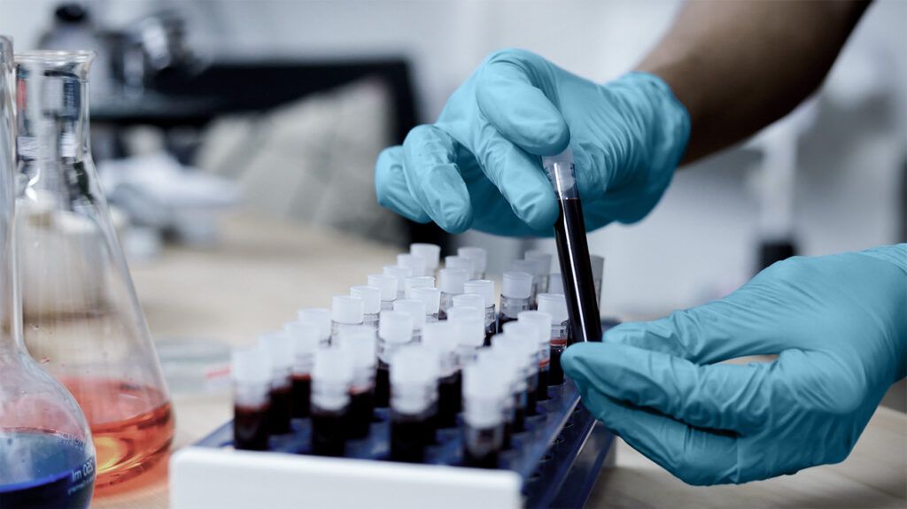
A new study analyzed blood samples from long COVID patients and healthy individuals. The findings suggest a dysregulation of the complement pathway, a part of the immune system, and the blood clotting pathways in individuals with long COVID at six months.
Changes in certain molecules in these immune and blood clotting pathways were predictive of the persistence of long COVID symptoms at 6 and 12 months, indicating the utility of these measures in developing diagnostic tools for long COVID.
The dysregulation of these immune and blood clotting pathways suggests that therapies targeting these pathways could help treat long COVID.
Approximately 10%-20%Trusted Source of individuals with a SARS-CoV-2 infection experience lingering symptoms beyond three months after symptom onset. These symptoms referred to as long COVID, can be debilitating, but there is a lack of diagnostic or therapeutic tools.
A new study published in Sciencefound that patients experiencing long COVID symptoms six months after the SARS-CoV-2 infection show dysregulation of the blood clotting or coagulation system and the complement pathway, which is a part of the immune system.
These changes in the coagulation and immune systems in long COVID patients were shown to predict the persistence of symptoms at six months. They may have the potential for the development of diagnostic tools. Moreover, therapeutics to counter the changes in the blood clotting and immune system could help alleviate long COVID symptoms.
Dr. Wolfram Ruf, Scientific Director at the Center for Thrombosis and Hemostasis (CTH), Johannes Gutenberg University, wrote in an accompanying editorial:
“Although therapeutic interventions with coagulation and complement inhibitors in acute COVID-19 produced mixed results, the pathological features specific for Long Covid suggest potential interventions for clinical testing.”
Long COVID refers to one or more symptoms that persist or develop after the acute phase of a SARS-CoV-2 infection. Common symptoms of long COVID include muscle weakness, fatigue, and brain fog.
Tissue damage, persistent inflammation, production of autoantibodies, and reactivation of latent virus reservoirs are some factors that have been hypothesizedTrusted Source to cause long COVID. However, the lack of knowledge about the precise mechanisms underlying long COVID has hampered the development of diagnostic tools and targeted therapies.
Several studies have shown that individuals with long COVID show immune system dysregulationTrusted Source. The present study further examined changes in the immune system associated with long COVID at six months.
The study included 39 healthy participants and 113 individuals with confirmed SARS-CoV-2 infection. During the 12-month follow-up period after the onset of a SARS-CoV-2 infection, 40 out of the 113 participants with an acute SARS-CoV-2 infection had at least one persistent symptom at the 6-month follow-up visit.
Serum samples were collected from the participants during the acute phase of the infection and six months after the infection. These serum samples were used to quantify changes in more than 6,500 proteins.
The participants with long COVID symptoms at six months showed changes in serum proteins belonging to the complement system compared with healthy individuals or those without long COVID at six months. The complement system is a part of the innate immune system, which serves as the first line of defense against germs.
The complement system activation helps elicit an immune response against pathogens or damaged tissue. During the activation of the complement pathway, the plasma proteins belonging to the complement system interact with each other to form a terminal complement complex. The terminal complement complex binds to the surface of or inserts into the membrane of pathogens and damaged cells to induce cell death or promote their removal by engulfment Trusted Sourceby phagocytes.
Among patients with long COVID at six months, the researchers found increased activation of the complement pathway during acute SARS-CoV-2 infection and at six months after diagnosis. Increased activation of the complement pathway and terminal complement complex formation in 6-month long COVID patients could lead to tissue damage.
The proteins in the complement system can be activated by three distinct pathways, each involving different types of molecules. The three complement activation pathways include the classical pathway, the alternative pathway, and the lectin pathway.
Individuals with long COVID at six months showed increased expression of molecules involved in forming the terminal complement complex via the activation of the classical and alternative pathways than those without long COVID or healthy patients.
In addition to the three complement activation pathways, thrombin, a protein that promotes blood coagulation, can also cause the activation of the complement pathway and lead to the formation of the terminal complement complex.
Patients with long COVID symptoms at the 6-month follow-up showed lower levels of antithrombin III, an enzyme that inhibits thrombin, during the acute phase and at six months after the onset of a SARS-CoV-2 infection than healthy individuals. The lower antithrombin III levels were accompanied by increased expression of markers of thrombosis, a state characterized by the formation of clots in the absence of bleeding.
Patients with long COVID at six months simultaneously showed increased markers for inflammation and those for thrombosis. The cooccurrence of inflammation and thrombosis is referred to as thromboinflammation.
Signs of thromboinflammation observed in individuals with long COVID at six months included destruction of red blood cells and dysfunction of endothelial cells that line blood vessels. Moreover, these patients also showed increased markers of tissue damage in the blood.
These changes associated with thromboinflammation reflect the dysregulation of the complement system in patients with 6-month long COVID. The dysregulation of the coagulation system in long COVID patients also underscores the need for cardiovascular health assessment.
The researchers found that changes in specific complement protein levels, coagulation system biomarkers, and age and body mass index predicted long COVID at 6 and 12 months.
MNT spoke with Dr. Hrishikesh Kulkarni, Assistant Professor of Medicine at Washington University School of Medicine, who was not involved in the study. Dr. Kulkarni said:
“By utilizing an unbiased screen and confirming it using distinct components of the membrane attack complex by which the complement system damages cells, the authors demonstrate that persistently increased complement activation is a key feature of Long COVID. Moreover, their models that incorporate the measurement of 2 protein ratios improve an already good clinical model comprising of age and body-mass index, most notably for 12-month Long COVID.”
The classical complement pathway is activated upon antibodies binding to viral proteins or autoantibodies in the body’s tissue. In the present study, the serum from patients with 6-month long COVID showed increased antibodies against cytomegalovirus, a type of herpesvirus.
This is consistent with evidence suggesting that long COVID symptoms may arise, in part, due to an inflammatory response to the reactivation of a prior herpesvirus infection. The persistence of SARS-CoV-2 in some tissues may also produce an immune response.
These results suggest that binding of the antibodies to proteins from a herpesvirus could contribute to the activation of the complement system. Besides explaining the increased complement activation, these results suggest that antivirals targeting the herpesvirus and SARS-CoV-2 could have the potential to ameliorate long COVID symptoms.
Researchers move closer to gene therapy solution for hearing loss
Hearing loss affects about 48 million Americans and 430 million people worldwide, with those numbers expected to grow as populations age.
More than 90 percent of individuals affected have sensorineural hearing loss, caused by damage to the inner ear and the destruction of the hair cells responsible for relaying sounds to the brain.
Hair cells cannot be regenerated in mammals, including humans, because unlike other cells in the body, any remaining hair cells in the inner ear cannot divide and other inner ear cells cannot convert themselves into new hair cells. Species like fish, birds, and reptiles, however, possess this ability
For this reason, effective hearing loss treatments for humans have eluded medicine, and the loss of hair cells, which can be caused by aging, noise exposure, and other factors, renders an individual’s hearing loss permanent.
Now, Harvard Medical School scientists at Mass Eye and Ear are hopeful they’ve developed a solution to address this longstanding limitation.
A research team led by Zheng-Yi Chen, an HMS associate professor of otolaryngology and associate scientist in the Eaton-Peabody Laboratories at Mass Eye and Ear, reported creating a drug-like cocktail of different molecules that successfully regenerated hair cells in a mouse model by reprogramming a series of genetic pathways within the inner ear.
The researchers hope their novel findings, publishedApril 17 in PNAS, could one day pave the way for clinical trials for a gene therapy that can be administered to people with hearing loss.
“These findings are extremely exciting because throughout the history of the hearing loss field, the ability to regenerate hair cells in an inner ear has been the holy grail,” said Chen. “We now have a drug-like cocktail that shows the feasibility of an approach that we can explore for future clinical applications.”
New approach to achieve hearing loss treatment
Previously, Chen’s research team studied zebrafish and chickens to uncover which pathways were responsible for inducing the cell division required to regenerate new hair cells. They discovered that two molecular signaling pathways, , were crucial to this process.
In a study published in 2019, the team showed for the first time that when these pathways were activated in adult transgenic mice, remaining inner ear cells could divide and develop characteristics of hair cells.
The new cells contained transduction channels that relay sound signals and the ability to form connections with auditory neurons — processes essential to hearing.
While an exciting discovery at the time, such an approach was not directly translatable to people, according to Chen. Unlike transgenic mice, humans cannot have Myc and Notch pathways turned on like a light switch.
A drug therapy, he explained, would have to be introduced to the inner ear to activate the Myc and Notch pathways.
Previous studies have shown that a chemical compound called valproic acid can activate Notch, however, no molecule exists to effectively activate Myc. That led the researchers to instead look for drug molecules that can alter the downstream pathways that turn on and off when Myc is activated.
Through single-cell RNA sequencing, they discovered that activating Myc and Notch led to a downstream effect in which two other pathways, Wnt and cAMP, became activated. Importantly, they found chemical compounds that can directly activate Wnt and cAMP.
They then used small biological molecules called small interfering RNAs (siRNAs) to remove genes downstream that suppressed the activation of the Myc pathway.
“Think about a brake when driving a car,” explained Chen. “If the brake is always engaged, you can’t drive. We found an siRNA that could remove the brake in this genetic pathway.”
The researchers then combined the chemical compounds and siRNA molecules into a drug-like cocktail. They delivered it to the inner ear of a normal adult mouse with damaged hair cells — an important distinction, as wild-type, non-transgenic mice would be more translatable to humans.
They further delivered the gene Atoh1 by a gene therapy approach that utilizes a harmless adenovirus into the cocktail-treated inner ear.
Remarkably, they found this drug-like cocktail combined with adenovirus turned on Myc and Notch, which led to the regeneration of new hair cells. They verified that the hair cells were functional through advanced imaging and other techniques.
Regenerating hair cells through gene therapy approach
Studies like Chen’s show the promise of gene therapy for treating incurable conditions like hearing loss. Last year, this research project was selected out of hundreds as one of the Disruptive Dozen gene and cell therapy technologies most likely to have a significant impact on health care over the next several years at the Mass General Brigham World Medical Innovation Forum.
Mass General Brigham recently launched its Gene and Cell Therapy Institute to help translate scientific discoveries made by researchers like Chen into first-in-human clinical trials and, ultimately, life-changing treatments for patients.
The researchers are conducting ongoing studies and refinements to this treatment approach in larger animal models, which are necessary before applying to initiate clinical trials.
They note that more research is needed to address limitations and challenges for delivering a treatment to the inner ear.
Scientists are examining different gene therapy and surgical methods, including an approach previously honed at Mass Eye and Ear, in which a different viral vector called an adeno-associated virus (AAV) was able to precisely and safely deliver gene therapy to the inner ear through a novel surgery.
Similar AAV-surgical approaches are currently used at Mass Eye and Ear in approved and experimental drug therapies for patients with inherited retinal disorders that can lead to blindness.
“My colleagues and I frequently are contacted by people with hearing loss who are desperate for effective treatments,” said Chen. “If we can combine a surgical procedure with a refined gene therapy delivery method, we hope we can achieve our number one goal of bringing a new treatment into the clinic.”
Authorship, funding, disclosures
Co-authors of the study include Yizhou Quan, Wei Wei, Volkan Ergin, PhD, Arun Prabhu Rameshbabu, Mingqian Huang, ChunJie Tian, Srinivas Vinod Saladi, and Artur Indzhykulian, of Mass Eye and Ear.
This study was funded by the National Institutes of Health (NIH grants R01DC006908, R01DC016875, R56DC006908, UG3TR002636, and R01DC017166), the Department of Defense (DOD grants W81XWH1810331, W81XWH2110957), the Fredrick and Ines Yeatts hair cell regeneration fellowship, Harvard Catalyst Translational Innovator Program The Five Senses, and the Mike Toth Head and Neck Cancer Center.
Chen is a co-founder and a SAB member of Salubritas Therapeutics, which is developing treatments for hearing loss including genome editing, inner ear regeneration, novel delivery, and gene therapy, and has ownership of over 5 percent equity. Quan and Chen are co-inventors on a patent application that has been filed based on this study. A patent application on the combination of small molecules/siRNAs in hair cell regeneration has also been filed.
Meeting a family affected by Usher syndrome type 1F has sent HMS researchers on a quest to save patients’ vision.
When neurobiologist David Corey showed up at a rare disease conference in 2017, he had no idea that he would enter a race against time to develop a treatment for it.
The conference was for Usher syndrome type 1F. Patients with this condition have a gene mutation that causes them to be born deaf and gradually lose their vision as they grow up. Corey, the Bertarelli Professor of Translational Medical Science in the Blavatnik Institute at Harvard Medical School, had devoted decades to studying the defective gene in a different context.
So when Corey happened upon an announcement for an Usher 1F conference in Boston, he knew he had to attend.
There, he introduced himself to Elliot Chaikof, chief of surgery at Beth Israel Deaconess Medical Center and the Johnson and Johnson professor of surgery at HMS, his wife Melissa, and their adult daughters, Rachel and Jessica, both of whom have Usher 1F. The Chaikofs had organized the conference through the nonprofit research collaborative they founded to find a treatment for the blindness part of the disease.
Meeting the Chaikofs — and especially Rachel and Jessica — stirred in Corey a powerful desire to help.
“We really felt that we know so much about this gene, if we don’t try to do something for the disease, who else is going to,” Corey said.
Six years later, the Corey lab has three candidate gene therapies for Usher 1F blindness. Each takes a different approach to correcting the disease-causing mutation.
The researchers are now testing the therapies in animal models and are confident that at least one will move to clinical trials in humans to become a successful treatment.
Defining the problem
Usher 1F is a particularly severe form of Usher syndrome, in which a gene mutation causes cells in the eye and ear to stop producing an essential protein. Rachel and Jessica’s gene mutation is most prevalent in the Ashkenazi Jewish community.
People with Usher 1F are usually born deaf and lacking the ability to balance. They develop an eye disease called retinitis pigmentosa, which causes a progressive loss of vision as the retina degenerates. Night vision often disappears first, followed by peripheral vision. For Jessica, this has meant giving up driving and getting a service dog to help her navigate the world around her. Eventually, people become completely blind.
“Everyone with Usher 1F has a unique experience, but one of the biggest challenges for me is slowly losing my independence as I lose my vision,” Jessica said.
Like many people with Usher 1F, Rachel and Jessica have benefited from cochlear implants, which have improved their ability to hear and communicate. However, the Chaikofs learned soon after their daughters were diagnosed that there was almost no research on the condition and virtually no therapies to treat it — so in 2013, they established Usher 1F Collaborative.
“Essentially there were zero research groups working on this particular problem — nothing, nobody,” Elliot said.
A call to action
While the Chaikofs were seeking answers, the Corey lab in the Department of Neurobiology at HMS was studying a protein called protocadherin-15 and its role in deafness.
The researchers figured out that protocadherin-15 in the inner ear helps sensory receptors called hair cells convert mechanical vibrations into electrical signals, which the brain interprets as sound. Without protocadherin-15, the conversion doesn’t happen, and the brain is unable to detect sound.
They also found protocadherin-15 in light-sensing cells, or photoreceptors, in the eye. However, they weren’t sure of its exact function there, nor did they know why people lacking the protein in their eyes lose their sight over time.
In the course of the research, Corey had become aware that in Usher 1F, a mutation in the gene that makes protocadherin-15 causes cells in the ear and eye to stop producing it.
Connecting with the Chaikofs gave Corey a new motivation for his research and reinvigorated the family’s quest for a cure.
“Because David understood the gene so well, he basically leapfrogged ahead of where the research was and hit the ground running,” said Melissa, who is chair of Usher 1F Collaborative.
“For the first time we felt as if we had someone who could truly make a difference on this particular problem,” Elliot added.
A daunting endeavor
The Corey lab chose to focus on the blindness aspect of Usher 1F, in part because patients are born profoundly deaf and lacking hair cells, making it unlikely that a therapy could restore their hearing. However, they are born with normal vision that gradually deteriorates, allowing time to intervene to preserve their sight.
The team found the perfect point person in Maryna Ivanchenko, an instructor in neurobiology at HMS who joined the lab to learn about gene therapy for deafness, but by serendipity is also an ophthalmologist and eye surgeon.
“Maryna has been absolutely central to this project because she understands both the hearing and the blindness aspects of this disease,” Corey said.
The researchers decided the way forward would be to design a gene therapy that either replaces or repairs the defective DNA that codes for protocadherin-15. Delivered inside eye cells, such a therapy would allow the cells to make the missing protein.
We really felt that we know so much about this gene, if we don’t try to do something for the disease, who else is going to.
David Corey
Bertarelli Professor of Translational Medical Science at HMS
But the team encountered a major challenge: Protocadherin-15 is a huge protein, and the DNA that codes for it is too large to fit inside the usual delivery vehicle — a small capsule made from a non-infectious virus.
With the clock ticking for Rachel, Jessica, and thousands of others with Usher 1F, the team knew that their best hope for quickly developing an effective gene therapy would be to work on three different potential solutions at the same time.
Three shots on goal
The logistics of such a project are daunting.
Because there is no good model for Usher 1F blindness in mice, Corey and colleagues started with a mouse model of deafness to test whether each therapy can restore protocadherin-15 production. They then moved to zebrafish, which offer a better model for progressive blindness. Testing continues in human eye cells grown in a dish, to be followed by tests in non-human primates. Only then can a therapy be deemed safe and effective enough for a human clinical trial.
“The project has been enormously complex because we’re testing three different strategies in two different organs and four different species,” Corey said. “Every time you add something, it multiplies the effort.”
Even so, the Corey lab — supported by funding from HMS and Usher 1F Collaborative — has made considerable progress on all three strategies:
The most promising so far is the mini gene — a shortened but still functional version of the protocadherin-15 gene that easily fits inside the viral capsule that carries the DNA into a cell. In a recent study, the researchers showed their mini gene can restore hearing in mice, and early experiments in zebrafish suggest that it can also restore vision.
The second strategy, called the dual approach, involves cutting the DNA in half. Each half is small enough to fit in the viral capsule. Once inside the cell, the halves reconnect, and can begin making the full-length protein. The approach has been used for other genes, Corey noted, and seems to be well suited for protocadherin-15. The researchers have tested this approach in mice and are starting to test it for safety in non-human primates.
The final strategy involves putting gene editing tools into the viral capsule instead of loading it with a replacement gene. Once inside the cell, the gene editors would correct the protocadherin-15 gene mutation. Many mutations cause the condition, but the researchers are designing the tools to fix the most common one, R245x. The researchers recently demonstrated that their approach can counteract hearing loss in mice.
Corey and his collaborators, including Marcos Sotomayor at The Ohio State University and Artur Indzhykulian, HMS assistant professor of otolaryngology head and neck surgery at Massachusetts Eye and Ear, hope at least one of these therapies will safely stop and perhaps even reverse vision loss in patients with Usher 1F.
“This is an outstanding example of how basic science can be translated to therapies,” Elliot said. “The progress that David has made in a very short period of time has been remarkable.”
The lab, whose work on Usher 1F has been supported by institutional programs including QFASTR and the Blavatnik Therapeutic Challenge Awards, plans to partner with a biotech company that will continue working on any therapies that succeed in initial testing.
Finding hope
If any of the strategies clear the hurdle of animal testing, the next step would be a clinical trial. Yet therein lies another challenge: Because Usher 1F is an orphan disease — meaning it affects fewer than 200,000 people in the United States — it will be hard to enroll enough people in a randomized controlled trial.
To get around this challenge, Usher 1F Collaborative has initiated a natural history study on vision loss in people with Usher 1F. The study will follow participants over four years to assess how their vision loss progresses naturally, essentially establishing the control group for a clinical trial ahead of time. That way, if and when a trial does happen, all participants can receive the therapy.
“We’re on a parallel track with David’s research so that when the natural history study is completed, we’ll be ready for the clinic,” Melissa said.
It’s not clear which, if any, of the therapies will succeed, nor when it will happen, but the possibility that they could make a tangible difference in people’s lives keeps Corey, Ivanchenko, and others motivated as they work long hours in the lab.
And the Corey lab’s three shots on goal have become many: Eight other groups at universities in the United States, Canada, and Australia are also working on treatments for Usher 1F, thanks to funding from the collaborative.
“There are a lot of patients and families whose world is about to go dark, and they need everyone’s best ideas,” Elliot said.
“I keep telling myself that there will be a time when I can finally stop letting my life revolve around Usher,” Jessica said. “Being able to see the research is what keeps me sane; it’s what helps me get through the day.”
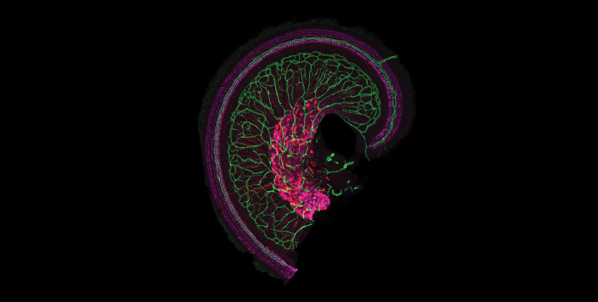
Study co-led by HMS scientist corrects gene mutation involved in inner ear function
Hair cells, shown in magenta, in the snail-shaped cochlea of the inner ear.
At a glance:
Trial treated six children aged 1 to 7 who had a mutation in the OTOF gene, which manufactures a protein important in transmitting signals from the ear to the brain.
The five participants who responded to treatment showed improvement in the ability to recognize sound as speech.
The gene therapy technique used in the study overcomes a roadblock presented by large genes and may prove useful in developing treatments for other forms of genetic deafness.
A novel gene therapy approach has given five children who were born deaf the ability to hear. The method, which overcomes a roadblock presented by large genes, may be useful in other treatments, according to researchers.
The work, conducted in Fudan, China, by a team co-led by Harvard Medical School researchers at Massachusetts Eye and Ear and by collaborators at Fudan University’s Eye & ENT Hospital, treated six children aged 1 to 7 who had a mutation of the OTOF gene, which manufactures a protein important in transmitting signals from the ear to the brain.
Five of the six children showed improvement in hearing over the 26-week trial, with four outcomes described by researchers as “robust.” With hearing a critical factor in language acquisition, researchers also measured speech perception — the ability to recognize sound as speech — and all five of those who responded to treatment showed improvement there.
“This really opens the door to developing other treatments for different kinds of genetic deafness,” said co-senior author Zheng-Yi Chen, HMS associate professor of otolaryngology head and neck surgery and a researcher at Mass Eye and Ear’s Eaton-Peabody Laboratories. Chen added that the study provides proof-of-concept that prior work in laboratory animals does translate to humans. “Now we can move forward in humans quickly. This has given us a real boost of confidence.”
Hearing loss affects more than 1.5 billion people worldwide, Chen said, including about 30 million cases of genetic impairment in children. Approximately 200,000 people worldwide are deaf due to a mutation in the OTOF gene. Those in the current trial had an OTOF mutation called DFNB9, which affects 2 to 8 percent of all cases of genetic deafness from birth.
The OTOF gene encodes the otoferlin protein, produced by cells in a snail-shaped part of the inner ear called the cochlea. In the cochlea, sound waves are translated into electric pulses carried by nerve cells to the brain, where they are interpreted as sound. Otoferlin plays a role in transmitting pulses from cochlear cells to the nerves and without it, sound is translated into electric signals but never reach the brain.
Shu, who was a postdoctoral fellow in Chen’s lab from 2010 to 2014, said the large response to the request for study participants reflects the need for improved treatment for congenital deafness for which, Chen pointed out, there are no approved drugs. Researchers ultimately screened 425 potential participants, enrolling just six.
Of the six, four had cochlear implants — which, with training, allow interpretation of speech and sound — in one ear while the two youngest participants, ages 1 and 2, had no implants. When the implants were switched off, all participants were completely deaf.
Before the trial began, researchers had to tackle a significant technical problem related to the size of the OTOF gene. The procedure called for the gene to be inserted into the cochlea using a type of virus researchers commonly use for this purpose.
The virus inserts the gene into the DNA of target cells, which then begin to manufacture the missing protein. The problem in this case is that the OTOF gene is too big for the virus to hold. Researchers got past this by dividing the gene into two, encapsulating the halves into separate viruses, and then injecting a mixture with both halves of the gene into the cochlea.
Though the viruses inserted the gene halves at different spots on the cells’ DNA, when those halves were expressed, cellular machinery assembled the complete protein, restoring the cells’ ability to transmit signals to the brain.
To get healthy copies of the OTOF gene into ear cells, the team used a technique like the one described here by HMS neurobiologist David Corey, whose lab aims to restore the hearing-related protein protocadherin-15.
The mixture was injected into the fluid of the inner ear, and the viruses made their way to the target cells as they would if they were a naturally occurring infection. Researchers had to wait for four to six weeks after injection to see the first signs that hearing was being restored.
In five of the six participants, the improvement was progressive. The three older children, with cochlear implants turned off, could understand and respond to speech by 26 weeks, with two able to recognize speech in a noisy room and have a telephone conversation.
The younger participants showed improvement in the ability to recognize speech but were too young for some tests. Anecdotally, Chen said the 1-year-old has been able to respond to stimuli and begun to verbalize simple first words, like “mama.” One of the six participants showed no improvement, which Chen and Shu said is poorly understood but may have been due to an immune reaction to the viral vector.
In some cases, Shu said, parents noticed the response even before the researchers conducted their first tests at four weeks.
“We first found out when the parents told us: When her mother called her, she turned back,” Shu said. “All of them are very hopeful. They were very, very excited, and all of them cried when they first found that their child can hear.”
The project, done in December 2022, was the first to make use of gene therapy to treat this condition, but several studies in various stages are targeting the same condition. This team and four others are expected to present their findings Feb. 3 at the Association for Research in Otolaryngology Annual Meeting.
Chen said that will hopefully accelerate work that, though it has taken years to reach this point, has recently seen rapid progress.
Next steps, he and Shu said, include monitoring the participants in this study, as well as beginning a new study with participants from more diverse backgrounds. If all goes well, Chen said, approval of the treatment by U.S. federal regulators could be as little as three to five years away.
“This is truly remarkable. When we tell the story, even for our colleagues, it brings a tear to the eye,” Chen said. “I’ve been working in this field for three decades, and I know how difficult it has been to come to this point. I also know we’re at the juncture of a great future.”
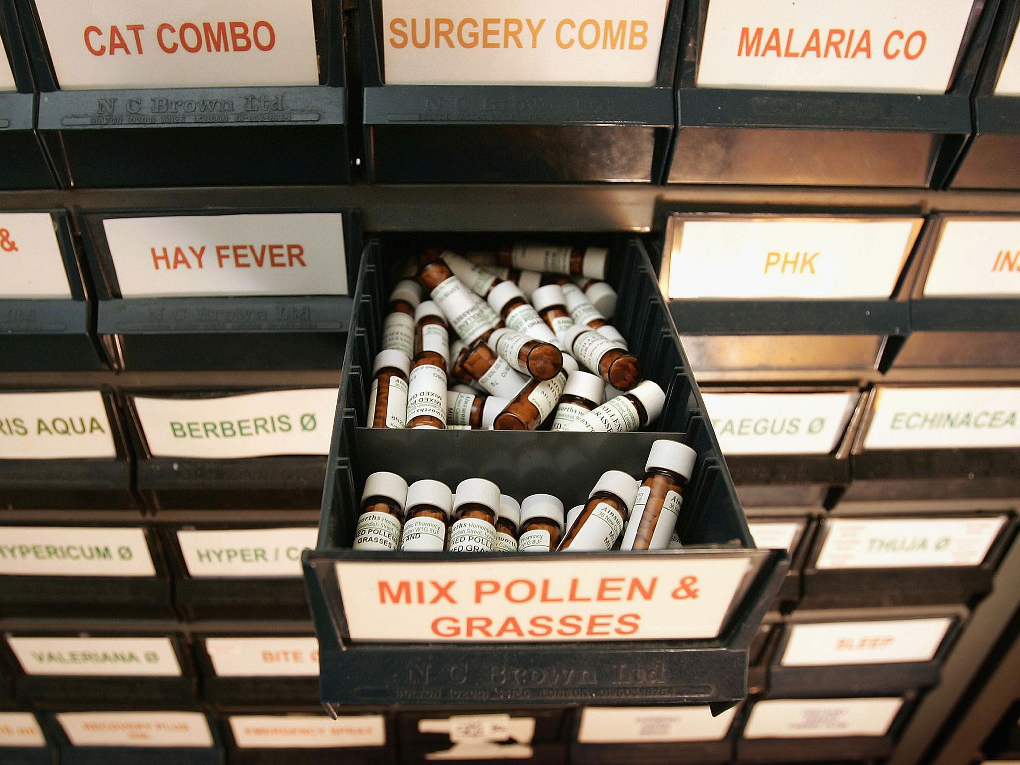
The Federal Trade Commission has demanded that producers of homeopathic treatments say on the label that they do not work
Tubes of homeopathic granules at Boiron laboratory in Brest, western France (Getty)
There is a huge market in the US for homeopathic remedies. In 2007 alone, it was estimated Americans spent more than $3bn on a controversial system of alternative medicine created in 1796 by Samuel Hahnemann, and which has long been dismissed by mainstream science.
Now, the US government is requiring that producers of such items ensure that if they want to claim they are effective treatments, then they need to make available the proof. Otherwise, they will need to point out that there is “no scientific evidence that the product works”.
“Homeopathy, which dates back to the late-eighteenth century, is based on the view that disease symptoms can be treated by minute doses of substances that produce similar symptoms when provided in larger doses to healthy people,” said a notice, filed earlier this month by the Federal Trade Commission.
“Many homeopathic products are diluted to such an extent that they no longer contain detectable levels of the initial substance. In general, homeopathic product claims are not based on modern scientific methods and are not accepted by modern medical experts, but homeopathy nevertheless has many adherents.”
Does Homeopathy Work?
Slate said there was near-unanimous mainstream scientific consensus that homeopathy’s purported mechanism of action – using ultra-highly diluted substances to allow “like to cure like” – runs counter to basic principles of chemistry, biology, and physics.
Health policy expert Timothy Caulfield recently said: “To believe homeopathy works … is to believe in magic.
Yet, reports suggest the so-called treatments are unlikely to disappear from the shelves of pharmacists’ shops.
The FTC said that a homeopathic drug claim that is not substantiated by competent and reliable scientific evidence “might not be deceptive if the advertisement or label where it appears effectively communicates that: 1) there is no scientific evidence that the product works; and 2) the product’s claims are based only on theories of homeopathy from the 1700s that are not accepted by most modern medical experts.”