Could dark chocolate have a blood pressure-lowering effect?


Pieces of dark chocolate
Dark chocolate may have blood pressure-lowering effects, research suggests.
  • Essential hypertension is high blood pressure that doesn’t have a known cause.
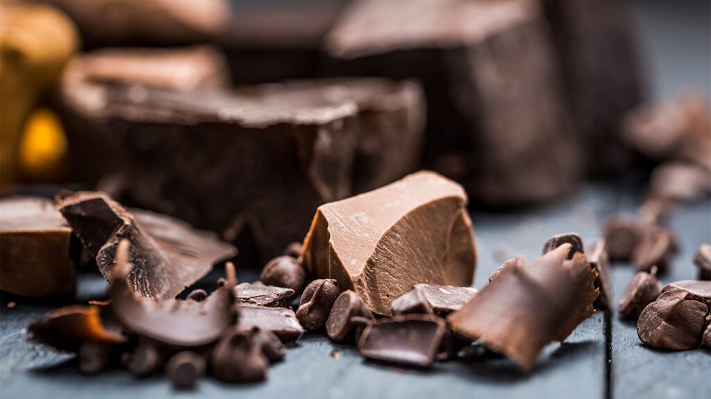
  • Dark chocolate is a popular food item that may offer valuable health benefits.
  • A Mendelian randomization study found that intake of dark chocolate was associated with a decreased risk for essential hypertension and possibly a reduced risk for blood clots.

High blood pressure can be dangerous to cardiovascular health. Prevention and management of high blood pressure can help ensure many positive health outcomes but researchers are still seeking to understand the best methods for preventing high blood pressure.

A recent studyTrusted Source published in Nature Scientific ReportsTrusted Source examined how the intake of dark chocolate may help decrease the risk of essential hypertension (high blood pressure).

The results also suggest that consuming dark chocolate may decrease blood clot risk, but researchers couldn’t establish a causal relationship.

The results point to the potential benefits of this food and the need for future research into its potential health benefits.

The dangers of hypertension and the benefits of dark chocolate

High blood pressureTrusted Source or hypertension is when the force blood exerts on the arteries in the body gets too high.

Essential hypertensionTrusted Source or high blood pressure involves high blood pressure that doesn’t have a known cause. Non-study author Dr. Rigved Tadwalkar, a board certified cardiologist at Providence Saint John’s Health Center in Santa Monica, CA, explained to Medical News Today:

“Essential hypertension is a highly prevalent medical condition characterized by elevated blood pressure levels without an apparent underlying cause. It is by far the most common cause of hypertension globally. Essential hypertension stands out as the predominant contributor to cardiovascular diseases, including coronary artery diseaseheart failure, and stroke.”

High blood pressure can be managed through lifestyle changes and certain medications. However, people can often take action to preventTrusted Source high blood pressure from occurring in the first place. In general, eating a healthy diet, maintaining a healthy weight, and exercising can help.

However, researchers are also looking at how eating specific foods may assist with high blood pressure prevention. Research like this could lead to people making more specific healthy food choices.

Dark chocolate is one food with many potential health benefits that researchers are interested in studying. Non-study author and registered dietitian nutritionist Karen Z. Berg explained to Medical News Today:

“For chocolate to be considered ‘dark chocolate’ it has to contain at least 50% cocoa solids, and many dark chocolate is made up of 70% or even 90% cocoa which leaves a lot less room for other additives like sugar. This is why dark chocolate tends to be more bitter than milk chocolate varieties.”

“Cocoa is rich in flavanols, so the higher the percentage of cocoa in your chocolate, the more health benefits are possible. [D]ark chocolate is also a good source of fiber, iron, magnesium, phosphorus and zinc. It is important to note that to get the most benefits, you want to choose more natural forms of cocoa. It is also important to note that the higher the percentage of cocoa you have, the more caffeine it will contain.”
— Karen Z. Berg

How dark chocolate lowers essential hypertension risk

Researchers wanted to understand more about how dark chocolate may help lower the risk for several cardiovascular diseases. One way researchers can look into possible causality as well is to use a technique called Mendelian randomizationTrusted Source.

This method involves looking at genetic differences to provide evidence that a particular intervention has a causal effect and helps eliminate the risk of reverse causation. While not perfect proof, it allows for safety and data collection that isn’t always possible with other studies due to ethical concerns.

Researchers of this study were able to use data from publicly available genome-wide association studies. They looked at dark chocolate intake and risk for several cardiovascular diseases, including essential hypertension, coronary heart disease, heart failure, stroke, blood clots beginning in the veins, and heart attack.

The analysis results were promising for the positives of dark chocolate intake. Researchers found that genetically predicted dark chocolate intake may help lower the risk for essential hypertension. The data further points to a possible causal relationship between dark chocolate intake and decreased risk for essential hypertension.

They also found a possible association between dark chocolate intake and a reduced risk for venous thromboembolismTrusted Source, which is when a blood clot forms in a vein.

Chocolate-derived treatments for hypertension?

Dr. Tadwalkar noted that the results held promise despite the study’s limitations.

“The study’s findings hold significant promise for the prevention of essential hypertension. If future research confirms the causal relationship, it could pave the way for not only dietary recommendations but also dark chocolate-derived bioactive compounds or extracts in the development of novel therapies for the prevention or management of essential hypertension. Ultimately, the study offers exciting possibilities for the future of essential hypertension prevention,” he explained to Medical News Today.

The researchers did not find any associations between dark chocolate intake and other cardiovascular diseases.

Dr. Cheng-Han Chen, a board certified interventional cardiologist and medical director of the Structural Heart Program at MemorialCare Saddleback Medical Center in Laguna Hills, CA, who was not involved in the study, commented with his thoughts on the study to MNT:

“Of the diseases studied, dark chocolate intake was specifically associated with reduced risk of hypertension, but not any other conditions. While this is a notable finding, its clinical implication is limited.”

“The clinical cardiovascular concern for hypertension would be on its potential impact on rates of resultant conditions such as heart attack and stroke, but no other associations between dark chocolate intake and other cardiovascular conditions were identified. I wouldn’t discourage dark chocolate intake based on this study, but I also wouldn’t recommend increasing dark chocolate intake based on this study alone,” Dr. Chen added.

Will eating more chocolate improve my heart health?

This research also presents particular challenges and limitations.

First, the sample size exposure data was small, which may have impacted the results. Second, they lost some of the single nucleotide polymorphisms summary-level data for stroke and heart failure, which could have affected the results. The research also utilized data from European ancestry, meaning the results cannot be generalized to other populations.

Their research did not allow for certain analyses, such as looking at the amount of dark chocolate intake or the risk for cardiovascular diseases based on factors like age or gender. There was also some potential bias because of sample overlap. Finally, there may have been some risk of confounding regarding coronary heart disease data.

Overall, the results point to the potential benefits of dark chocolate for preventing essential hypertension. It also opens the door for future research in this area.

Dr. Tadwalkar noted the following:

“Several exciting research avenues remain open in the wake of studies like this one. Next steps include unraveling the precise mechanisms by which dark chocolate consumption influences cardiovascular health. This could involve utilizing advanced genetic techniques to understand how dark chocolate intake might influence gene expression patterns relevant to cardiovascular health.”

“Also, further research could delve into the potential impact of dark chocolate on other cardiovascular endpoints, such as atherosclerotic plaque formation and progression, cardiac function and remodeling, as well as blood clotting and fibrinolysis,” he added.

How To Become a Morning Person


Break up with your snooze button by shifting your bedtime and establishing a consistent nighttime routine

person sitting on bed stretching

It’s not easy being a night owl. Whether you’re dragging yourself to an 8 a.m. meeting or trying to deliver your kids to school without earning them a tardy slip, being a night owl who operates on the rest of society’s schedule can leave you short on shut-eye.

So, how can a night owl become an early bird? Psychologist and behavioral sleep medicine specialist Michelle Drerup, PsyD, DBSM, offers advice on how to become a morning person so you can finally break up with your snooze button. 

How to become a morning person

Though it may feel like your night owl ways are set in stone, Dr. Drerup says your sleep-related habits and behaviors — known as sleep hygiene — can make a massive and meaningful impact on your body’s natural tendencies. 

“By making behavioral changes, you may be able to shift your sleep schedule preferences,” she says. Here’s how.

1. Shift your bedtime

To become a morning person, you first need to become an early-to-bed person. But how? Count back from the time your alarm rings (or the time you want it to ring), aiming for a total of seven to nine hours a night. That will become your new target bedtime — eventually.

“It’s important to adjust your sleep time gradually,” Dr. Drerup says. If you’re used to turning in well after midnight, willing yourself to suddenly fall asleep at 10 p.m. is sure to backfire. 

Instead, aim to go to bed 15 or 20 minutes earlier than usual for a few days. Then, push it back another 15 minutes for several more days, and so on until you reach your bedtime goal.

2. Develop a bedtime routine

quiet bedtime routine is the key to helping you fall asleep earlier (which is the key to helping you wake up earlier).

At least an hour before lights out, dim the lights and power down your electronics. Find something soothing to do, like taking a warm bath, reading a book or listening to a (not-too-stimulating) podcast. That means not watching an action film or spending an hour doomscrolling on social media right before you tuck in. 

“Anything that activates our brain as we’re trying to wind down can keep us going,” Dr. Drerup shares. “Give yourself time to wind down and prepare your mind for bed.” 

3. Rely on natural light

Your body’s circadian rhythm — aka your internal clock — has pretty particular needs.

“Our circadian rhythms are responsive to light and dark,” Dr. Drerup explains. Being exposed to bright light first thing in the morning helps you feel more alert, and it also helps shift your internal rhythm toward an earlier wake time. 

Natural light is best, so get outside or open your bedroom window. If you can’t get outside or your room is deprived of natural light (darn you, basement apartment!), try a light therapy lamp that mimics the spectrum of natural light.

Being exposed to light during the rest of the day is important, too. It’s one of the ways your body knows when it’s time to sleep and when you’re supposed to be awake.

“During the daytime, try to get outside and get some natural light on days when there’s sunlight,” Dr. Drerup suggests. “That helps keep your circadian rhythm on track.

4. Move your alarm clock

Hitting snooze is all too tempting, so remove that option. Try putting your alarm clock across the room so you have to get up to turn it off. 

Some apps make it even harder to sleep in by forcing you to engage in mentally stimulating activities like solving a puzzle to stop the beeping.

“Do whatever works to keep you from hitting snooze,” Dr. Drerup advises.

5. Get moving in the morning

If you’re a night owl, an early morning jog might sound like punishment. But if you can get yourself into the habit, exercising in the morning can give you energy to jump-start your day.

6. Make mornings more pleasant

Try to schedule something to look forward to in the morning so that getting up feels like less of a slog, Dr. Drerup suggests.

Perhaps a hot cup of coffee, sipped in silence, while you take on the daily crossword puzzle? Maybe eating a healthy breakfast while you read a book? Knowing that something pleasant awaits can help you take that first painful step out of bed. 

7. Get high-quality sleep

Sleep isn’t just about quantity; it’s also about quality. It can feel impossible to get up in the morning if you’re tossing and turning and waking up all night long.

Everything from your diet and stress levels to your partner’s tendency to snore can get in the way of a good night’s sleep, so make sure you’re taking steps not just to go to sleep but to stay asleep. You may need to: 

It may seem like an endless list, but making just a few changes can make a world of a difference in terms of the quality of your sleep.

8. Be consistent

Routine is key, Dr. Drerup says, and deviating from it too much can throw things back into disarray.

“Try to keep a consistent wake time,” she continues. “Whatever schedule you’re keeping, be consistent with it.”

That doesn’t mean you have to be inflexible with your sleep schedule. If you want to stay out late at a Friday night event or catch some extra ZZZs on vacation, that’s OK — every once in a while.

“Life happens,” Dr. Drerup acknowledges. “But try to limit these types of exceptions to your new schedule, or they’ll snowball and push you back toward your old schedule again.”

9. Maintain your motivation

When you feel tempted to hit snooze and go back to sleep, it’s helpful to remember why you’re trying to make this change in the first place. Make sure you identify your reasons so that you have to remind yourself of them.

Do you want to become a morning person so you can be more productive (or just less of a zombie) at work? So you can spend more time with your family on weekends? So you don’t wake up at noon feeling like you’ve wasted half the day?

“Thinking about your reasons can help keep you motivated,” Dr. Drerup says. 

Why some people just aren’t morning people

Your natural sleep/wake cycle, or circadian rhythm, plays a big role in your sleep schedule — and it can vary a lot from person to person.

“Our internal rhythm influences when we start to feel sleepy and when we’re inclined to wake up,” Dr. Drerup states.

Your sleep chronotype matters

People fall into different groups, or chronotypes, depending on whether they feel most awake and alert in the morning, in the evening or somewhere in between — and unfortunately, you can’t pick your chronotype. Genetics can play a part in whether you identify as a night owl or a morning lark.

But no chronotype is inherently better or worse than another. As Dr. Drerup points out, there’s nothing wrong with staying up late and sleeping in: “If that schedule fits with your lifestyle and your obligations, it’s not necessary to change it,” she says.

The trouble comes when your late bedtime clashes with your early morning obligations. If you’re regularly getting less than the recommended seven to nine hours of sleep a night, your health and well-being can suffer.

Can you adjust our circadian rhythm?

Adopting (and really enforcing) healthy sleep habits can help you reset your circadian rhythm and change your sleep/wake schedule. But if you’ve made these changes and are still struggling to drag yourself out of bed, consider seeing a sleep specialist.

“We can help figure out if there are barriers keeping you from making these behavior changes, or if you might have an underlying sleep disorder,” Dr. Drerup says. This could include, for example, a circadian rhythm disorder.

While shifting your schedule takes some effort, it’ll make it easier to accomplish that first task of the day: waking up.

An Infant with Inconsolable Crying and Weakness


res360

Is infant botulism most often caused by exposure to honey?

Signs and symptoms of infant botulism include irritability, constipation, features of bulbar palsies (a weak cry, ptosis in both eyes, and poor feeding), lethargy, weakness, and respiratory difficulties. Bulbar and facial muscle weakness may mask the clinical signs typically associated with respiratory distress, such as facial grimacing or nasal flaring. Read the NEJM Case Records of the Massachusetts General Hospital here.

Clinical Pearls

Q: What are the different ways that Clostridium botulinum may be transmitted to humans?

A: The neurotoxins produced mostly by C. botulinum are the drivers of the clinical illness known as botulism. There are multiple C. botulinum transmission categories: the bacterial spores or toxins can be introduced into the body through food consumption (infant botulism, foodborne botulism, or adult intestinal toxemia), through contamination of an infected wound (wound botulism), or iatrogenically (botulism associated with cosmetics or migraine treatment). Overall, botulism is a rare disease in the United States, and infant botulism is the most common transmission category.

Q: Is infant botulism most often caused by exposure to honey?

A: In patients with infant botulism, the ingestion of spores may result in colonization and germination, followed by toxin production and subsequent toxemia. Clearly defined food exposures, such as exposures to honey or corn syrup, account for only a minority of cases. In fact, multiple factors affect the acquisition of the clinical disease, with infants’ gastrointestinal tracts being particularly susceptible. Often, there is a history involving rural living, dust production, or nearby soil perturbation. The incubation period after exposure to spores may be a few days to a few weeks.

Morning Report Questions

Q: Describe the effect of botulinum neurotoxin on the nervous system.

A: After sporulation, the enteric botulinum neurotoxin enters the systemic circulation and interrupts the normal signal transmission at the neuromuscular junction. The enteric botulinum neurotoxin irreversibly binds a specific receptor on the presynaptic peripheral cholinergic nerve terminal and then enters the cell, breaking down a critical protein associated with exocytosis and interrupting the release of the neurotransmitter acetylcholine into the intersynaptic space. Once cleaved, this exocytosis-associated protein complex requires up to 4 weeks to regenerate, which explains the protracted course of the illness.

Q: Is there an antitoxin to treat infant botulism?

A: The diagnosis of botulism is typically made with the detection of enteric botulinum neurotoxin in stool. This test is generally available only through local departments of public health, and it may take several days for the results to become available. Therefore, it is not appropriate to delay the administration of the antitoxin while waiting for the diagnosis to be microbiologically confirmed. Early recognition of the illness and prompt administration of medication, preferably within 7 days after symptom onset, are imperative to increase the likelihood of a good outcome. Infant botulism is treated with human-derived anti–botulism toxin antibodies (infant botulism immune globulin, known as BabyBIG). BabyBIG antibodies do not penetrate the neuron, so it is critical to administer the antitoxin as soon as possible, before enteric botulinum neurotoxin enters the cell.

PROTECTing β-Cell Function in Children with Type 1 Diabetes Mellitus


res360

Type 1 diabetes mellitus (T1DM) is a chronic disease that is primarily diagnosed in childhood and marked by the autoimmune destruction of pancreatic insulin-producing β-cells. Histopathologic studies in the 1960s showed that by the time the disease becomes clinically apparent, fewer than 10% of β-cells remain. One third of pediatric patients with T1DM first present with diabetic ketoacidosis, which can be life-threatening. Diagnosis and treatment of T1DM before the pancreas accrues major damage might mitigate some of the complications described.

Teplizumab is an anti-CD3 monoclonal antibody that has the potential to modulate the CD8+ T cells that induce β-cell destruction. In the randomized, placebo-controlled TN10 trial published in NEJM in 2019, 14 daily infusions of teplizumab delayed the time to diagnosis of diabetes by a median of 24 months in children aged 8 years and older with stage 2 diabetes (defined as the presence of ≥2 autoantibodies and evidence of dysglycemia but not yet meeting diagnostic criteria for diabetes mellitus). If teplizumab delayed the progression of prediabetic dysglycemia in the TN10 trial, could it also delay the progression of diabetes after diagnosis?

Investigators examined this question in the international, phase 3, randomized, placebo-controlled PROTECT study in 328 predominantly white children aged 8–17 years with a recent (mean, 5.3 weeks) diagnosis of overt (stage 3) T1DM. The children had relatively nascent disease marked by robust stimulated C-peptide levels and a mean glycated hemoglobin of 9.00%. Participants were randomized to receive two 12-day courses of daily teplizumab or placebo infusions 26 weeks apart. The teplizumab protocol was modified from the single 14-day course in the TN-10 trial based on the observation that teplizumab’s effect on immune cells was short-lived.

Teplizumab was efficacious in preserving β-cell function at week 78 (the primary endpoint), as measured by the mean change from baseline in C-peptide area under the concentration-time curve (AUC) in children with newly diagnosed T1DM. Secondary endpoints, including required insulin doses, glycated hemoglobin levels, time in target glucose range, and hypoglycemic events did not differ significantly between groups. The safety data were reassuring; two episodes of cytokine release syndrome in the treatment group and one case of Epstein-Barr virus infection in each group.

Although questions remain about the clinical utility of teplizumab in patients with newly diagnosed T1DM, let us hope that we are turning the page to a new chapter in T1DM care and not just entering a “honeymoon” period of drug development that will rapidly fade.

Cannabis Heightens Workout Enjoyment – But Does It Boost Performance?


Recent research indicates that cannabis can enhance the enjoyment and motivation for exercise, particularly in casual contexts rather than performance-focused activities. The study, involving runners who used either THC or CBD before exercising, found that while cannabis use increased positive feelings during exercise, THC made physical efforts feel more challenging. Credit: SciTechDaily.com

8 in 10 cannabis users report combining marijuana with exercise, saying it boosts motivation and mood, eases pain.

A bit of weed before a workout can boost motivation and make exercise more enjoyable. But if performance is the goal, it may be best to skip that joint.

That’s the takeaway of the first-ever study to examine how legal, commercially available cannabis shapes how exercise feels.

The study of 42 runners, published recently in the journal Sports Medicine, comes almost exactly 10 years after Colorado became the first state to commence legal sales of recreational marijuana, at a time when cannabis users increasingly report mixing it with workouts.

Boulder-based ultrarunner Heather Mashhoodi runs on the treadmill in 2021 as part of a study exploring how cannabis influences exercise. First author Laurel Gibson, left, takes notes. Study participants used cannabis on their own at home before being picked up and driven to the lab for testing. Credit: Patrick Campbell/CU Boulder

“The bottom-line finding is that cannabis before exercise seems to increase positive mood and enjoyment during exercise, whether you use THC or CBD. But THC products specifically may make exercise feel more effortful,” said first author Laurel Gibson, a research fellow with the University of Colorado Boulder’s Center for Health and Addiction: Neuroscience, Genes and Environment (CU Change).

The findings, and previous research by the team, seem to defy long-held stereotypes that associate cannabis with “couch-lock” and instead raise an intriguing question: Could the plant play a role in getting people moving?

“We have an epidemic of sedentary lifestyle in this country, and we need new tools to try to get people to move their bodies in ways that are enjoyable,” said senior author Angela Bryan, a professor of psychology and neuroscience and co-director of CU Change. “If cannabis is one of those tools, we need to explore it, keeping in mind both the harms and the benefits.”

Why do people mix weed and workouts?

When researchers asked study participants, here’s what they said:

  • 90.5% It increases enjoyment
  • 69% It decreases pain
  • 59.5% It increases focus
  • 57.1% It increases motivation
  • 45.2% It makes time go by faster
  • 28.6% It improves performance

‘A First-of-a-Kind Study’

In one previous survey of cannabis users, Bryan’s research group found that a whopping 80% had used before or shortly after exercise. Yet very little research has been done at the intersection of the two.

For the study, Bryan and Gibson recruited 42 Boulder-area volunteers who already run while using cannabis.

After a baseline session, in which the researchers took fitness measurements and survey data, they assigned participants to go to a dispensary and pick up either a designated flower strain that contained mostly cannabidiol (CBD) or a Tetrahydrocannabinol (THC) -dominant strain.

THC and CBD are active ingredients in cannabis, with THC known to be more intoxicating.

Boulder-based ultrarunner Heather Mashhoodi runs on the treadmill in 2021 as part of a study exploring how cannabis influences exercise. First author Laurel Gibson takes notes in the background. Study participants used cannabis on their own at home before being picked up and driven to the lab for testing. Credit: Patrick Campbell/CU Boulder

On one follow-up visit, volunteers ran on a treadmill at a moderate pace for 30 minutes, answering questions periodically to assess how motivated they felt, how much they were enjoying themselves, how hard the workout felt, how quickly time seemed to pass, and their pain levels.

On another visit, they repeated this test after using cannabis.

Federal law prohibits the possession or distribution of marijuana on college campuses, so the runners used it at home, before being picked up in a mobile laboratory, a.k.a the ‘CannaVan,’ and brought to the lab.

The runners also wore a safety belt on the treadmill.

‘Not a Performance-Enhancing Drug’

Across the board, participants reported greater enjoyment and more intense euphoria, or ‘runner’s high,’ when exercising after using cannabis.

Surpisingly, this heightened mood was even greater in the CBD group than in the THC group, suggesting athletes may be able to get some of the benefits to mood without the impairment that can come with THC.

Participants in the THC group also reported that the same intensity of running felt significantly harder during the cannabis run than the sober run.

This may be because THC increases heart rate, Bryan said.

In a previous study conducted remotely, she and Gibson found that while runners felt more enjoyment under the influence of cannabis, they ran 31 seconds per mile slower.

“It is pretty clear from our research that cannabis is not a performance-enhancing drug,” said Bryan.

Notably, numerous elite athletes—including U.S. sprinter Sha’Carri Richardson— have been prohibited from competing in recent years after testing positive for cannabis.

An NCAA committee recently recommended that it be removed from its list of banned substances.

A Different Kind of Runner’s High

Why does cannabis make exercise feel better?

While natural, pain-killing endorphins have long been credited with the famous “runner’s high,” newer research suggests that this is a myth: Instead, naturally produced brain chemicals known as endogenous cannabinoids are likely at play, kicking in after an extended period of exercise to produce euphoria and alertness.

“The reality is, some people will never experience the runner’s high,” Gibson notes.

By consuming CBD or THC, cannabinoids that bind to the same receptors as the cannabinoids our brain makes naturally, athletes might be able to tap into that high with a shorter workout or enhance it during a long one, she said.

Athletes considering using cannabis should be aware that it can come with risks — including dizziness and loss of balance— and it’s not for everyone.

For someone gunning for a fast 5k or marathon PR, it doesn’t really make sense to use beforehand, Bryan said.

But for an ultrarunner just trying to get through the grind of a double-digit training run, it might.

As a public health researcher, Bryan is most interested in how it could potentially impact those who struggle to exercise at all, either because they can’t get motivated, it hurts, or they just don’t like it.

“Is there a world where taking a low-dose gummie before they go for that walk might help? It’s too early to make broad recommendations but it’s worth exploring,” she said.

Scientists Have Proven That Severe COVID-19 Is a Thrombotic Disease.


Severe COVID-19 causes early lung capillary thrombosis, leading to respiratory distress, with studies emphasizing prompt anti-coagulation treatment to mitigate complications.

Scientists from the University of São Paulo have discovered that severe COVID-19 is primarily caused by damage to the small blood vessels in the lungs, a result of SARS-CoV-2 infection.

Blood clot formation (thrombosis) in the small blood vessels of the lungs is an early result of severe COVID-19, often occurring before the breathing difficulties caused by widespread damage to the air sacs, according to a Brazilian study reported in an article published in the Journal of Applied Physiology. Post-mortem examinations of nine individuals who passed away from severe COVID-19 revealed a distinct pattern of changes in lung blood vessel structure and thrombosis.

For the first time, the article describes sub-cellular aspects of the endothelial damage and associated thrombotic phenomena caused by the infection. It notes the impact of acute inflammation on lung microvascular circulation as the key factor in severe COVID-19, contributing to a deeper understanding of the pathophysiology of the disease and the development of novel therapeutic strategies.

“This study furnished the final proof of what we’d been pointing out since the very start of the pandemic – that severe COVID-19 is a thrombotic disease. The virus SARS-CoV-2 has a tropism for [is attracted to] the endothelium, the layer of cells that lines blood vessels. When it invades endothelial cells, it first affects microvascular circulation. The problem starts in the capillaries of the lungs [the tiny blood vessels that surround the alveoli], followed by clotting in the larger vessels that can reach any other organ,” said pulmonologist Elnara Negri, first author of the article and a professor at the University of São Paulo’s Medical School (FM-USP). She was one of the first researchers in the world to reach the conclusion that severe COVID-19 is a thrombotic disease.

The researchers at USP analyzed lung tissue from nine patients who died from COVID-19. Credit: Elia Caldini

In the study, which was supported by FAPESP, the researchers used transmission and scanning electron microscopy to observe the effects of the virus on lung endothelial cells from severe COVID-19 patients who died at Hospital das Clínicas, the hospital complex operated by FM-USP.

All nine samples obtained by minimally invasive autopsies displayed a high prevalence of thrombotic microangiopathy – microscopic blood clots in small arteries and capillaries that can lead to organ damage and ischemic tissue injury. The samples came from patients who were hospitalized between March and May 2020, required intubation and intensive care, and died owing to refractory hypoxemia and acute respiratory failure.

It is worth noting that none of the patients included in the study was treated with anti-coagulants, as this was not part of the COVID-19 treatment protocol at the time. Nor were any COVID-19 vaccines available in the period.

Endothelial glycocalyx shedding

Negri explained that the endothelium is itself lined by a gel-like layer of glycoproteins called the glycocalyx, which acts as a barrier to regulate the access of macromolecules and blood cells to the endothelial surface. This barrier prevents clotting in blood vessels by inhibiting platelet interaction with the endothelium.

“Previous studies conducted by Helena Nader at UNIFESP [the Federal University of São Paulo] showed that SARS-CoV-2 invades cells mainly by binding to the receptor ACE-2 [a protein on the surface of various cell types, including epithelial and endothelial cells in the respiratory system] but before that, it binds to heparan sulfate [a polysaccharide], a major component of the glycocalyx in endothelial cells. When it invades the endothelium, it triggers shedding and destruction of the glycocalyx, resulting in tissue exposure and intravascular clotting. The process starts in the microcirculation,” Negri explained.

Because the virus initially acts on the pulmonary microcirculation, contrast examinations performed during the pandemic to investigate the presence of blood clots in larger vessels in severe COVID-19 patients failed to detect the problem at any early stage, she added. However, endothelial dysfunction is a key phenomenon in COVID-19 since it is directly associated with the activation of the inflammatory response that is characteristic of the disease.

“Massive viral invasion and destruction of the endothelium break down the endothelial barrier and impair the recruitment of circulating immune cells, activating pathways associated with thrombogenesis and inflammation,” she said.

In the study, the researchers found that endothelial injury tended to precede two common processes in cases of respiratory distress: significant alveolar-capillary membrane leakage, and intra-alveolar accumulation of fibrin (associated with blood clotting and wound healing).

A study by the same group at FM-USP, led by Thais Mauad and including transcriptomics (analysis of all RNA transcripts, coding, and non-coding), showed that several pathways associated with blood clotting and platelet activation had been activated prior to inflammation in the lungs of patients with alveolar damage.

The analysis also confirmed that the clotting was not typical of the usual process triggered by the activation of coagulation factors. “In COVID-19, the clotting is due to endothelial injury and exacerbated by NETosis [an immune mechanism involving programmed cell death via formation of neutrophil extracellular traps or NETs], dysmorphic red blood cells and platelet activation, all of which makes the blood thicker and causes many complications,” Negri said.

When the blood is thick and highly thrombogenic, she added, the patient must be kept hydrated, whereas diffuse alveolar damage in acute respiratory distress syndromes due to other causes requires reduced hydration. “Also, the timing and rigorous control of anti-coagulation are fundamental,” she stressed.

Another study by the same group of researchers, including Marisa Dolhnikoff and Elia Caldini, showed lung damage in severe COVID-19 to be associated with the degree of NETosis: the higher the level of NETs in lung tissue obtained by autopsy, the more the lungs were damaged.

Negri said she began to suspect there was a link between COVID-19 and thrombosis early in the pandemic when she noticed a phenomenon recalling her experience some 30 years ago with patients who had microvascular clotting after open-heart surgery with extracorporeal circulation and a bubble oxygenator, no longer used because it causes endothelial damage.

“It was a widely used technique 30 years ago, but it causes lung injury very similar to that seen in COVID-19. So I’d already seen it. Besides the pulmonary injury, another similarity is the occurrence of peripheral thrombotic phenomena, such as red toes, for example,” she said.

“As severe COVID-19 sets in, the drop in blood oxygen levels is secondary to pulmonary capillary thrombosis. Initially, there’s no buildup of fluid in the lungs, which aren’t ‘saturated’ and don’t lose their compliance or elasticity. This means the lungs in early severe COVID-19 patients don’t look like sponges full of liquid, as they do in acute respiratory distress syndrome [ARDS] patients. On the contrary, the respiratory failure associated with severe COVID-19 involves dehydration of the lungs. The alveoli fill with air but the oxygen can’t enter the bloodstream because of capillary clotting. This leads to what we call ‘happy hypoxia’, where patients don’t experience shortness of breath and aren’t aware their oxygen saturation is dangerously low.”

While observing the intubation of a severe COVID-19 patient, Negri realized the treatment of such cases should be entirely different from what it was at the start of the pandemic. “The secret to treating severe COVID-19 patients is keeping them hydrated and using anti-coagulant at the right dose, meaning the dose required in the hospital environment at the onset of oxygen desaturation, i.e. low levels of oxygen in the blood,” she said. “After that, the therapeutic dose of anti-coagulant must be calculated daily on the basis of blood work, always in the hospital environment to avoid any risk of bleeding. Prophylaxis is required for an average of four to six weeks after discharge because that’s how long the endothelium takes to regenerate.”

This hydration and anti-coagulation protocol is needed because, in contrast with other kinds of ARDS in which oxygen in the lungs is prevented from entering the bloodstream mainly by alveolar inflammation, lung capillary endothelial damage is the main obstacle in early severe COVID-19, she explained.

“No one knew about this difference between COVID-19 and other types of ARDS at the very start of the pandemic. Indeed, this is why so many Italian patients died in ICUs [intensive care units], for example. The treatment protocol used then was different,” she recalled.

In 2020, before the study was reported in the Journal of Applied Physiology, Negri and her group had already observed that the use of the anti-coagulant heparin improved oxygen saturation in critical patients. In 2021, in collaboration with colleagues in several countries, they conducted a randomized clinical trial in which they succeeded in demonstrating that treatment with heparin reduced severe COVID-19 mortality. The findings were published in the British Medical Journal.

“That study helped bring about a global change in COVID-19 treatment guidelines by showing that COVID-19 mortality risk fell 78% when anti-coagulation was started in patients who needed oxygen supplementation but weren’t yet in intensive care,” Negri said.

Endothelial dysfunction should be reversed without delay in severe COVID-19, using an anti-coagulant, she explained. “Blood clotting has to be stopped as soon as possible in order to avert the development of acute respiratory distress and other consequences of the disease, such as the problems now known as long COVID,” she said.

An article recently published in Nature Medicine by researchers affiliated with institutions in the United Kingdom reinforces the thrombotic nature of the disease, reporting a study in which the only long COVID prognostic markers identified were fibrinogen and D-dimer, proteins associated with coagulation.

“The study shows that long COVID results from inadequately treated thrombosis. The microcirculatory problem can persist in several organs, including the brain, heart, and muscles, as if the patient were having small heart attacks,” Negri said.

The Future of Sustainable Energy? Scientists Create First-Ever Battery Using Hemoglobin


Researchers at the University of Cordoba, in collaboration with other institutions, have developed a new type of battery using hemoglobin as a catalyst in zinc-air batteries. This biocompatible battery can function for up to 30 days and offers several advantages, such as sustainability and suitability for use in human body devices. Despite its non-rechargeable nature, this innovation marks a significant step towards environmentally friendly battery alternatives, addressing the limitations of current lithium-ion batteries. (Artist’s Concept.) Credit: SciTechDaily.com

Researchers at the Chemical Institute for Energy and the Environment (IQUEMA) at the University of Cordoba have developed a battery that employs hemoglobin to facilitate electrochemical reactions, maintaining functionality for approximately 20 to 30 days.

Hemoglobin is a protein present in red blood cells and is responsible for conveying oxygen from the lungs to the different tissues of the body (and then transferring carbon dioxide the other way around). It has a very high affinity for oxygen and is fundamental for life, but, what if it were also a key element for a type of electrochemical device in which oxygen also plays an important role, such as zinc-air batteries?

This is what the Physical Chemistry (FQM-204) and Inorganic Chemistry (FQM-175) groups at the University of Córdoba (UCO) wanted to verify and develop, together with a team from the Polytechnic University of Cartagena, after study by the University of Oxford and a Final Degree Project at the UCO demonstrated that hemoglobin featured promising properties for the reduction and oxidation (redox) process by which energy is generated in this type of system.

The research team of the University of Cordoba. Credit: University of Cordoba

Thus, the research team developed, through a Proof of Concept project, the first biocompatible battery (which is not harmful to the body) using hemoglobin in the electrochemical reaction that transforms chemical energy into electrical energy.

The Mechanism and Advantages of the Hemoglobin Battery

Using zinc-air batteries, one of the most sustainable alternatives to those that currently dominate the market (lithium-ion batteries), hemoglobin would function as a catalyst in such batteries. That is, it is a protein that is responsible for facilitating the electrochemical reaction, called the Oxygen Reduction Reaction (ORR), causing, after the air enters the battery, oxygen to be reduced and transformed into water in one of the parts of the battery (the cathode or positive pole), releasing electrons that pass to the other part of the battery (the anode or negative pole), where zinc oxidation occurs.

As UCO researcher Manuel Cano Luna explains: “To be a good catalyst in the oxygen reduction reaction, the catalyst has to have two properties: it needs to quickly absorb oxygen molecules, and form water molecules relatively easily. And hemoglobin met those requirements.” In fact, through this process, the team managed to get their prototype biocompatible battery to work with 0.165 milligrams of hemoglobin for between 20 and 30 days.

In addition to strong performance, the battery prototype they have developed boasts other advantages. First of all, zinc-air batteries are more sustainable and can withstand adverse atmospheric conditions, unlike other batteries affected by humidity and requiring an inert atmosphere for their manufacture.

Secondly, as Cano Luna argues, “the use of hemoglobin as a biocompatible catalyst is quite promising as regards the use of this type of battery in devices that are integrated into the human body,” such as pacemakers. In fact, the battery operates at pH 7.4, which is a pH similar to that of blood. In addition, since hemoglobin is present in almost all mammals, protein of animal origin could also be used.

Challenges and Future Directions

The battery they have developed has some room for improvement, however. The main one is that it is a primary battery, so it only discharges electrical energy. Also, it is not rechargeable. Therefore, the team is already taking the next steps to find another biological protein that can transform water into oxygen and, thus, recharge the battery. In addition, the batteries would only work in the presence of oxygen, so they could not be used in space.

The study, published in the journal Energy & Fuels, opens the door to new functional alternatives for batteries in a context in which more and more mobile devices are expected, and in which there is a rising commitment to renewable energies, such that it is necessary to have devices that store excess electrical energy in the form of chemical energy. Most importantly, the most common batteries today, lithium-ion, are saddled with the problems of lithium’s scarcity and its environmental impact as hazardous waste.

Nanoscale Power Plants: Turning Heat Into Power With Graphene Ribbons


Mickael Perrin’s pioneering work in quantum electronics focuses on generating electricity with minimal loss and improving energy efficiency in electronics, using groundbreaking applications of graphene nanoribbons. His research, recognized by prestigious awards, aims to revolutionize the practical application of quantum technologies.

Quantum physicist Mickael Perrin uses graphene ribbons to build nanoscale power plants that turn waste heat from electrical equipment into electricity.

When Mickael Perrin started out on his scientific career 12 years ago, he had no way of knowing he was conducting research in an area that would be attracting wide public interest only a few years later: quantum electronics.

“At the time, physicists were just starting to talk about the potential of quantum technologies and quantum computers,” he recalls. “Today there are dozens of start-ups in this area, and governments and companies are investing billions in developing the technology further. We are now seeing the first applications in computer science, cryptography, communications, and sensors.”

Perrin’s research is opening up another field of application: electricity production using quantum effects with almost zero energy loss. To achieve this, the 36-year-old scientist combines two usually separate disciplines of physics: thermodynamics and quantum mechanics.

Recognizing Excellence

In the past year, the quality of Perrin’s research and its potential for future applications has brought him two awards: he received not only one of the ERC Starting Grants that are so highly sought-after by young researchers, but also an Eccellenza Professorial Fellowship of the Swiss National Science Foundation (SNS)F. He now leads a research group of nine at Empa as well as being an Assistant Professor of Quantum Electronics at ETH Zurich.

A Journey Through Physics

Perrin tells us that he never regarded himself as having a natural gift for mathematics. “It was mainly curiosity that pushed me in the direction of physics. I wanted to gain a better understanding of how the world around us works, and physics offers excellent tools for doing just that.” After finishing high school in Amsterdam, he began a degree in applied physics at Delft University of Technology (TU Delft) in 2005. Right from the start, Perrin was more interested in concrete applications than theory.

It was while studying under Herre van der Zant, a pioneer in the field of quantum electronics, that Perrin first experienced the fascination of engineering tiny devices at microscale and nanoscale. He soon recognized the endless possibilities presented by molecular electronics, since circuits have completely different characteristics depending on the molecules and materials selected, and can be used as transistors, diodes, or sensors.

The Challenge of Nanoscale Engineering

While studying for his doctorate, Perrin spent a great deal of time in the nanolab cleanroom at TU Delft – constantly enveloped in a white full-body overall to prevent the miniature electronics from being contaminated by hairs or dust particles. The cleanroom provided the technological infrastructure to build machines a few nanometres in size (around 10,000 times smaller than the diameter of a human hair).

“As a general rule, the smaller the structure you want to build, the bigger and more expensive the machine you will need to do so,” explains Perrin. Lithography machines, for example, are used to pattern complex mini-circuits on microchips. “Nanofabrication and experimental physics require a lot of creativity and patience, because something nearly always goes wrong,” says Perrin. “Yet it’s the strange and unexpected results that often turn out to be the most exciting.”

Graphene – A Miracle Material

A year after completing his doctorate, Perrin obtained a post at Empa in the laboratory of Michel Calame, an expert in integrating quantum materials into nano devices. Since then, Perrin – a French and Swiss national – has lived in Dübendorf with his partner and two daughters.

“Switzerland was a good choice for me for several reasons,” he says. “The research infrastructure is unparalleled.” Empa, ETH Zurich and the IBM Research Center in Rüschlikon provide him with everything he needs in order to produce nanostructures, as well as the measuring instruments to test them.

“Also, I’m an outdoor type. I love the mountains, and often go walking and skiing with my family.” Perrin is a keen rock climber, too. He sometimes takes himself off climbing in remote valleys for weeks at a time, often in France, which is his family’s country of origin.

At Empa this young researcher had the freedom to continue experimenting with nanomaterials. A certain material soon attracted his particular attention: graphene nanoribbons, a material made from carbon atoms that is as thin as the individual atoms. These nanoribbons are manufactured with the greatest precision by Roman Fasel’s group at Empa. Perrin was able to show that these ribbons have unique properties and can be used for a whole raft of quantum technologies.

At the same time, he began to take a close interest in converting heat into electrical energy. In 2018 it was in fact proved that quantum effects can be used to efficiently convert thermal energy into electricity.

Up to now, the problem has been that these desirable physical properties appear only at very low temperatures – close to absolute zero (0 Kelvin; -273°C). This is of little relevance to potential future applications such as in smartphones or minisensors. Perrin had the idea of circumventing this problem by using graphene nanoribbons. Their specific physical properties mean that temperature has a much smaller impact on the quantum effects – and thus the desired thermoelectric effects – than is the case with other materials.

His group at Empa was soon able to demonstrate that the quantum effects of graphene nanoribbons are largely preserved even at 250 Kelvin, i.e. -23°C. In the future, the system is expected to work at room temperature, too.

Future Challenges and Ambitions

There are still many challenges to overcome before the technology will enable our smartphones to use less power. Extreme miniaturization means that special components keep being required to ensure that the built systems actually work.

Perrin, together with colleagues from China, the UK, and Switzerland, recently showed that carbon nanotubes just one nanometre in diameter can be integrated into those systems as electrodes. However, Perrin estimates that it will take at least another 15 years before these delicate and highly complicated materials can be manufactured at scale and incorporated in devices.

“My aim is to work out the fundamental basis for applying this technology. Only then will we be able to gauge its potential for practical uses.”

Revolution in AI: New Brain-Like Transistor Mimics Human.


Researchers have developed a novel synaptic transistor that mimics the human brain’s integrated processing and memory capabilities. This device operates at room temperature, is energy-efficient, and can perform complex cognitive tasks such as associative learning, making it a significant advancement in the field of artificial intelligence. Credit: Xiaodong Yan/Northwestern University

A transistor conducts energy-efficient associative learning at room temperature.

Drawing on the intricate workings of the human brain, a team of researchers from Northwestern University, Boston College, and the Massachusetts Institute of Technology (MIT) has created an innovative synaptic transistor.

This advanced device not only processes but also stores information, mirroring the multifunctional nature of the human brain. Recent experiments by the team have shown that this transistor goes beyond simple machine-learning tasks to categorize data and is capable of performing associative learning.

Although previous studies have leveraged similar strategies to develop brain-like computing devices, those transistors cannot function outside cryogenic temperatures. The new device, by contrast, is stable at room temperatures. It also operates at fast speeds, consumes very little energy and retains stored information even when power is removed, making it ideal for real-world applications.

The study was recently published in the journal Nature.

Mimicking the Brain’s Efficiency

“The brain has a fundamentally different architecture than a digital computer,” said Northwestern’s Mark C. Hersam, who co-led the research. “In a digital computer, data move back and forth between a microprocessor and memory, which consumes a lot of energy and creates a bottleneck when attempting to perform multiple tasks at the same time. On the other hand, in the brain, memory and information processing are co-located and fully integrated, resulting in orders of magnitude higher energy efficiency. Our synaptic transistor similarly achieves concurrent memory and information processing functionality to more faithfully mimic the brain.”

Hersam is the Walter P. Murphy Professor of Materials Science and Engineering at Northwestern’s McCormick School of Engineering. He also is chair of the department of materials science and engineering, director of the Materials Research Science and Engineering Center, and member of the International Institute for Nanotechnology. Hersam co-led the research with Qiong Ma of Boston College and Pablo Jarillo-Herrero of MIT.

Driving Forces Behind the Development

Recent advances in artificial intelligence (AI) have motivated researchers to develop computers that operate more like the human brain. Conventional, digital computing systems have separate processing and storage units, causing data-intensive tasks to devour large amounts of energy. With smart devices continuously collecting vast quantities of data, researchers are scrambling to uncover new ways to process it all without consuming an increasing amount of power. Currently, the memory resistor, or “memristor,” is the most well-developed technology that can perform combined processing and memory functions. But memristors still suffer from energy-costly switching.

“For several decades, the paradigm in electronics has been to build everything out of transistors and use the same silicon architecture,” Hersam said. “Significant progress has been made by simply packing more and more transistors into integrated circuits. You cannot deny the success of that strategy, but it comes at the cost of high power consumption, especially in the current era of big data where digital computing is on track to overwhelm the grid. We have to rethink computing hardware, especially for AI and machine-learning tasks.”

Innovative Design Using Moiré Patterns

To rethink this paradigm, Hersam and his team explored new advances in the physics of moiré patterns, a type of geometrical design that arises when two patterns are layered on top of one another. When two-dimensional materials are stacked, new properties emerge that do not exist in one layer alone. And when those layers are twisted to form a moiré pattern, unprecedented tunability of electronic properties becomes possible.

For the new device, the researchers combined two different types of atomically thin materials: bilayer graphene and hexagonal boron nitride. When stacked and purposefully twisted, the materials formed a moiré pattern. By rotating one layer relative to the other, the researchers could achieve different electronic properties in each graphene layer even though they are separated by only atomic-scale dimensions. With the right choice of twist, researchers harnessed moiré physics for neuromorphic functionality at room temperature.

“With twist as a new design parameter, the number of permutations is vast,” Hersam said. “Graphene and hexagonal boron nitride are very similar structurally but just different enough that you get exceptionally strong moiré effects.”

Advanced Capabilities and Testing

To test the transistor, Hersam and his team trained it to recognize similar — but not identical — patterns. Just earlier this month, Hersam introduced a new nanoelectronic device capable of analyzing and categorizing data in an energy-efficient manner, but his new synaptic transistor takes machine learning and AI one leap further.

“If AI is meant to mimic human thought, one of the lowest-level tasks would be to classify data, which is simply sorting into bins,” Hersam said. “Our goal is to advance AI technology in the direction of higher-level thinking. Real-world conditions are often more complicated than current AI algorithms can handle, so we tested our new devices under more complicated conditions to verify their advanced capabilities.”

First, the researchers showed the device one pattern: 000 (three zeros in a row). Then, they asked the AI to identify similar patterns, such as 111 or 101. “If we trained it to detect 000 and then gave it 111 and 101, it knows 111 is more similar to 000 than 101,” Hersam explained. “000 and 111 are not exactly the same, but both are three digits in a row. Recognizing that similarity is a higher-level form of cognition known as associative learning.”

In experiments, the new synaptic transistor successfully recognized similar patterns, displaying its associative memory. Even when the researchers threw curveballs — like giving it incomplete patterns — it still successfully demonstrated associative learning.

“Current AI can be easy to confuse, which can cause major problems in certain contexts,” Hersam said. “Imagine if you are using a self-driving vehicle, and the weather conditions deteriorate. The vehicle might not be able to interpret the more complicated sensor data as well as a human driver could. But even when we gave our transistor imperfect input, it could still identify the correct response.”

Eating More Oily Fish Could Reduce Your Risk of Heart Disease


A study from Karolinska Institutet indicates that people with a family history of cardiovascular diseases can reduce their risk by consuming more oily fish, which are high in omega-3 fatty acids. This finding is based on the analysis of data from over 40,000 individuals.

A recent study conducted by Karolinska Institutet published in the journal Circulation suggests that individuals with a family history of cardiovascular disease might benefit from increasing their intake of oily fish.

Oily fish, like salmon, mackerel, herring, and sardines, are rich in omega-3 fatty acids, specifically eicosapentaenoic acid (EPA) and docosahexaenoic acid (DHA). These essential fatty acids, vital for various bodily functions, cannot be synthesized by the body and must be acquired through diet. Research consistently highlights the importance of omega-3 in everyone’s diet.

Study Focus on Cardiovascular Disease and Diet

Now a large international study shows that it is likely to be particularly important for people with a family history of cardiovascular disease. The cardiovascular diseases that researchers have looked at are fatal and non-fatal coronary heart disease such as unstable angina, heart attack and cardiac arrest, and cerebral infarction (stroke). 

“Cardiovascular disease is to some extent hereditary, as shown by twin studies, but it has been difficult to identify the controlling genes. A strong hypothesis is therefore that it is a combination of genetics and environment,” says Karin Leander, senior lecturer and associate professor of epidemiology at the Institute of Environmental Medicine, Unit for Cardiovascular and Nutritional Epidemiology, Karolinska Institutet, and research leader of the study. 

Therefore, she and her research colleagues examined the effect of the interaction between family history and dietary intake. In the study, they pooled data from over 40,000 people without cardiovascular disease.

Findings of the Study

During the follow-up period, nearly 8,000 of these suffered from cardiovascular disease. In their analysis, the researchers were able to show that those who had both cardiovascular disease in a close relative such as a parent or sibling, and also low levels of the omega-3 fatty acids EPA/DHA, had an increased risk of cardiovascular disease of over 40 percent. The elevated risk for those who ‘only’ had cardiovascular disease in the immediate family was 25 percent.

“The study suggests that those with a family history of cardiovascular disease have more to gain from eating more oily fish than others,” says Karin Leander. 

Objective Measurements and New Knowledge

The levels of EPA/DHA were measured in all study participants. Since these fatty acids cannot be produced in the body, the levels are a reliable measure of the dietary intake of oily fish, according to Karin Leander. 

“The fact that the measurements of fatty acids in blood and tissue are objective, as opposed to self-reported data on eating habits, is an important advantage,” she says. 

So, despite being an observational study in an area where there are already plenty of randomized clinical trials, these findings represent completely new knowledge, according to Karin Leander. 

“We are the first to study the effect of the combination of family history and fatty fish intake using fatty acid measurements,” she says.