How Sensory Experiences Shape Neurons


Summary: A new study introduces BARseq—a rapid, cost-effective method for mapping brain cells, revealing new insights into how our brains are structured at a cellular level. Researchers used BARseq to classify millions of neurons across multiple mouse brains, discovering unique ‘cellular signatures’ that define each brain region.

The study also highlighted how sensory deprivation, such as loss of sight, can significantly reorganize these neuronal structures, underscoring the importance of sensory experiences in shaping the brain. This new tool not only advances our understanding of brain architecture but also opens up possibilities for exploring brain changes associated with diseases.

Key Facts:

  1. BARseq technology enables rapid and extensive mapping of neurons across the brain, identifying distinct cellular signatures unique to each brain region.
  2. Sensory experiences, particularly vision, play a critical role in maintaining and shaping the distinct cellular identities of different brain areas.
  3. The BARseq method is both more affordable and faster than previous brain mapping technologies, allowing broader accessibility for researchers to conduct advanced brain studies.

Source: Allen Institute

Scientists have long known that our brains are organized into specialized areas, each responsible for distinct tasks. The visual cortex processes what we see, for instance, while the motor cortex governs movement.  But how these regions form—and how their neural building blocks differ—remain a mystery. 

A study published today in Nature sheds new light on the brain’s cellular landscape. Researchers at the Allen Institute for Brain Science used an advanced method called BARseq to swiftly classify and map millions of neurons across nine mouse brains.

They discovered that while brain regions share the same types of neurons, the specific combination of these cells gives each area a distinct ‘signature,’ akin to a cellular ID card.

The team further explored how sensory inputs influence these cellular signatures. They discovered that mice deprived of sight experienced a major reorganization of cell types within the visual cortex, which blurred the distinctions with neighboring areas.

These shifts were not confined to the visual area but occurred across half of cortical regions, though to a lesser extent.

The study underscores the pivotal role of sensory experiences in shaping and maintaining each brain region’s unique cellular identity.

“BARseq lets us see with unprecedented precision how sensory inputs affect brain development,” said Xiaoyin Chen, Ph.D., the study’s co-lead author and an Assistant Investigator at the Allen Institute.

“These broad changes illustrate how important vision is in shaping our brains, even at the most basic level.”

A powerful new brain mapping tool

Previously, capturing single-cell data across multiple brains was challenging, said Mara Rue, Ph.D., co-lead author and a Scientist at the Allen Institute. But BARseq is cheaper and less time-consuming than similar mapping technologies, she said, enabling researchers to examine and compare brain-wide molecular architecture across multiple individuals.

BARseq tags individual brain cells with unique RNA ‘barcodes’ to track their connections across the brain. This data, combined with gene expression analysis, allows scientists to pinpoint and identify vast numbers of neurons in tissue slices.

For this study, the researchers used BARseq as a standalone method to rapidly analyze gene expression in intact tissue samples. In just three weeks, the researchers mapped more than 9 million cells from eight brains.

The scale and speed of BARseq provides scientists with a powerful new tool to delve deeper into the intricacies of the brain, Chen said. 

“BARseq allows us to move beyond mapping what a ‘model’ or ‘standard’ brain looks like and start to use it as a tool to understand how brains change and vary,” Chen said. “With this throughput, we can now ask these questions in a very systematic way, something unthinkable with other techniques.”

Chen and Rue emphasized that the BARseq method is freely available. They hope their study encourages other researchers to use it to investigate the brain’s organizational principles or zoom in on cell types associated with disease.

“This isn’t something that only the big labs can do,” Rue said. “Our study is a proof of principle that BARseq allows a wide range of people in the field to use spatial transcriptomics to answer their own questions.” 


Abstract

Whole-cortex in situ sequencing reveals input-dependent area identity

The cerebral cortex is composed of neuronal types with diverse gene expression that are organized into specialized cortical areas. These areas, each with characteristic cytoarchitecture, connectivity and neuronal activity, are wired into modular networks.

However, it remains unclear whether these spatial organizations are reflected in neuronal transcriptomic signatures and how such signatures are established in development.

Here we used BARseq, a high-throughput in situ sequencing technique, to interrogate the expression of 104 cell-type marker genes in 10.3 million cells, including 4,194,658 cortical neurons over nine mouse forebrain hemispheres, at cellular resolution. De novo clustering of gene expression in single neurons revealed transcriptomic types consistent with previous single-cell RNA sequencing studies. The composition of transcriptomic types is highly predictive of cortical area identity.

Moreover, areas with similar compositions of transcriptomic types, which we defined as cortical modules, overlap with areas that are highly connected, suggesting that the same modular organization is reflected in both transcriptomic signatures and connectivity.

To explore how the transcriptomic profiles of cortical neurons depend on development, we assessed cell-type distributions after neonatal binocular enucleation.

Notably, binocular enucleation caused the shifting of the cell-type compositional profiles of visual areas towards neighbouring cortical areas within the same module, suggesting that peripheral inputs sharpen the distinct transcriptomic identities of areas within cortical modules.

Enabled by the high throughput, low cost and reproducibility of BARseq, our study provides a proof of principle for the use of large-scale in situ sequencing to both reveal brain-wide molecular architecture and understand its development.

New cause of neuron death in Alzheimer’s discovered.


Short, toxic RNAs kill brain cells and may allow Alzheimer’s to develop

Alzheimer’s disease, which is expected to have affected about 6.7 million patients in the U.S. in 2023, results in a substantial loss of brain cells. But the events that cause neuron death are poorly understood.

A new Northwestern Medicine study shows that RNA interference may play a key role in Alzheimer’s. For the first time, scientists have identified short strands of toxic RNAs that contribute to brain cell death and DNA damage in Alzheimer’s and aged brains. Short strands of protective RNAs are decreased during aging, the scientists report, which may allow Alzheimer’s to develop.

The study also found that older individuals with a superior memory capacity (known as SuperAgers) have higher amounts of protective short RNA strands in their brain cells. SuperAgers are individuals aged 80 and older with a memory capacity of individuals 20 to 30 years younger.

“Nobody has ever connected the activities of RNAs to Alzheimer’s,” said corresponding study author Marcus Peter, the Tom D. Spies Professor of Cancer Metabolism at Northwestern University Feinberg School of Medicine. “We found that in aging brain cells, the balance between toxic and protective sRNAs shifts toward toxic ones.”

Relevance beyond Alzheimer’s disease

The Northwestern discovery may have relevance beyond Alzheimer’s. “Our data provide a new explanation for why, in almost all neurodegenerative diseases, affected individuals have decades of symptom free life and then the disease starts to set in gradually as cells lose their protection with age,” Peter said.

New avenue for treatment

The findings also point to a new way for treating Alzheimer’s and potentially other neurodegenerative diseases.

Alzheimer’s is characterized by a progressive occurrence of amyloid-beta plaques, tau neurofibrillary tangles, scarring and ultimate brain cell death.

“The overwhelming investment in Alzheimer’s drug discovery has been focused on two mechanisms: reducing amyloid plaque load in the brain — which is the hallmark of Alzheimer’s diagnosis and 70 to 80% of the effort — and preventing tau phosphorylation or tangles,” Peter said. “However, treatments aimed at reducing amyloid plaques have not yet resulted in an effective treatment that is well tolerated.

“Our data support the idea that stabilizing or increasing the amount of protective short RNAs in the brain could be an entirely new approach to halt or delay Alzheimer’s or neurodegeneration in general.”

Such drugs exist, Peter said, but they would need to be tested in animal models and improved.

The next step in Peter’s research is to determine in different animal and cellular models (as well as in brains from Alzheimer’s patients) the exact contribution of toxic sRNAs to the cell death seen in the disease and screen for better compounds that would selectively increase the level of protective sRNAs or block the action of the toxic ones.

What are toxic and protective short RNAs?  

All our gene information is stored in form of DNA in the nucleus of every cell. To turn this gene information into the building blocks of life, DNA needs to be converted into RNA which is used by cell machinery to produce proteins. RNA is essential for most biological functions.

In addition to these long coding RNAs, there are large numbers of short RNAs (sRNAs), which do not code for proteins. They have other critical functions in the cell. One class of such sRNAs suppresses long coding RNAs through a process called RNA interference that results in the silencing of the proteins that the long RNAs code for.

Peter and colleagues have now identified very short sequences present in some of these sRNAs that when present can kill cells by blocking production of proteins required for cells to survive resulting in cell death. Their data suggest that these toxic sRNAs are involved in the death of neurons which contributes to the development of Alzheimer’s disease.

The toxic sRNAs are normally inhibited by protective sRNAs. One type of sRNA is called microRNAs. While microRNAs play multiple important regulatory roles in cells, they are also the main species of protective sRNAs. They are the equivalent of guards that prevent the toxic sRNAs from entering the cellular machinery that executes RNA interference. But the guards’ numbers decrease with aging, thus allowing the toxic sRNAs to damage the cells.

Key findings

  • The amount of protective sRNAs is reduced in the aging brain.
  • Adding back protective miRNAs partially protects brain cells engineered to produce less protective sRNAs from cell death induced by amyloid beta fragments (which trigger Alzheimer’s).
  • Enhancing the activity of the protein that increases the amount of protective microRNAs partially inhibits cell death of brain cells induced by amyloid beta fragments and completely blocks DNA damage (also seen in Alzheimer’s patients.)

How the study worked:

Scientists analyzed the brains of Alzheimer’s disease mouse models, the brains of young and old mice, induced pluripotent stem cell-derived neurons from normal individuals (both young and aged) and from Alzheimer’s patients, the brains of a group of older individuals over 80 with memory capacity equivalent to individuals 50 to 60 years old, and multiple human brain-derived neuron-like cell lines treated with amyloid beta fragments, a trigger of Alzheimer’s.

Brain Cancer Cells Imitate Neurons, Evading Treatment Strategies


Summary: Researchers uncover glioblastoma’s treatment resistance secrets using advanced proteomics. By analyzing tumor proteins and their modifications, the team discovered that glioblastoma cells transform into neuron-like states to resist initial therapies.

This groundbreaking approach identified the kinase BRAF as a potential target, leading to successful tests of a BRAF inhibitor in mice models. The research paves the way for precision therapies against glioblastoma and other resistant cancers.

Key Facts:

  1. Glioblastoma cells transition to neuron-like states to evade treatment, mimicking normal brain cells.
  2. Proteomics analysis led to the identification of the kinase BRAF as a potential target.
  3. A BRAF inhibitor, vemurafenib, showed promise in knocking down treatment-resistant glioblastoma cells.

Source: University of Miami

Certain cancers are more difficult to treat because they contain cells that are highly skilled at evading drugs or our immune systems by disguising themselves as healthy cells.

Glioblastoma, for example, an incurable brain cancer, is characterized by cells that can mimic human neurons, even growing axons and making active connections with healthy brain neurons. This cancer is usually deadly – average survival time is just over one year from diagnosis – because it almost always recurs after initial treatment and recurrent tumors are always resistant to therapy.

This shows a skull and neurons.
“These platforms can provide a landscape of alterations in individual tumors that you cannot get from genetics alone,” he added.

But now, a new study by researchers at Sylvester Comprehensive Cancer Center at the University of Miami Miller School of Medicine and collaborating organizations provides insight into this neuron mimicry and potential therapies to prevent treatment resistance. Their work appears Jan. 11 in the journal Cancer Cell.

“Our findings were made possible by a unique approach to studying glioblastoma,” explained Antonio Iavarone, M.D., deputy director at Sylvester who led the study with Jong Bae Park, Ph.D., of the National Cancer Center in Korea. Iavarone noted that they used a platform designed to study glioblastoma cells’ full set of proteins, also known as the proteome, and certain modifications on those proteins indicating enzyme activity in cells.

“These platforms can provide a landscape of alterations in individual tumors that you cannot get from genetics alone,” he added.

Largest dataset to date

The research team assembled  what became the largest dataset of its kind, featuring matched tumor samples from 123 glioblastoma patients both at diagnosis and then recurrence after initial therapy. By studying the tumors’ proteomes and protein modifications in the samples, researchers were able to detect important changes not previously seen in similar cancer studies that examined the tumors’ genomes or transcriptomes, the set of RNA molecules in cancer cells.

This study represents the first time scientists have used proteomics to study glioblastoma’s transition from treatable to treatment-resistant, according to the researchers. By looking at cancer proteins and their modifications, namely a specific modification known as phosphorylation, they demonstrated that before treatment, glioblastoma cells were in a proliferative state where the cells expend energy toward replicating themselves.

Many chemotherapies work by targeting the cell functions in self-replication, as cancer cells typically grow faster than healthy cells. But once tumors recurred in glioblastoma patients months later, the cells looked very different – and more like healthy neurons.

The researchers asserted that there is something about this replication-to-neuronal transition that helps cancer cells evade being killed by the initial glioblastoma treatment, usually a combination of chemotherapy, radiation and surgery.

“The tumor cells actually resemble normal brain cells,” said Simona Migliozzi, Ph.D., an assistant scientist at Sylvester and one of the study’s lead authors. “Why? Because tumor cells want to survive, they want to live, and they’re able to acquire therapy resistance by mimicking the normal brain.”

Finding glioblastoma weaknesses

The authors then used their new dataset to identify potential therapies that could kill these resistant cancers, focusing on enzymes known as kinases that are responsible for phosphorylating other proteins. Migliozzi and colleagues deployed a machine-learning approach they had developed previously to find the most active kinases in the neuron-like glioblastomas. Kinases are important for many different cellular functions and are key targets for many FDA-approved cancer drugs.

One kinase stood out: BRAF. Gene encoding for this kinase is commonly mutated in some cancers, including melanoma, but in glioblastoma, BRAF protein levels increase without corresponding gene changes. The researchers would not have made this important discovery without examining the cancer proteome.

They then tested an existing BRAF inhibitor, vemurafenib, on treatment-resistant glioblastoma cells in a petri dish and a patient-derived xenograft tumor in mice. In both cases, the drug, used in combination with the chemotherapy drug temozolomide, knocked down the formerly resistant tumors. In the mouse model, the BRAF inhibitor extended the animals’ survival over chemotherapy alone.

Future plans

Iavarone believes their artificial-intelligence algorithm to predict glioblastoma’s most active kinase can be applied to other cancer types. He and his researchers are working to develop a clinical test that would use AI to identify therapeutic weaknesses in a variety of cancers by finding each tumor’s most active kinase and treating it with an existing kinase inhibitor.

Presently, Iavarone and colleagues are discussing plans for a clinical trial testing vemurafenib or another BRAF inhibitor drug for glioblastoma. They plan to treat trial patients with the inhibitor from the start to prevent the cancers from transitioning to the resistant state.

“Proteomics give us a much more direct prediction of protein activity,” Iavarone said. “We hope this analysis can be seamlessly translated into the clinic as a next-generation precision therapy for this very challenging disease and other resistant cancers as well.”

Breakthrough study discovers that psychedelics breach our neurons


New research shows psychedelics activate receptors inside brain cells that other compounds, like serotonin, cannot.

The clinical evidence for using psychedelics to treat major depressive disorder, PTSD, addiction, and other mental health conditions is building.

But despite the growing pile of data, we do not know just how psychedelics might be helping. (This isn’t unusual, by the way — we still don’t really know why most antidepressants work, just that they do.)

One theory behind conditions like depression is that they’re caused by the breakdown of connections between brain cells. 

Researchers have found, in multiple studies, that psychedelics can increase connections between cortical neurons — specifically, they spark growth of the tendril-y antennae on neurons, called dendrites, that catch signals from other brain cells. In theory, this may mean new connections being formed and strengthened, helping the brain to rewire itself.

Now, surprising new research out of UC Davis may have finally explained why psychedelics spark dendrite growth when other drugs, which activate the same targets in your neurons, do not — and it may be the key to their therapeutic value.

It’s a discovery that could be “potentially paradigm changing,” Bryan Roth, a distinguished professor of pharmacology at UNC-Chapel Hill who was not involved with the research, tells Freethink.

Many psychedelics and the neurotransmitter serotonin both activate the same receptor in brain cells: 5-HT2A.

Rewiring the brain: The first clues about how psychedelics may be physically altering neurons came from ketamine, which researcher Ron Duman’s lab at Yale showed could promote dendritic growth in mice. Subsequent research tied this growth to ketamine’s antidepressant effects.

In 2018, David E. Olson’s lab at UC Davis published a paper in Cell Reports showing that a variety of psychedelics (“like pretty much all the major ones you’ve heard of,” he says) were effective at promoting the growth of the cortical neuron dendrites. 

Those findings have since been backed up and expanded upon by multiple labs and in multiple models, including neurons in a dish, in fruit flies, in mice, and, in 2021, in pigs.

Here’s the thing, though: all the research led to a very odd realization. If psychedelics spur neuronal growth, why is that the many other drugs that interact with the same receptor — including serotonin itself — do not?

“The goal of this study,” Olson says, “was to solve an enigma.”

Serotonin and psychedelics: Despite the many mysteries of how psychedelics do what they do, there has long been a consensus among scientists that one particular receptor on brain cells, called “5-HT2A,” is key.

The natural home for the neurotransmitter serotonin, 5-HT2A is activated by a variety of pharmaceutical drugs used to treat depression, migraines, and psychosis — as well as all of the popular psychedelic drugs: magic mushrooms, LSD, MDMA, DMT, ketamine.

“There is a ton of evidence that suggests that activation of the 5-HT2A receptor is what leads to the hallucinogenic effects of the drugs,” Olson, director of UC Davis’ Institute for Psychedelics and Neurotherapeutics and co-founder of Delix Therapeutics, tells Freethink.

Which raises the question again: since other drugs hitting the receptor don’t cause hallucinations, or neural growth, what’s special about psychedelics?

Now, new research by Olson and his colleagues, published in Science, shows that psychedelics, unlike serotonin, appear to act on 5-HT2A receptors contained within brain cells — not just the receptors on the outside.

Sparking growth in neurons may be key to psychedelics’ therapeutic ability. But why do these 5-HT2A activating drugs cause growth, when serotonin does not?

Location, location, location: The receptors in 5-HT2A’s family exist on the outside of most types of cells in the human body. Receptors like this are designed to help cells interact with molecules in the cell’s environment, doing any number of different functions.

But some older studies had suggested that cortical neurons had a unique arrangement, with 5-HT2A receptors on the inside, as well.

What Olson’s lab found was that serotonin does a very poor job of crossing the membranes of cortical neurons, making it incapable of activating the 5-HT2A receptors inside of them. 

But psychedelics like psilocin and DMT are “greasier” than serotonin, allowing them to slip through the membrane and activate the receptors. 

“Maybe the reason that serotonin cannot produce cortical neuron growth via the 5-HT2A receptor is because maybe it can’t actually access the 5-HT2A receptor,” Olson says.

The team tested their theory by artificially helping serotonin get into brain cells. Using methods like jolts of electricity and transport proteins to get serotonin into the cell, they were able to activate the interior receptors and cause neuron growth in vitro — like what they saw with psychedelics.

When they tested it on mice, they saw both neural growth and antidepressant effects in the animals.

(As an aside, it’s also long been known that the greasier a psychedelic, the less of it you need to cause an effect — this could possibly explain why.)

“It seems to overturn a lot about what we think should be true about how these drugs work,” Cornell neuroscientist Alex Kwan, who was not involved in the research, told ScienceNews. “Everybody, including myself, thought that [psychedelics] act on receptors that are on the cell surface.”

Serotonin cannot activate 5-HT2A receptors located inside of certain brain cells — but psychedelics can.

The upshot: It’s important to note that the study does not prove that activating 5-HT2A on the inside of cells or neural growth are responsible for the therapeutic effects of psychedelics. 

But it does suggest they play a role, Olson says, and studies that knocked out the 5-HT2A receptor in mice have found that it eliminated the hallucinogenic, and some of the therapeutic, effects of psychedelics.

Finding out more about the basic pharmacological action of psychedelics could have important implications for drug development.

“Once we know the aspects of the drug’s effects that are beneficial, then we could potentially develop more targeted therapeutics,” Olson says.

Drugs that are more narrowly targeted, perhaps, could lead to higher efficacy and fewer side effects, unlocking a world of benefits for more patients in need.

Alzheimer’s: Vitamin B supplementation could slow aging of neurons


Scientists are trying to see if supplements can have an impact on brain aging. Extreme Media/Getty Images

  • Aging can cause cognitive decline due to changes that happen in our brain cells; however, it is not clear how much of this is intrinsic or due to diseases such as Alzheimer’s.
  • In order to improve energy metabolism in the brain, a group of scientists looked at the effect of supplementing a group of adults with a form of vitamin B3.
  • The researchers found that the supplement nicotinamide riboside was converted into a molecule involved in energy metabolism in neurons.
  • They also observed a small but significant decrease in the levels of amyloid beta protein in neurons, following supplementation.

Age isn’t just a number, and aging mechanisms affect us at a cellular level. The reason why some people age faster than others has been the focus of much recent research.

One condition for which age is a risk factor is dementia. About one-third of people who are over the age of 85 have some form of dementiaTrusted Source.

As humans are living longer, the number of people with dementia in the population is also growing, and the World Health OrganisationTrusted Source reports there are currently more than 55 million people living with dementia worldwide, and nearly 10 million new cases every year.

Despite this high prevalence, the mechanisms and risk factors underlying dementia are poorly understood.

The prevailing understanding is that Alzheimer’s disease is thought to be underpinned by the presence of clumps of certain forms of a protein called beta-amyloid between neurons, or nerve cells, in the brain. This is thought to affect their ability to signal, causing the cognitive decline seen in individuals with the condition.

However, it is important to note there is still significant debate over this mechanism, and how much of an impact it has on the development of Alzheimer’s disease, as well as its suitability as a potential target for treatment.

One theory is that the decline in cognition observed in people with Alzheimer’s disease is due to the disruption of typical energy production and metabolism in the brain.

A recent paper published in Aging Cellon the subject explores whether vitamin B could help offset this disruption.

Alzheimer’s and energy metabolism in the brain

The brain is hugely energy dependent, and uses up to 20% of oxygen and therefore calories, of those used by the whole body, despite making up just 2% of its mass. This energy metabolism is understood to be disrupted in the brains of people with Alzheimer’s disease.

One way this can be disrupted is when nerve cells in the brain become insulin resistant. Insulin resistance means that the cells do not take up glucose for energy as they should. When this occurs in the brain energy metabolism, signalling and immune response functions are all affected negatively.

This can occur in individuals who have type 2 diabetesTrusted Source, which is characterised by insulin resistance, and there is a correlation between the condition and Alzheimer’s disease, though it is unclear why.

Dr. Kellie Middleton, orthopaedic surgeon at Northside Hospital, Atlanta, Georgia, who was not involved in the study, explained to Medical News Today:

“Neurodegeneration is a term used to describe the progressive loss of nerve cells in the brain and spinal cord, leading to problems with memory, cognition, movement, and other neurological functions. It can be caused by genetic or [underlying medical conditions including] aging, diabetes, stroke, Parkinson’s disease, Alzheimer’s disease, or traumatic brain injury.”

“Biochemical pathways are known to be associated with various forms of neurodegeneration, and research into these pathways is ongoing. For example, studies have highlighted connections between imbalances in energy metabolism, oxidative stress, inflammation, and mitochondrial dysfunction with the development of neurological diseases,” she continued.

If the brain cells can’t produce the energy they need to be able to function, then they can’t signal, and if nerve cells in the brain can’t signal effectively then cognition will be affected. Whether this is a cause of the disease or a symptom is unclear, said Dr. Christopher Martens, director of the Delaware Center for Cognitive Aging Research, and lead author of the current study.

“One of the main challenges with Alzheimer’s disease is the disruption of energy metabolism in the brain, which may actually contribute to the development of the disease.”
— Dr. Christopher Martens

Replenishing NAD+

Dr. Martens and his team looked at the role of a particular molecule involved in energy metabolism, called nicotinamide adenine dinucleotide, or NAD+.

“NAD+ is essential for cells to create energy and there is strong evidence from animal studies that aging and metabolic dysfunction results in a depletion of NAD+ within cells. Therefore, there is strong rationale that replenishing the NAD+ within the brain could have a positive effect on brain function,” he explained to MNT.

In order to do this a cohort of 10 adults were given a form of vitamin B3 called nicotinamide riboside as a supplement, as this molecule is a precursor for NAD+. This means that the body converts it into NAD+.

A group of 12 other adults received a placebo. Neither group knew whether they were receiving the supplement or a placebo.

In order to measure whether or not taking 500mg of the supplement twice a day for six weeks actually increased NAD+ in neurons, researchers measured the NAD+ in extracellular vesicles that are present in the neurons and end up in the blood. They extracted these from blood samples and found a small, but significant difference.

These results were previously published in 2018 in NatureTrusted Source.

In addition to this finding, the team have now published data showing that changes in levels of NAD+ and its precursors were correlated with changes in the presence of insulin-signalling proteins and molecules involved in inflammation, also thought to play a role in the development of Alzheimer’s and dementia.

While decreases in the tau and amyloid proteins, thought to be involved in the development of Alzheimer’s disease, were not significant when comparing all supplemented participants to placebo, a small but significant change was observed in the levels of these marker proteins in the extracellular vesicles of a sub-set of the supplemented participants who responded.

Can vitamins cross the blood-brain barrier?

Still, it is unclear if the supplement had crossed the blood-brain barrier and that these changes took place in brain cells.

“We don’t have definitive proof that the supplement itself crosses the blood-brain barrier, especially not from our data. What we do know is that taking the supplement results in an increase in NAD+ within tiny vesicles that likely originated in the brain and other neural tissue,” Dr. Martens told MNT.

“This is one of the big challenges in the field [d]etermining whether the compound can reach its intended target. [A]lthough we do not have direct evidence, the results of our study suggest that it is having an effect on the brain and also changing the metabolism of molecular pathways known to be involved in Alzheimer’s disease,” he added.

Next steps in research

This was the next step for the team said Dr. Martens.

“This is something we are actively testing now in my laboratory in a follow-up trial in older adults with mild cognitive impairment, but first we wanted to understand whether we could detect an increase in NAD+ in brain tissue after taking the supplement,” he said.

“We did this using small vesicles found in the blood that we are quite confident originated in neurons. What’s really interesting is that we also found changes in more established markers of Alzheimer’s disease (e.g., amyloid beta) after taking the supplement,” he added.

“While there are some promising therapeutic strategies being explored for neurodegenerative diseases, more research is needed to fully understand their potential benefit.”
— Dr. Kellie Middleton

How to Generate New Neurons in the Brain


Summary: After discovering the importance of cell metabolism in neurogenesis, researchers were able to increase the number of neurons in the brains of adult and elderly mice.

Source: University of Geneva

Some areas of the adult brain contain quiescent, or dormant, neural stem cells that can potentially be reactivated to form new neurons. However, the transition from quiescence to proliferation is still poorly understood.

A team led by scientists from the Universities of Geneva (UNIGE) and Lausanne (UNIL) has discovered the importance of cell metabolism in this process and identified how to wake up these neural stem cells and reactivate them.

Biologists succeeded in increasing the number of new neurons in the brain of adult and even elderly mice.

These results, promising for the treatment of neurodegenerative diseases, are to be discovered in the journal Science Advances.

Stem cells have the unique ability to continuously produce copies of themselves and give rise to differentiated cells with more specialized functions. Neural stem cells (NSCs) are responsible for building the brain during embryonic development, generating all the cells of the central nervous system, including neurons.


Neurogenesis capacity decreases with age 

Surprisingly, NSCs persist in certain brain regions even after the brain is fully formed and can make new neurons throughout life. This biological phenomenon, called adult neurogenesis, is important for specific functions such as learning and memory processes. However, in the adult brain, these stem cells become more silent or ‘‘dormant’’ and reduce their capacity for renewal and differentiation.

As a result, neurogenesis decreases significantly with age.

The laboratories of Jean-Claude Martinou, Emeritus Professor in the Department of Molecular and Cellular Biology at the UNIGE Faculty of Science, and Marlen Knobloch, Associate Professor in the Department of Biomedical Sciences at the UNIL Faculty of Biology and Medicine, have uncovered a metabolic mechanism by which adult NSCs can emerge from their dormant state and become active.

‘‘We found that mitochondria, the energy-producing organelles within cells, are involved in regulating the level of activation of adult NSCs,’’ explains Francesco Petrelli, research fellow at UNIL and co-first author of the study with Valentina Scandella.

This shows newly produced neurons in the dentate gyrus
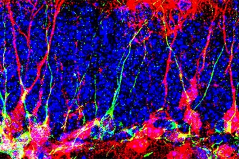
Newly produced neurons (red) in the dentate gyrus with cell nuclei (blue) and a marker for immature neurons (green).

The mitochondrial pyruvate transporter (MPC), a protein complex discovered eleven years ago in Professor Martinou’s group, plays a particular role in this regulation. Its activity influences the metabolic options a cell can use.

By knowing the metabolic pathways that distinguish active cells from dormant cells, scientists can wake up dormant cells by modifying their mitochondrial metabolism.


New perspectives 

Biologists have blocked MPC activity by using chemical inhibitors or by generating mutant mice for the Mpc1gene. Using these pharmacological and genetic approaches, the scientists were able to activate dormant NSCs and thus generate new neurons in the brains of adult and even aged mice.

‘‘With this work, we show that redirection of metabolic pathways can directly influence the activity state of adult NSCs and consequently the number of new neurons generated,’’ summarizes Professor Knobloch, co-lead author of the study.

‘‘These results shed new light on the role of cell metabolism in the regulation of neurogenesis. In the long term, these results could lead to potential treatments for conditions such as depression or neurodegenerative diseases’’, concludes Jean-Claude Martinou, co-lead author of the study.

Abstract

Mitochondrial pyruvate metabolism regulates the activation of quiescent adult neural stem cells

Cellular metabolism is important for adult neural stem/progenitor cell (NSPC) behavior. However, its role in the transition from quiescence to proliferation is not fully understood.

We here show that the mitochondrial pyruvate carrier (MPC) plays a crucial and unexpected part in this process. MPC transports pyruvate into mitochondria, linking cytosolic glycolysis to mitochondrial tricarboxylic acid cycle and oxidative phosphorylation. Despite its metabolic key function, the role of MPC in NSPCs has not been addressed.

We show that quiescent NSPCs have an active mitochondrial metabolism and express high levels of MPC. Pharmacological MPC inhibition increases aspartate and triggers NSPC activation.

Furthermore, genetic Mpc1 ablation in vitro and in vivo also activates NSPCs, which differentiate into mature neurons, leading to overall increased hippocampal neurogenesis in adult and aged mice.

These findings highlight the importance of metabolism for NSPC regulation and identify an important pathway through which mitochondrial pyruvate import controls NSPC quiescence and activation.

Temperature-Regulating Neurons Identified, Hint at Treatment Strategies for Heat Stroke, Obesity


The results of a study in rodents carried out by scientists at Nagoya University suggest that a group of neurons, called EP3 neurons, in the preoptic area (POA) of the brain play a key role in regulating body temperature in mammals. The discovery could pave the way for the development of technology that artificially adjusts body temperature, as a way of helping to treat conditions such as heat stroke, or hypothermia, and potentially obesity.

“On top of that, this technology could lead to new strategies for survival of people in hotter global environments, which are becoming a serious worldwide problem,” said research lead Kazuhiro Nakamura, PhD. The scientists reported on their study in Science Advances, in a paper titled, “Prostaglandin EP3 receptor–expressing preoptic neurons bidirectionally control body temperature via tonic GABAergic signaling.”

Thermoregulation is a physiological function that is fundamental to homeostasis in mammals, the authors noted. “Body core temperature is maintained within a control range by autonomous regulation of the balance between heat production within the body and heat loss to the environment.” In humans and many other mammals, body temperature is regulated at around 37°C (98.6°F), which optimizes all regulatory functions. But when body temperature noticeably deviates from the normal range, these functions can be impaired, potentially leading to heat stroke, hypothermia, and, in the worst case, death. These conditions might possibly be treated if body temperature could be artificially adjusted to remain within the normal range.

The brain’s temperature regulation center resides in the preoptic area, a part of the hypothalamus that controls the body’s vital functions. For example, when the preoptic area receives signals from a mediator called prostaglandin E (PGE2) that is produced in response to infections, this area releases a command to raise body temperature to fight against viruses, bacteria, and other disease-causing organisms. “The POA receives and integrates thermosensory (cool- and warm-sensory) neural signals from skin thermoreceptors and a pyrogenic humoral signal mediated by prostaglandin E2 (PGE2), which is produced in response to infections,” the team noted.

However, it is still unclear exactly which neurons in the preoptic area release commands to increase or decrease body temperature. To identify such neurons, Nakamura, together with Yoshiko Nakamura, PhD, and colleagues at Nagoya University, in collaboration with Hiroyuki Hioki, PhD, at Juntendo University, designed a study in rodents. They focused on how EP3 neurons in the preoptic area that express the EP3 subtype of PGE2 receptor may be involved in regulating body temperature. “… we investigated the physiological role of POAEP3R neurons in the central circuit mechanisms of thermoregulation and fever,” the team explained.

Nakamura and colleagues started by looking at how the activity of EP3 neurons in the preoptic area varied in response to changes in ambient temperature. “We first examined activation of POAEP3R neurons in response to ambient thermal challenges and then histochemically and physiologically determined the neurotransmitter phenotype of POAEP3R neurons,” they noted. A comfortable environmental temperature for rats is around 28°C. For two hours, the researchers exposed the rats to cold (4°C), room (24°C), and hot (36°C) temperatures. Results showed that exposure to 36°C activated EP3 neurons, while exposure to 4°C and 24°C did not. “These observations indicate that the POAEP3R neuronal group includes a substantial subpopulation of warming-activated neurons but not cooling-activated neurons,” the investigators suggested.

The group then observed nerve fibers of EP3 neurons in the preoptic area to identify where the signals from EP3 neurons are transmitted. Their observations revealed that nerve fibers are distributed to various brain regions, particularly to the dorsomedial hypothalamus (DMH), which activates the sympathetic nervous system. Their analysis also showed that the substance that EP3 neurons use for signal transmission to DMH is gamma-aminobutyric acid (GABA), a major inhibitor of neuronal excitation. “Although many POAEP3R neuronal cell bodies express a glutamatergic messenger RNA marker, their axons in the DMH predominantly release γ-aminobutyric acid (GABA), and their GABAergic terminals are increased by chronic heat exposure,” they stated. The combined results, the team further wrote, “… demonstrate that POAEP3R neuron–derived axons predominantly form GABAergic synapses onto DMH neurons.”

To further investigate the role of EP3 neurons in temperature regulation, researchers artificially manipulated their activity using a chemogenetic approach. They found that activating the neurons led to a decrease in body temperature, whereas suppressing their activity led to their increase. “Chemogenetic stimulation of POAEP3R neurons at room temperature reduces body temperature by enhancing heat dissipation, whereas inhibition of them elicits hyperthermia involving brown fat thermogenesis, mimicking fever,” they noted.

Rodent experiments to investigate neuronal control of body temperature
In hot environments, EP3 neurons in the preoptic area continually send inhibitory signals with GABA to suppress sympathetic outflows to defend body temperature from ambient heat. In cold environments or during infections, EP3 neurons are inhibited and therefore, sympathetic pathways are activated to increase heat production and inhibit heat loss to prevent hypothermia or to develop fever. The activity level of EP3 neurons is a critical determinant of body temperature. [© 2022 Yoshiko Nakamura]

The combined study results demonstrated that EP3 neurons in the preoptic area play a key role in regulating body temperature by releasing GABA to send inhibitory signals to DMH neurons to control sympathetic responses. “The present study demonstrates that POAEP3R neurons, a target of PGE2 for its pyrogenic action, play a pivotal role in the preoptic efferent control of central sympathetic outflow for basal thermoregulation,” the team wrote. “Our study shows strong evidence that POAEP3R neurons provide tonic GABAergic inhibitory signaling to sympathoexcitatory efferent pathways as a fundamental determinant of body temperature for thermal homeostasis and fever.”

Lead author Nakamura further suggested, “Probably, EP3 neurons in the preoptic area can precisely regulate the signal strength to fine-tune body temperature. For example, in a hot environment, signals are augmented to suppress sympathetic outputs, resulting in increased blood flow in the skin to facilitate the radiation of the body’s heat to prevent heat stroke. However, in a cold environment, signals are reduced to activate sympathetic outputs, which promote heat production in brown adipose tissue and other organs to prevent hypothermia. Furthermore, at the time of infection, PGE2 acts on EP3 neurons to suppress their activity, resulting in activation of sympathetic outputs to develop fever.”

This study’s findings could pave the way for the development of a technology that artificially adjusts body temperature, which might then be applied to a wide range of medical fields. Interestingly, this technology may also be helpful in the treatment of obesity, by keeping body temperature slightly higher than normal to promote fat burning.

“On top of that, this technology could lead to new strategies for survival of people in hotter global environments, which are becoming a serious worldwide problem,” said Nakamura.

Beyond Neurons: How Astrocytes Contribute to Brain Disorders


Summary: Study reveals how a molecule produced by astrocytes interferes with normal neuron development in a range of neurodevelopmental disorders.

Source: Salk Institute

Neurons often get most of the credit for keeping our brains sharp and functioning—as well as most of the blame when it comes to brain diseases. But star-shaped cells called astrocytes, another abundant cell in the human brain, may bear the brunt of the responsibility for exacerbating the symptoms of some neurodevelopmental disorders.

Salk Institute scientists have now identified a molecule produced by astrocytes that interferes with normal neuron development in Rett, fragile X and Down syndromes.

As the team reports in Nature Neuroscience on August 30, 2022, blocking the molecule reduces the signs of disease in mice brains.  

“These findings are part of a new push to look at how all the cells in the brain, not just neurons, interact in neurodevelopmental disorders,” says Associate Professor Nicola Allen, who led the new study. “This opens the door to potential therapeutics to treat these disorders by targeting astrocytes.”

In recent years, scientists have discovered that astrocytes play key roles in brain development and disease. Isolated neurons, for instance, don’t form connections and communicate unless astrocytes are present. If astrocytes affected by disease are mixed with healthy neurons, the neurons begin showing signs of disease. Similarly, if neurons affected by neurodevelopmental disorders are exposed to healthy astrocytes, their function improves.

However, researchers haven’t been able to pin down what molecules from astrocytes are responsible.

In the new study, Allen and colleagues isolated astrocytes and neurons from the developing brains of mice with genetic mutations causing Rett, fragile X or Down syndrome or from healthy animals. Then they determined the levels of 1,235 different proteins produced by each set of astrocytes. They found hundreds of proteins present at higher or lower levels in each disease, with 120 proteins in common between all three diseases—88 at higher-than-usual levels, and 32 at lower-than-usual levels.

“From a basic science perspective, it’s fascinating that there are so many changes seen in astrocyte protein secretion in these genetic disorders—and more importantly, that so many of those changes overlap between the disorders,” says Alison Caldwell, first author of the paper and a former graduate student in Allen’s lab. “To me, this highlights how important astrocytes are for normal neuronal development.”

One molecule stood out to the scientists. They knew that insulin-like growth factor (IGF) could sometimes reduce symptoms of disease in mice with neurodevelopmental disorders. Researchers had long assumed the treatment worked because diseased neurons weren’t producing enough IGF. But they found a different explanation—astrocytes impacted by Rett, fragile X or Down syndrome make high levels of Igfbp2, a protein that blocks IGF.

This shows astrocytes
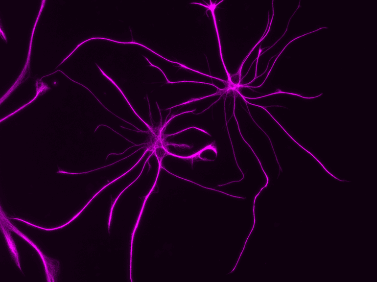
Salk researchers studied the molecules produced by astrocytes, like those pictured, to understand how the cells play a role in neurodevelopmental disorders.

“It turns out that neurons are making plenty of IGF, but it can’t get where it needs to be because these molecules made by astrocytes are interfering with it,” says Allen.

The group went on to show that excess Igfbp2 produced by astrocytes is responsible for slowing the growth of neurons and that blocking Igfbp2 made by Rett syndrome astrocytes enhanced neuron growth. Moreover, when mice with Rett syndrome were treated with antibodies blocking Igfbp2, signs of disease in the brain were lessened.

“We still have a long way to go to get a therapy based on this to humans, but we think it has promise,” says Allen. “Rather than giving an IGF treatment that has actions throughout the whole body, it makes sense to target Igfbp2 in the brain, where we want IGF to act.”

Allen’s lab group is planning follow-up studies on other proteins they identified in diseased astrocytes, as well as future experiments to better understand Igfbp2.

Other authors included Laura Sancho, James Deng, Alexandra Bosworth, Audrey Miglietta, Jolene Diedrich and Maxim Shokhirev of Salk.

Funding: The work was supported in part by Autism Speaks (Dennis Weatherstone Predoctoral Fellowship), the Chapman Foundation, the National Institute of Child Health and Human Development (F30HD106699), the Chan Zuckerberg Initiative, the Hearst Foundation and the Pew Foundation.

Aberrant astrocyte protein secretion contributes to altered neuronal development in multiple models of neurodevelopmental disorders

Astrocytes negatively impact neuronal development in many models of neurodevelopmental disorders (NDs); however, how they do this, and if mechanisms are shared across disorders, is not known.

In this study, we developed a cell culture system to ask how astrocyte protein secretion and gene expression change in three mouse models of genetic NDs (Rett, Fragile X and Down syndromes).

ND astrocytes increase release of Igfbp2, a secreted inhibitor of insulin-like growth factor (IGF). IGF rescues neuronal deficits in many NDs, and we found that blocking Igfbp2 partially rescues inhibitory effects of Rett syndrome astrocytes, suggesting that increased astrocyte Igfbp2 contributes to decreased IGF signaling in NDs.

We identified that increased BMP signaling is upstream of protein secretion changes, including Igfbp2, and blocking BMP signaling in Fragile X and Rett syndrome astrocytes reverses inhibitory effects on neurite outgrowth.

This work provides a resource of astrocyte-secreted proteins in health and ND models and identifies novel targets for intervention in diverse NDs.

Researchers Have Discovered a Population of Neurons That Light up Whenever We See Images of Food


Summary: Images of food stimulate a newly discovered population of food-responsive neurons in the ventral visual stream. Researchers believe there may be an evolutionary reason for this neural population that may reflect the significance of food in human culture.

Source: MIT

A gooey slice of pizza. A pile of crispy French fries. Ice cream dripping down a cone on a hot summer day. When you look at any of these foods, a specialized part of your visual cortex lights up, according to a new study from MIT neuroscientists.

This newly discovered population of food-responsive neurons is located in the ventral visual stream, alongside populations that respond specifically to faces, bodies, places, and words. The unexpected finding may reflect the special significance of food in human culture, the researchers say. 

“Food is central to human social interactions and cultural practices. It’s not just sustenance,” says Nancy Kanwisher, the Walter A. Rosenblith Professor of Cognitive Neuroscience and a member of MIT’s McGovern Institute for Brain Research and Center for Brains, Minds, and Machines. “Food is core to so many elements of our cultural identity, religious practice, and social interactions, and many other things that humans do.”

The findings, based on an analysis of a large public database of human brain responses to a set of 10,000 images, raise many additional questions about how and why this neural population develops. In future studies, the researchers hope to explore how people’s responses to certain foods might differ depending on their likes and dislikes, or their familiarity with certain types of food.

MIT postdoc Meenakshi Khosla is the lead author of the paper, along with MIT research scientist N. Apurva Ratan Murty. The study appears today in the journal Current Biology.

Visual categories

More than 20 years ago, while studying the ventral visual stream, the part of the brain that recognizes objects, Kanwisher discovered cortical regions that respond selectively to faces. Later, she and other scientists discovered other regions that respond selectively to places, bodies, or words. Most of those areas were discovered when researchers specifically set out to look for them. However, that hypothesis-driven approach can limit what you end up finding, Kanwisher says.

“There could be other things that we might not think to look for,” she says. “And even when we find something, how do we know that that’s actually part of the basic dominant structure of that pathway, and not something we found just because we were looking for it?”

To try to uncover the fundamental structure of the ventral visual stream, Kanwisher and Khosla decided to analyze a large, publicly available dataset of full-brain functional magnetic resonance imaging (fMRI) responses from eight human subjects as they viewed thousands of images.

“We wanted to see when we apply a data-driven, hypothesis-free strategy, what kinds of selectivities pop up, and whether those are consistent with what had been discovered before. A second goal was to see if we could discover novel selectivities that either haven’t been hypothesized before, or that have remained hidden due to the lower spatial resolution of fMRI data,” Khosla says.

To do that, the researchers applied a mathematical method that allows them to discover neural populations that can’t be identified from traditional fMRI data. An fMRI image is made up of many voxels — three-dimensional units that represent a cube of brain tissue.

Each voxel contains hundreds of thousands of neurons, and if some of those neurons belong to smaller populations that respond to one type of visual input, their responses may be drowned out by other populations within the same voxel.

The new analytical method, which Kanwisher’s lab has previously used on fMRI data from the auditory cortex, can tease out responses of neural populations within each voxel of fMRI data.

Using this approach, the researchers found four populations that corresponded to previously identified clusters that respond to faces, places, bodies, and words. “That tells us that this method works, and it tells us that the things that we found before are not just obscure properties of that pathway, but major, dominant properties,” Kanwisher says.

Intriguingly, a fifth population also emerged, and this one appeared to be selective for images of food.

“We were first quite puzzled by this because food is not a visually homogenous category,” Khosla says. “Things like apples and corn and pasta all look so unlike each other, yet we found a single population that responds similarly to all these diverse food items.”

The food-specific population, which the researchers call the ventral food component (VFC), appears to be spread across two clusters of neurons, located on either side of the FFA. The fact that the food-specific populations are spread out between other category-specific populations may help explain why they have not been seen before, the researchers say.

“We think that food selectivity had been harder to characterize before because the populations that are selective for food are intermingled with other nearby populations that have distinct responses to other stimulus attributes. The low spatial resolution of fMRI prevents us from seeing this selectivity because the responses of different neural population get mixed in a voxel,” Khosla says.

This shows a range of different dishes
MIT neuroscientists have discovered a population of food-responsive neurons located in the ventral visual stream.

“The technique which the researchers used to identify category-sensitive cells or areas is impressive, and it recovered known category-sensitive systems, making the food category findings most impressive,” says Paul Rozin, a professor of psychology at the University of Pennsylvania, who was not involved in the study.

“I can’t imagine a way for the brain to reliably identify the diversity of foods based on sensory features. That makes this all the more fascinating, and likely to clue us in about something really new.”

Food vs non-food

The researchers also used the data to train a computational model of the VFC, based on previous models Murty had developed for the brain’s face and place recognition areas. This allowed the researchers to run additional experiments and predict the responses of the VFC. In one experiment, they fed the model matched images of food and non-food items that looked very similar — for example, a banana and a yellow crescent moon.

“Those matched stimuli have very similar visual properties, but the main attribute in which they differ is edible versus inedible,” Khosla says. “We could feed those arbitrary stimuli through the predictive model and see whether it would still respond more to food than non-food, without having to collect the fMRI data.”

They could also use the computational model to analyze much larger datasets, consisting of millions of images. Those simulations helped to confirm that the VFC is highly selective for images of food.

From their analysis of the human fMRI data, the researchers found that in some subjects, the VFC responded slightly more to processed foods such as pizza than unprocessed foods like apples. In the future they hope to explore how factors such as familiarity and like or dislike of a particular food might affect individuals’ responses to that food.

They also hope to study when and how this region becomes specialized during early childhood, and what other parts of the brain it communicates with. Another question is whether this food-selective population will be seen in other animals such as monkeys, who do not attach the cultural significance to food that humans do.

Study finds a striking difference between neurons of humans and other mammals.


https://bigthink.com/neuropsych/human-and-mammal-neurons/?utm_medium=Social&utm_source=Facebook#Echobox=1654791889