New Research Points to Causes for Brain Disorders with No Obvious Injury


Functional neurological disorders are very real, and medical compassion is an important part of treatment

New Research Points to Causes for Brain Disorders with No Obvious Injury
A picture of a human brain taken by a positron emission tomography scanner, also called PET scan, is seen on a screen on January 9, 2019, at the Regional and University Hospital Center of Brest in France.

“Stop faking!” Imagine hearing those words moments after your doctor diagnosed you with, say, a stroke or a brain tumor. That sounds absurd but for many people diagnosed with a condition called functional neurological disorder (FND), this is exactly what happens.

Although the disorder is not well known to many people, FND is actually one of the most common conditions that neurologists like myself encounter. In it, abnormal brain functioning causes symptoms to appear. FND comes in many forms, with symptoms that can include seizures, feelings of weakness and movement disorders. People may lose consciousness or their ability to move or walk. Or they may experience abnormal tremors or tics. It can be highly disabling and just as costly as structural neurological conditions such as amyotrophic lateral sclerosis (ALS), also known as Lou Gehrig’s disease, multiple sclerosis and Parkinson’s disease.

[Read more about new findings on functional neurological disorder]

Although men can develop FND, young to middle-aged women receive this diagnosis most frequently. And during the first two years of the COVID pandemic, FND briefly made international headlines when functional tic-like behaviors spread with social media usage, particularly among adolescent girls.

So why would a doctor or other medical professional accuse someone who has lost control of their limbs or has experienced a seizure of faking their symptoms? Unfortunately, many such professionals have a poor or outdated understanding of FND, despite the frequency with which they encounter it. Because nothing is structurally wrong with the brain—there’s no injury noted on clinical testing, for instance—they may write your symptoms off as “all in your head” or dismiss them as psychological. That response, recent research shows, can harm a patient who is already suffering. Fortunately, there is another path forward, rooted in sensitivity, respect and new evidence-based approaches.

Historically, FND was called “conversion disorder.” The term came from the belief that traumatic stress “converted” into functional neurological symptoms via psychological mechanisms. This is no longer how we understand FND. Stress and trauma can play a part. In fact, some researchers believe the unique global stressors our society faced during the COVID pandemic increased some people’s susceptibility to the condition. But not every person with FND has experienced a traumatic event.

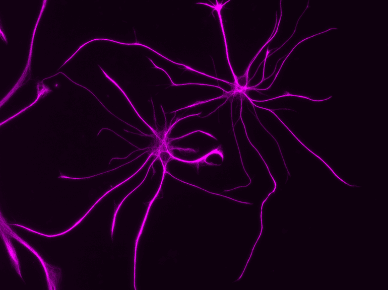
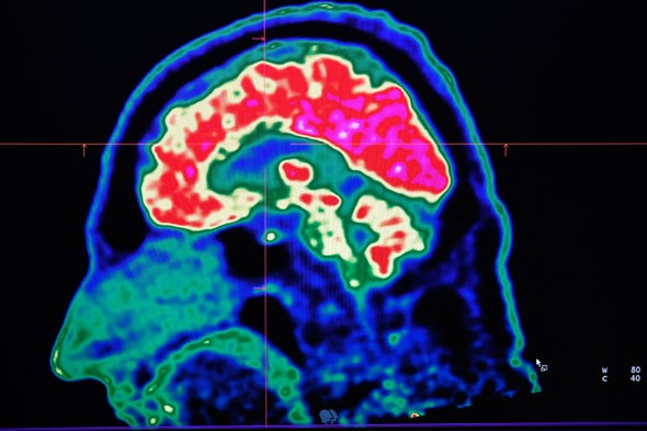
Instead recent advances in brain imaging suggest that FND is caused by abnormalities in the functioning of brain networks. Some experts use the analogy that the brain’s hardware (or structure) is unchanged, but the software (or processing) is malfunctioning. For example, studies suggest that, in FND, the neurological networks—electrical and chemical signaling pathways between groups of neurons or larger brain regions—that affect our reactions to traumatic stress, emotional regulation, sensorimotor functioning, attentional processing, body awareness and self-agency are not functioning together, as is typically expected. These networks include limbic system structures, such as the amygdala, which are important in our brain’s processing of emotions and stress.

Neuroimaging underscores that people are not “faking” anything. Scientists have found decreased activity in areas that influence whether a patient’s symptoms feel under their control. There are also abnormalities in the connections between brain areas responsible for interpreting internal physical sensations and motor planning. Simply put, one of the hallmark features of FND is that patients feel their symptoms are involuntary. By contrast, as a research team at the University of Calgary in Alberta explored in a paper published last November, patients with the structural neurological condition Tourette’s syndrome report some degree of control in suppressing their tics.

Clinicians are also finding better ways to diagnose FND. In the past, neurologists considered conversion disorder to be a diagnosis of exclusion, meaning a diagnosis was made after ruling out structural neurological abnormality through examination, radiological imaging, laboratory studies and neurophysiological testing such as electroencephalography (EEG). As a result, many patients with FND felt their doctor had told them what they didn’t have, not what they did have.

But in the past decade neurologists have developed diagnostic criteria to determine which symptoms are linked to functional brain abnormalities. These emphasize characteristic “positive,” or “rule-in,” findings based on a neurologist’s physical examination, which can predict FND as the basis for a patient’s symptoms. A combination of a thorough neurological examination, EEG, brain imaging and laboratory testing can show whether a person’s symptoms are consistent with a structural brain pathology—for instance, a stroke or a brain tumor—or a functional condition such as FND.

Together, these advances in the diagnosis and understanding of FND mean that doctors are in a better position than ever to identify and understand this disorder. Nevertheless, many patients still have the disorienting, distressing experience of being treated with dismissal or disbelief by medical professionals

This reaction has damaging consequences. In January a collaboration of researchers at the University of Sheffield in England, Arizona State University and the Northeast Regional Epilepsy Group laid out case studies and other evidence that clinicians’ unsupportive response to their patients may contribute to a sense of shame in people who are already suffering psychologically from their functional symptoms. In fact, being prone to shame may itself be an additional risk factor for FND.

This connection to shame and stigma takes on an even greater weight when we consider that minoritized groups such as LGBTQ+ community members may be at an increased risk for functional disorders. A person experiencing stressors—such as discrimination, bias and stigma—because of their minoritized identity can internalize feelings of shame when their psychosocial support systems and coping mechanisms are inadequate or overwhelmed. If someone in this situation has FND, receiving treatment from a doctor who lacks empathy or a current understanding of the condition only makes things worse. Telling a patient their condition is “in their head” contributes to medical misinformation and further stigmatizes patients with these disorders.

But this problem can be addressed. Researchers have found that how empathetically a doctor informs their patient about an FND diagnosis influences that patient’s likelihood to accept the diagnosis and successfully complete treatment. And appropriate treatment works. Therapy may combine psychoeducation, medication for any coexisting mental health conditions, psychotherapy and physiotherapy. Outcomes for people who receive sensitive and appropriate care are actually very good.

This year my colleagues and I will publish our observations on the treatment of LGBTQ+ people with FND. Our preliminary findings are promising. Most patients had improvement or complete resolution of their functional symptoms following treatment. In some of our patients, these results can be really important. We have treated patients with functional blindness who then regained the ability to see. And we have watched those in wheelchairs regain the ability to walk. In short, care and compassion can be powerful medicine.

Beyond Neurons: How Astrocytes Contribute to Brain Disorders


Summary: Study reveals how a molecule produced by astrocytes interferes with normal neuron development in a range of neurodevelopmental disorders.

Source: Salk Institute

Neurons often get most of the credit for keeping our brains sharp and functioning—as well as most of the blame when it comes to brain diseases. But star-shaped cells called astrocytes, another abundant cell in the human brain, may bear the brunt of the responsibility for exacerbating the symptoms of some neurodevelopmental disorders.

Salk Institute scientists have now identified a molecule produced by astrocytes that interferes with normal neuron development in Rett, fragile X and Down syndromes.

As the team reports in Nature Neuroscience on August 30, 2022, blocking the molecule reduces the signs of disease in mice brains.  

“These findings are part of a new push to look at how all the cells in the brain, not just neurons, interact in neurodevelopmental disorders,” says Associate Professor Nicola Allen, who led the new study. “This opens the door to potential therapeutics to treat these disorders by targeting astrocytes.”

In recent years, scientists have discovered that astrocytes play key roles in brain development and disease. Isolated neurons, for instance, don’t form connections and communicate unless astrocytes are present. If astrocytes affected by disease are mixed with healthy neurons, the neurons begin showing signs of disease. Similarly, if neurons affected by neurodevelopmental disorders are exposed to healthy astrocytes, their function improves.

However, researchers haven’t been able to pin down what molecules from astrocytes are responsible.

In the new study, Allen and colleagues isolated astrocytes and neurons from the developing brains of mice with genetic mutations causing Rett, fragile X or Down syndrome or from healthy animals. Then they determined the levels of 1,235 different proteins produced by each set of astrocytes. They found hundreds of proteins present at higher or lower levels in each disease, with 120 proteins in common between all three diseases—88 at higher-than-usual levels, and 32 at lower-than-usual levels.

“From a basic science perspective, it’s fascinating that there are so many changes seen in astrocyte protein secretion in these genetic disorders—and more importantly, that so many of those changes overlap between the disorders,” says Alison Caldwell, first author of the paper and a former graduate student in Allen’s lab. “To me, this highlights how important astrocytes are for normal neuronal development.”

One molecule stood out to the scientists. They knew that insulin-like growth factor (IGF) could sometimes reduce symptoms of disease in mice with neurodevelopmental disorders. Researchers had long assumed the treatment worked because diseased neurons weren’t producing enough IGF. But they found a different explanation—astrocytes impacted by Rett, fragile X or Down syndrome make high levels of Igfbp2, a protein that blocks IGF.

This shows astrocytes
Salk researchers studied the molecules produced by astrocytes, like those pictured, to understand how the cells play a role in neurodevelopmental disorders.

“It turns out that neurons are making plenty of IGF, but it can’t get where it needs to be because these molecules made by astrocytes are interfering with it,” says Allen.

The group went on to show that excess Igfbp2 produced by astrocytes is responsible for slowing the growth of neurons and that blocking Igfbp2 made by Rett syndrome astrocytes enhanced neuron growth. Moreover, when mice with Rett syndrome were treated with antibodies blocking Igfbp2, signs of disease in the brain were lessened.

“We still have a long way to go to get a therapy based on this to humans, but we think it has promise,” says Allen. “Rather than giving an IGF treatment that has actions throughout the whole body, it makes sense to target Igfbp2 in the brain, where we want IGF to act.”

Allen’s lab group is planning follow-up studies on other proteins they identified in diseased astrocytes, as well as future experiments to better understand Igfbp2.

Other authors included Laura Sancho, James Deng, Alexandra Bosworth, Audrey Miglietta, Jolene Diedrich and Maxim Shokhirev of Salk.

Funding: The work was supported in part by Autism Speaks (Dennis Weatherstone Predoctoral Fellowship), the Chapman Foundation, the National Institute of Child Health and Human Development (F30HD106699), the Chan Zuckerberg Initiative, the Hearst Foundation and the Pew Foundation.

Aberrant astrocyte protein secretion contributes to altered neuronal development in multiple models of neurodevelopmental disorders

Astrocytes negatively impact neuronal development in many models of neurodevelopmental disorders (NDs); however, how they do this, and if mechanisms are shared across disorders, is not known.

In this study, we developed a cell culture system to ask how astrocyte protein secretion and gene expression change in three mouse models of genetic NDs (Rett, Fragile X and Down syndromes).

ND astrocytes increase release of Igfbp2, a secreted inhibitor of insulin-like growth factor (IGF). IGF rescues neuronal deficits in many NDs, and we found that blocking Igfbp2 partially rescues inhibitory effects of Rett syndrome astrocytes, suggesting that increased astrocyte Igfbp2 contributes to decreased IGF signaling in NDs.

We identified that increased BMP signaling is upstream of protein secretion changes, including Igfbp2, and blocking BMP signaling in Fragile X and Rett syndrome astrocytes reverses inhibitory effects on neurite outgrowth.

This work provides a resource of astrocyte-secreted proteins in health and ND models and identifies novel targets for intervention in diverse NDs.

Newly discovered state of memory could help explain learning and brain disorders.


Memory researchers have shone light into a cognitive limbo. A new memory—the name of someone you’ve just met, for example—is held for seconds in so-called working memory, as your brain’s neurons continue to fire. If the person is important to you, the name will over a few days enter your long-term memory, preserved by permanently altered neural connections. But where does it go during the in-between hours, when it has left your standard working memory and is not yet embedded in long-term memory?

In Science, a research team shows that memories can be resurrected from this limbo. Their observations point to a new form of working memory, which they dub prioritized long-term memory, that exists without elevated neural activity. Consistent with other recent work, the study suggests that information can somehow be held among the synapses that connect neurons, even after conventional working memory has faded.

“This is a really fundamental find—it’s like the dark matter of memory,” says Geoffrey Woodman, a cognitive neuroscientist at Vanderbilt University in Nashville who was not involved with the work. “It’s hard to really see it or measure it in any clear way, but it has to be out there. Otherwise, things would fly apart.”

Cognitive neuroscientist Nathan Rose and colleagues at the University of Wisconsin (UW) in Madison initially had subjects watch a series of slides showing faces, words, or dots moving in one direction. They tracked the resulting neural activity using functional magnetic resonance imaging (fMRI) and, with the help of a machine learning algorithm, showed they could classify the brain activity associated with each item. Then the subjects viewed the items in combination—a word and face, for example—but were cued to focus on just one item. At first, the brain signatures of both items showed up, as measured in this round with electroencephalography (EEG). But neural activity for the uncued item quickly dropped to baseline, as if it had been forgotten, whereas the EEG signature of the cued item remained, a sign that it was still in working memory. Yet subjects could still quickly recall the uncued item when prompted to remember it a few seconds later.

Rose, who recently left UW for the University of Notre Dame in South Bend, Indiana, and his colleagues then turned to transcranial magnetic stimulation (TMS), a noninvasive method that uses rapidly changing magnetic fields to deliver a pulse of electrical current to the brain. They had subjects perform the same cued memory task, then applied a broad TMS pulse just after the signature of the uncued memory item had faded. The appropriate neural activity for that “forgotten” item spiked, showing the memory was reactivated into immediate consciousness from its latent state. What’s more, when the TMS directly targeted the brain areas that were initially active for the uncued item, the reactivation response was even stronger.

The study doesn’t address how synapses or other neuronal features can hold this second level of working memory, or how much information it can store. “It’s a primitive early step in understanding how we bring things into mind,” says UW cognitive neuroscientist Bradley Postle, a study co-author.

Woodman agrees. “Good studies tend to bring to light more questions than they answer,” he says. “This work absolutely does that.” Ultimately, he says, this new memory state could have a range of practical implications, from helping college students learn more efficiently to assisting people with memory-related neurological conditions such as amnesia, epilepsy, and schizophrenia.