A woman underwent colonoscopy with biopsy sampling that revealed a tiny neuroendocrine tumor (NET) about 5 cm from the anus. After several months, the biopsy sampling site healed completely, making the identification of the residual NET difficult. The specific location of the lesion could not be confirmed even under magnifying endoscopy and EUS. When a higher concentration of methylene blue was injected into the submucosa, the residual lesion was exposed. The endoscopist then inferred the likely location of the lesion on magnifying endoscopy on the basis of the marker and the location of the lesion after injection of methylene blue. Finally, the residual lesion was successfully removed, and the postoperative pathology confirmed that the resected lesion was a residual NET measuring 1.5 × .5 mm.
A 69-year-old woman underwent colonoscopy with biopsy sampling that revealed a small neuroendocrine tumor (NET) G1 about 5 cm from the anus. After a few months, the patient decided to undergo endoscopic resection to remove the lesion. However, the biopsy sampling wound healed completely, and the residual lesion was extremely tiny, making the identification of the residual NET more difficult even with magnifying endoscopy and EUS (Fig. 1A and B).
Figure 1The tiny lesion of residual neuroendocrine tumor (NET) under endoscopy and pathology. A, No obvious manifestation for the residual lesion or biopsy sampling scar was observed during ordinary endoscopy. B, An indeterminate hypoechoic lesion in the lamina propria of the mucosa was found 5 cm from the anus (black arrow). C, After injection of methylene blue, a yellowish smooth lesion was noted (black arrow). D, Pathologic findings indicated the lesion was a NET 1.5 × .5 mm in size (as measured by a pathologic section scale). E, The possible location of the residual lesion under magnifying endoscopy (black arrow).View Large Image Figure ViewerDownload Hi-res image Download (PPT)
Because the patient decided to undergo endoscopic resection of the lesion and injection of methylene blue is a routine procedure in endoscopic resection, the endoscopist attempted to inject methylene blue into the suspicious lesion to identify it. Surprisingly, when a colored solution of methylene blue (diluted it to approximately .2% using normal saline solution) was injected into the submucosa of the rectum, the residual lesion was exposed. It appeared as a flat bulge, 5 × 5 mm in size, with a smooth surface, and a relatively yellowish color (Fig. 1C). Subsequently, the endoscopist performed EMR to remove the lesion in its entirety. Finally, the postoperative pathology confirmed that the resected lesion was a residual NET measuring 1.5 × .5 mm (Fig. 1D). In addition, the endoscopist reviewed the entire examination process after the operation and judged the possible location of the lesion under magnifying endoscopy before injection of methylene blue according to the marker made under magnifying endoscopy and the location of the lesion after the injection of methylene blue (Fig. 1E, arrow). Through the above process (Fig. 2), we found that tiny lesions hidden in the lamina propria, such as small residual lesions after biopsy sampling, could be easily visualized by injecting a colored solution into the submucosa for enhancing the color contrast between the lesion and background mucosa.
Figure 2The procedure flowchart of injecting methylene blue to identify a tiny lesion of the residual neuroendocrine tumor (NET). A, A tiny NET was revealed by the colonoscopy with biopsy sampling. B, After several months, the biopsy sampling wound healed completely, and the mucosa was smooth. C, Injecting a higher concentration of methylene blue into the submucosa exposed the residual lesion. D, The residual lesion could be identified by increasing the color contrast between the lesion and background mucosa. EP, Epithelium; MM, muscularis mucosa; SM, submucosa; MP, muscularis propria.
Nearly two-thirds of trial participants with advanced neuroblastoma responded to an experimental CAR T-cell therapy.
Investigators reported no cases of high-grade treatment-related toxicities during the study.
A novel chimeric antigen receptor T-cell therapy induced clinically impactful antitumor responses in 63% of younger patients with relapsed or refractory, high-risk neuroblastoma, results of a phase 1/phase 2 trial showed.
The findings, published in TheNew England Journal of Medicine, suggest the investigational agent also has a manageable safety profile similar to other CAR T-cell therapies, researchers noted.
Data derived from Del Bufalo F, et al. N Engl J Med. 2023;doi:10.1056/NEJMoa2210859.
Franco Locatelli
“Our results indicate that CAR T cells can be an effective therapeutic option, not only in patients with hematologic malignancies, but also in patients with solid tumors,” Franco Locatelli, MD, PhD, professor of pediatrics at Catholic University of the Sacred Heart and director of the department of pediatric hematology and oncology at IRCCS Ospedale Pediatrico Bambino Gesù in Rome, told Healio.
Background
Previous study has firmly established that the disialoganglioside GD2 is highly expressed on the surface of neuroblastoma tumor cells, making it an attractive therapeutic target, according to Locatelli.
A team of researchers from Italy developed a third-generation, gene edited, autologous CAR T-cell therapy (GD2-CART01) that targets GD2. The novel agent incorporates two co-stimulatory domains — CD28 and 4-1BB — with the aim of increasing its potency and efficacy, Locatelli noted.
“We also added to the construct the sequence coding for a suicide gene — or safety switch — to improve the safety profile of the approach,” he said. “Indeed, this suicide gene can be activated in case of severe or life-threatening toxicity not controlled by pharmacologic therapies.”
Methodology
Locatelli and colleagues conducted a single-center phase 1/phase 2 dose-escalation study to determine the safety, feasibility and recommended phase 2 dose of GD2-CART01 among patients aged 1 to 25 years with relapsed or refractory, high-risk neuroblastoma.
Twenty-seven patients (median age, 6.7 years; range, 2.7-18.6; 67% male) received a single infusion of GD2-CART01 at one of three dose levels (3 × 10⁶, 6 × 10⁶ or 10 × 106 CAR T cells/kg).
The study included 12 patients with refractory disease, 14 with relapsed disease and one who had a complete response after first-line therapy.
Researchers reported successfully manufacturing CAR T cells for all study participants.
Key findings
Investigators reported no dose-limiting toxicities during the phase 1 portion of the study. They established a recommended phase 2 dose of 10 × 106 CAR T cells/kg.
Twenty patients (74%) experienced cytokine release syndrome, with 95% of cases being either grade 1 or grade 2.
Suicide gene activation occurred for one patient using two infusions of rimiducid, resulting in “rapid elimination of circulating GD2-CART01” cells, the investigators wrote.
Infusion of GD2-CART01 cells resulted in clinical responses in 17 patients, for an overall response rate of 63% — nine complete responses and eight partial responses.
Patients who received the recommended phase 2 dose of GD2-CART01 had a 3-year OS rate of 60% and 3-year EFS rate of 36%.
Clinical implications
“We will work to increase the proportion of patients responding to GD2-CART01 and to prolong the durability of the response,” Locatelli told Healio.
Future studies are also planned to evaluate GD2-CART01 as earlier therapy for patients with neuroblastoma, according to Locatelli.
“We expect that this will translate into even better results,” he said.
Locatelli noted that the treatment development and ensuing clinical trial occurred entirely within the academic setting, without commercial support.
“This observation emphasizes that academic institutions may play a major role in the development of the most advanced and innovative therapies,” he said.
The study results suggest an overall favorable safety profile for GD2-CART01, in addition to encouraging objective response rates and durability for treatment of relapsed or refractory neuroblastoma, Oladapo O. Yeku, MD, PhD, of Mass General Cancer Center, and Dan L. Longo, MD, of Harvard Medical School, wrote in an accompanying editorial.
“As with all solid-tumor cancers, understanding the resistance mechanisms in patients who did not have a response could provide insight for the design of future clinical trials,” they wrote. “[Locatelli] and colleagues found that the tumors in patients who did not have a response retained expression of GD2, a finding that raises the question of whether immunosuppressive cytokines, myeloid-derived suppressor cells, T-regulatory cells or tumor-associated macrophages contribute to a lack of efficacy in these patients.”
Franco Locatelli, MD, PhD, can be reached at Department of Pediatric Hematology and Oncology, IRCCS Ospedale Pediatrico Bambino Gesu, Piazza Sant’Onofrio, 4, 00165 Rome, Italy; email: franco.locatelli@opbg.net.
Perspective
Robbie Majzner, MD
There has long been skepticism about whether CAR T cells could demonstrate the same depth of responses in solid tumors as they have in hematologic malignancies. This exciting study by Locatelli and colleagues stands out as the largest series of CAR T-cell responses in a solid tumor to date.
Approximately 60% of patients experienced an objective response to GD2-directed CAR T cells, with half of those being complete responses. This work clearly and irrefutably demonstrates that CAR T cells can eradicate solid tumors in patients.
The researchers should be congratulated on their success, which builds on previous work engineering and testing GD2-directed CARs at Baylor College of Medicine and Texas Children’s Hospital starting in the 2000s. The first CAR T cell ever tested in a child was a first-generation GD2-directed CAR that demonstrated safety and some clinical efficacy, including complete responses. However, multiple additional GD2-CAR constructs and clinical trials failed to improve on this approach, until now.
The researchers’ success suggests that iterative engineering and testing of CAR constructs in small clinical trials will be key to advancing the field. It is essential to recognize the role of academic physicians and scientists in this process, as all of this work was carried out by pediatric oncologists at academic centers, often funded by grants and philanthropy.
GD2 is expressed on normal peripheral nerves, and anti-GD2 antibodies are associated with severe pain in the clinic. However, GD2-directed CAR T cells did not cause any pain or other on-target neurotoxicities in patients, even when they eradicated GD2-expressing tumors. GD2 is expressed at much higher levels on cancer compared [with] normal nerves. Therefore, CAR T cells — like other cancer drugs — demonstrate a therapeutic window, and some expression of target antigens on normal tissue may be tolerable, opening the door to engineering CARs against a host of antigens that are highly differentially expressed.
This trial has several limitations that should be noted. First, neuroblastoma is a highly heterogeneous cancer that ranges from indolent to aggressive, and results of any single-arm, single-institution trial need to be interpreted carefully. Most durable responses on the trial were seen in patients with low-burden disease, often restricted to the bone or bone marrow. These sites may be more accessible to CAR T cells than bulky tumors, hinting that current CAR designs will be most effective when patients are in a minimal residual disease state. Finally, only half of the population of patients in this trial had received anti-GD2 monoclonal antibodies, with no indication how many received combined anti-GD2 antibody and chemotherapy, considered the de facto standard of care for relapsed and refractory patients in the United States.
Despite these limitations, this is a landmark study that shows that CAR T cells are poised to eventually alter the treatment paradigm for immunologically cold tumors, which includes most pediatric cancers.
Caffeine boosts your energy and mood and makes you more alert. That can sometimes be helpful, especially in the morning or when you’re trying to work. Though your body doesn’t store it, caffeine can affect you for up to 6 hours after you swallow it. But more is not always better. Too much can push you over the line from alert to jittery and anxious.
Interferes With Sleep
2/11
Too much caffeine can make it hard to nod off when you go to bed at night. Even moderate amounts can cause insomnia in some people, especially if you have it too close to bedtime. The effects may be worse as you age. Avoid caffeine in the afternoon and evening if you notice it affects your sleep. And remember, it’s not just in tea and coffee. It’s also in chocolate, energy drinks, and other prepackaged foods and drinks.
Raises Heart Rate
3/11
Caffeine is a stimulant and may cause your heart to beat a little faster as it wakes you up. For most folks that’s not a problem. But if you have too much caffeine or you’re overly sensitive, your pulse may go up too much or stay high too long. It may even feel like your heart beats in a weird rhythm, sometimes called heart palpitations. Some people say it feels as if their ticker skips a beat.
Causes a Headache
4/11
If you have caffeine every day, whether in a pill, energy bar, or cup of coffee, you build up tolerance. Then, without your daily dose, you might get a “rebound” headache. It may be worse if you quit caffeine completely and all at once. You’ll find your head will feel better if you reduce caffeine a little at a time.
Makes You Pee
5/11
Caffeine is a diuretic, which means it can make you pee more. Around 300 milligrams of caffeine — the amount in three cups of coffee — is enough to do it if you’re not used to it. Water loss is minor and is unlikely to cause dehydration as long as you are otherwise healthy. The diuretic effect can fade if you get the same regular daily dose of caffeine because you build up tolerance.
Boosts Sports Performance
6/11
If you do “endurance” sports, like running, biking, or swimming, caffeine might help you go faster and with less muscle pain. It seems to work best in a non-liquid form, like a pill, taken about an hour before you exercise so that your body can absorb it completely. Around 200 to 400 milligrams (2 to 4 cups of coffee) should do it. More than that doesn’t seem to help further.
Helps You Recover From a Workout
7/11
Some studies show that caffeine can help your body recover more quickly after hard exercise by making and restocking a stored form of fuel called glycogen. It seems to do this best if you combine it with carbohydrates, like in certain sports gels, sports bars, and drinks. Just take care not to overdo the caffeine, which could have the opposite effect on recovery or performance if it interferes with your sleep.
Raises Blood Pressure
8/11
Though the reason isn’t clear, caffeine can spike your blood pressure for a short while and sometimes over the long term as well. It could be that it blocks a hormone that keeps your arteries wide and pressure down. Or it might cause your body to release more adrenaline, a hormone that raises blood pressure. Talk to your doctor about how much caffeine you can have if you have high blood pressure or heart problems.
Protects Against Disease
9/11
Caffeine seems to help prevent gallstones and inflammation, among other medical problems. Some studies show that regular caffeine might help keep away certain neurological diseases, like Parkinson’s, Alzheimer’s, Huntington’s, multiple sclerosis, and epilepsy. More research is needed.
Affects Menopause
10/11
Women pass through menopause around age 50, a phase that brings an end to their monthly cycle. Caffeine can worsen the sudden body heat and sweats, known as hot flashes, that often happen at this time of life. The symptoms can go on for 10 years or more. Your doctor may be able to help you with hormone therapy if they get in the way of your everyday routine.
How Much Caffeine Is Too Much?
11/11
It depends on your weight, diet, medications, and overall health. Up to about 400 milligrams of caffeine a day is OK for most healthy adults. Over 600 milligrams daily is probably too much. But some people are just more sensitive to it. If you notice stomach problems, headaches, muscle twitches, or heart palpitations, it might be time to cut back. Talk to your doctor about how much is safe if you’re pregnant or have heart trouble.
As human life expectancy has increased, so has the number of people living to 100 years of age or older.
Researchers have found that centenarians have a unique immune cell composition and activity, giving them an immune system that helps them live longer.
Scientists believe these findings may be used to develop healthy aging therapeutics.
The life expectancy of humans on our planet has more than doubled since 1900. Global life expectancy has increased from 31 years in 1900 to 73.2 years in 2023, and is expected to further increase to 77.1 years in 2050.
Also increasing is the number of people reaching the age of 100 or more. Known as centenariansTrusted Source, researchers estimate there were about 450,000 centenarians globally in 2015, with that number projected to increase to 3.7 million in 2050.
PreviousTrusted Source research in the early 2000s estimated that globally, the number of people living to 100 years or older would more than quintuple between 2005 and 2030.
One thing still unknown is what allows some people to live into their 100s, while others do not.
Led by researchers from Tufts Medical Center and Boston University School of Medicine, a new study is helping to answer this question by finding that centenarians possess a unique immune cell composition and activity, giving them a highly-functional immune system and allowing them to live longer.
Scientists believe these findings could be used potentially to develop healthy aging therapeutics.
As we age, all parts of the body experience changes, including the immune system.
According to Dr. Scott Kaiser, a geriatrician, and director of Geriatric Cognitive Health for the Pacific Neuroscience Institute in Santa Monica, California, there are two main concepts when it comes to how the immune system changes as we get older.
“One is immunosenescence and that’s the age-related process of immune dysfunction,” he explained to Medical News Today.
“So changes in our immune system composition and function over time can lead to poor immune function in older people. And that’s closely related to people’s vulnerability to infection, autoimmune disease, and even various types of cancer,” he said.
“And then there’s this issue of inflammagingTrusted Source, which is a term that’s been used to describe age-related increases in inflammation because of high levels of pro-inflammatory markers in the blood and different tissues in the body. That’s a strong risk factor for all sorts of diseases, including neurodegenerative processes like Alzheimer’s disease, for example,” Dr. Kaiser continued.
“So there’s a lot to look at in terms of the immune function over time and how our immune systems changes with age may either make us more vulnerable or protect us,” he added.
According to Dr. Tanya Karagiannis, a senior bioinformatician at the Center for Quantitative Methods and Data Science in the Institute for Clinical Research and Health Policy Studies at Tufts Medical Center, and lead author of this study, she and her team decided to study the immune systems of centenarians because with age comes changes in our immune systems including in their function and cell makeup, and these changes can lead to aging-related diseases.
“Many centenarians experience delays in the onset of aging-related disease and this suggests the presence of an elite immunity that continues to remain highly functional even at extreme old age,” she told Medical News Today.
“We used single-cell data and applied new computational methods to analyze immune cells that circulate through the immune system across the human lifespan. We looked at differences in the presence of specific immune cell types across younger ages and extreme old age and found cell type-specific changes in aging and extreme old age,” Dr. Karagiannis explained.
“We also took the same cell types and explored the differences in gene expressionTrusted Source across ages to discover different gene expression patterns of extreme longevity that change with age but also are unique to extreme old age,” she added.
Upon analysis, the researchers confirmed observations made in previous studies of aging that identified unique cell type-specific compositional and transcriptional changes only found in centenarians that reflect normal immune response.
They also found centenarians had cell type signatures specific to exceptional longevity in both genes with age-related changes and genes expressed uniquely in centenarians.
“We were not as surprised to find genes that change with age in centenarians since they are an aging population. What was surprising was the different aging patterns we identified including genes that were aging-specific in which expression levels changed with age but not in extreme longevity across various cell populations,” Dr. Karagiannis said.
“Our findings can provide a foundation to explore potential drivers of extreme old age that could lead to the discovery of healthy aging therapeutics. We would like to explore longitudinal changes in immune cells of centenarians and younger aged individuals to help better define the protective drivers of extreme longevity that provides the beneficial health outcome observed in these individuals,” she continued.
After reviewing this study, Dr. Kaiser told Medical News Today he found this study interesting as it actually looked at people who have aged extremely well, who have defied age, so to speak, and then looked at what’s going on in them to see if we can learn anything.
“The potential lessons here are in what makes us more resilient,” he explained.
“Looking at these people who had extreme longevity, living into 100s and even beyond, and figure out what is the nature, what is the characteristic of their immune system so that we could better understand what might be going on, and then figure out how that could be translated into potential therapies for other people, so that more people can enjoy that.” — Dr. Kaiser
MNT also spoke withKathleen Cameron, senior director of theNational Council on Aging’s Center for Healthy Aging, about this study.
She said understanding the immune changes that come with aging is important to help people live longer. And a lot of people want to live longer if they can also be healthy.
“If we can determine what is creating this immune resilience for those who live over 100, that can lead to treatments that can help people live longer. Or, if there are certain healthy behaviors that lead to this resilience, that would also help us,” Cameron continued.
However, she said this is all very preliminary research, as this study was small, and it should lead to other studies to help healthcare practitioners better understand this immune resilience.
“More research is needed to understand the effect these immune patterns have on longevity. Is there something in the centenarians’ family history or other things that happened in their life, exposure to certain things that might have changed their immune system? We don’t know that from this study. Knowing more about this could lead to new therapies or new ways to improve the immune system.” — Kathleen Cameron
Researchers investigated the link between sleep problems and stroke risk.
They found that sleep problems increased stroke incidence by as much as five times.
Further studies are needed to confirm the results.
Quality sleep is essentialTrusted Source for health. Sleep problems range from too short or too long sleep duration, difficulty falling and staying asleep, and symptoms such as snoring, snorting, and pauses in breathing.
Knowing more about how sleep problems may affect stroke risk could aid the development of preventative strategies.
Recently, researchers investigated the link between sleep problems and the incidence of acute strokeTrusted Source.
They found that the more sleep problems individuals had, the more likely they were to experience stroke. The study paper appears in the journal Neurology.
Dr. Adi Iyer, a neurosurgeon and neurointerventional surgeon at Pacific Neuroscience Institute at Providence Saint John’s Health Center in Santa Monica, CA, not involved in the study, told Medical News Today that, “[w]hile there may be independent physiological changes that occur with poor sleep predisposing to stroke, it is likely that overall sleep represents an epiphenomenon of known stroke risk factors such as obesity, older age, alcohol use, etc.”
“Sleep may be a modifiable risk factor for stroke and clinicians should assess patients’ sleep quality and duration,” he added.
For the study, the researchers analyzed healthcare data from 1,799 participants who had experienced an ischemic stroke — the most common kind of stroke, in which a blood clot blocks an artery leading to the brain.
They also analyzed data from 439 people who experienced an intracerebral hemorrhage (ICH) — bleeding in brain tissue — and 4,496 controls who were matched according to age and sex. Participants were an average of 62 years old.
Finally, the researchers asked the participants about their sleep behaviors, including sleep duration and sleep quality, in the month before experiencing their stroke.
In the end, the researchers found that multiple sleep problems were linked to increased stroke incidence.
More specifically, those who slept less than 5 hours per night were three times more likely to have a stroke than those who slept for 7 hours. Meanwhile, stroke risk was doubled for those sleeping more than 9 hours per night compared to those sleeping 7 hours.
Sleep apnea — pauses in breathing multiple times per hour — was linked to a three times higher stroke risk. Napping for an hour or more was also linked to an 88% higher stroke risk when compared to no napping at all.
The researchers also found that those who snored were 91% more likely to have a stroke than those who did not, and that those who snorted were almost three times more likely to have a stroke than those who did not.
They further found that the more sleep problems individuals had, the more likely they were to experience stroke or ICH.
“Not only do our results suggest that individual sleep problems may increase a person’s risk of stroke but having more than five of these symptoms may lead to five times the risk of stroke compared to those who do not have any sleep problems,” warns Dr. Christine McCarthy of the University of Galway in Ireland, the leading author and corresponding author of the study.
The results remained valid even after the researchers controlled for potentially confounding factors, including depression, alcohol use, and physical activity.
MNT spoke with Dr. Thomas Kilkenny, director of the Institute Sleep Medicine at Staten Island University Hospital, not involved in the study, to understand more about the link between sleep problems and stroke risk.
He said that 80% of adult sleep is non-REM sleep, and that during this time, the cardiovascular system is controlled by the autonomic nervous system which reduces blood pressure, heart rate, and cardiovascular strain. These factors, he noted, have a protective effect on cardiovascular health.
He added that sleep disruption, including sleep apnea, insomnia, and shift work, may impair this protective effect by reducing time spent in non-REM sleep. He added: “When this occurs, there is a decrease in the [cardiovascular] recovery but also a significant increase [in] stress on the cardiovascular system.”
“Poor sleep quality, through [a] reduction in non-REM sleep, also activates a multitude of other mechanisms including intermittent hypoxia injury, blood pressure swings, cardiac arrhythmia, inflammation, insulin resistance, stress hormone activation, and hypercoagulability, all of which have the potential to provoke cardiovascular diseases including strokes.”
– Dr. Thomas Kilkenny
Dr. McCarthy also noted that “[s]leep is increasingly being recognized as the third pillar of health, along with diet and exercise, although it has not been as thoroughly researched.”
Nevertheless, she cautioned that it is difficult to tell whether sleep problems cause stroke risk factors or vice versa.
“For example,” she noted, “increased alcohol intake may cause disruptions in sleep quality, but impairments in sleep quality may result in increased use of alcohol as a sedative. Future interventional research is required to determine causal associations.”
When reflecting on their study’s limitations, Dr. McCarthy noted that one factor to keep in mind is potential bias, as”people reported their own symptoms of sleep problems, so the information may not have been accurate, subject to recall and misclassification bias.”
Dr. Kilkenny added that “[t]he major limitation is that the study findings are just associations and not a direct cause and effect.”
“This means that people with poor sleep quality are more likely to have a stroke, not necessarily that poor sleep directly causes the stroke,” he said.
“Further studies are needed. Likewise having insomnia, taking long naps, or being a long sleeper may be a sign of another health disorder that could be triggering the cardiovascular disease,” noted Dr. Kilkenny.
When asked about the study’s implications of the study, Dr. Kilkenny said it should be an “awakening” to patients and physicians on the importance of good quality sleep.
“The study results also give physicians an incentive to inquire about sleep habits when seeing patients in the office. Sleep is rarely discussed routinely in the office and physicians may be missing an opportunity to improve their patient’s health by picking up sleep disturbances sooner,” he added.
Dr. McCarthy noted:
“Our findings suggest that sleep disturbance symptoms may represent either risk factors for stroke, and/ or their presence identifies people at increased risk of stroke. The study does not show that sleeping problems cause stroke. It only shows an association. Given our findings, future studies should look at interventions for sleep problems, and their potential to reduce stroke risk.”
Researchers from Harvard Medical School (HMS) and Massachusetts General Hospital (MGH) show that the approved leukemia drug venetoclax helps large animals accept transplanted stem cells and organs, like kidneys. The research team, led by Hajime Sasaki, MD, PhD, and Takayuki Hirose, MD, PhD, demonstrated that selective inhibition of B cell lymphoma 2 (Bcl-2) promoted the tolerance of hematopoietic stem cell (HSC) transplantation and renal allograft in a nonhuman primate model despite using only half of the dose of total body irradiation previously required. Selective inhibition of Bcl-2 is therefore a new strategy to improve stem cell transplants and lessen the toxic effects of conditioning regimens, which are a major obstacle to the widespread use of stem cell transplantation in clinical settings.
“This method (brief inhibition of Bcl-2) makes bone marrow transplantation easier,” senior author Tatsuo Kawai, MD, PhD, told GEN. “The patient does not need to be admitted for bone marrow transplantation, which may expand bone marrow transplantation applications for many benign diseases, such as induction of tolerance for transplant organs.”
HSC transplantation (HSCT) has become the standard of care for both cancerous and noncancerous diseases of the blood and bone marrow, like autoimmune diseases, genetic disorders, and making the body accept a transplant. However, the wider clinical application of HSCT has been limited by the nonselective conditioning therapies associated with this approach.
In order for HSCT to work, the recipient must go through a harsh conditioning process that includes total body irradiation or chemotherapy drugs like cyclophosphamide, busulfan, or fludarabine. These treatments often have serious side effects like a lower-than-normal number of red and white blood cells and platelets in the blood (pancytopenia), infections, infertility, and even death.
Transient HSC chimerism induction has led to long-term immunosuppression-free renal allograft survival in humans, but pancytopenia and other side effects of the HSCT conditioning regimen have kept this method from being used more often. Therefore, developing a safer conditioning regimen without myelosuppressive complications is critically important.
In 2013, research done at the University of Zürich by Pietro E. Cippà and Thomas Fehr (who are both authors of the Science Translational Medicine article) showed that blocking B cell lymphoma 2 (Bcl-2) promotes mixed chimerism in mice without the need for traditional myelosuppressive conditioning. However, this study used a non-clinically available Bcl-2 inhibitor (Bcl-2i), ABT-737, which inhibits not only Bcl-2 but also BclxL.
Since venetoclax, which selectively inhibits Bcl-2 alone, is the only Bcl-2i approved by the U.S. Food and Drug Administration (FDA), the research team from HMS and MGH evaluated the effect of venetoclax on peripheral lymphocytes and bone marrow progenitors in a non-human primate model and determined its efficacy in inducing mixed chimerism and renal allograft tolerance. The research article shows that Bcl-2i causes dose-dependent cell death in vitro and causes HSCs and multipotent progenitors (MPPs) to be partially deleted in vivo. This partial removal of HSCs and MPPs led to a big increase in hematopoietic chimerism, which made it possible to cut the amount of radiation given to the whole body in half. However, Bcl-2 inhibition by itself was not sufficient to completely eradicate TBI.
Promoting mixed chimerism by Bcl-2i resulted in improved immunosuppression-free renal allograft survival. In the group receiving the original protocol with total body irradiation but without Bcl-2i, one of the eight animals experienced acute rejection, and three of the eight developed chronic rejection, reducing long-term, rejection-free allograft survival to 50%. When the total body irradiation dose was decreased to half, four of the five recipients lost their kidney allografts because of acute rejection. By adding Bcl-2i to the regimen, superior rejection-free allograft survival was consistently achieved with a higher degree of lymphoid chimerism, which has been found to be relevant to allograft tolerance.
This approach not only removes a major hurdle from the wider application of our approach for the induction of renal allograft tolerance but also advances the design of less toxic conditioning regimens that can be used in conjunction with HSCT to treat a wide variety of disorders.
When we say pure sleep, we mean deep, peaceful, quality sleep without the use of drugs. Lack of quality deep sleep is linked to a host of problems including depression, obesity, poor nutrient absorption and assimilation, and overall poor health. If your quality of sleep improves, you will wake up more easily feeling more refreshed, have more stamina and energy during the day, and quite likely need less sleep at night. We need deep, quality, pure sleep. On the next page we give you ten tips for getting a better night’s sleep.
1. Don’t Go to Sleep on a Full Stomach
Your body needs energy to digest food. Going to sleep on a full stomach is bad for your digestion and can lower your quality of sleep. And you shouldn’t be eating enough to have a “full” stomach anyway.
2. Cut out Alcohol and Cut out Caffeine and Other Artificial Stimulants
While alcohol may decrease the time it takes you to fall asleep, it actually increases the number of times you awaken in the latter half of the night. If your sleep isn’t restful, alcohol (beer, wine, or hard liquor) may be the cause.
Artificial stimulants can affect your adrenal glands, which can affect your energy levels during the day and your sleep at night.
3. Exercise
Exercise during the day can help you sleep more soundly at night.
4. Eat a Healthy Diet
There are many ways an unhealthy diet can affect your quality of sleep. Eating healthy can give you more energy and a better night’s sleep.
5. Get More Sunlight
Whether you sleep at night like most people or sleep during the day, you need sunlight. Your body needs sunlight to produce vitamin D (which is actually a hormone). Getting some sun on your bare skin can increase your energy and elevate your mood. Most people do not get enough sunlight. Lack of sunlight contributes to irritability, depression, low energy levels, and a desire for more sleep.
6. Make Sure You Are Sleeping in the Dark
If you work at night and sleep during the day you need to take the steps to block light out of your room. Make your room as dark as possible. Invest in blackout shades or blackout curtains.
7. Make Your Bed Comfortable
High quality sheets with a high thread count feel good on your skin and make sleeping easier. Or maybe you prefer the touch of flannel. Comfort is important. Comfort decreases needless tossing and turning.
You also need comfortable, quality pillows that cradle your head rather than raise it. Your spine should lie straight.
A poor mattress will make it more difficult to get a good night’s sleep. Make sure your mattress is well supported with no lumps or dips.
8. Make Your Bed Organic
Sleep with all natural (not synthetic) materials. Better yet, sleep with only organic, toxin free materials. Your skin needs to breathe. Synthetic fibers do not facilitate air circulation.
9. Stop Snoring
Breath Right ® can help. If you are overweight, you are more likely to snore, too. Snoring can cause you to wake, and it certainly decreases your air flow.
10. Try a Humidifier
If you are running your air conditioner or your heater, the air in your house may be dry. If you wake with a sore throat, a dry nose, or have nose bleeds, you might benefit from more humidity. Be sure to change the water daily.
11. Eat a Grapefruit Before Bed
Don’t skip that white bitter skin of the fruit. And get a little bit of rind from the peel too. It’ll help you sleep soundly.
Despite the stream of “good news” about vitamin D, the supplement’s prospects to prevent mental health disorders in adults remain dim
In February scientists reported that vitamin D was associated with a reduced risk of suicide attempts among U.S. veterans. The study compared more than 600,000 veterans who took various doses of vitamin D with an equal number of those who did not ingest the supplements. Taking vitamin D, they concluded, was linked with a 45 to 48 percent overall reduction in the risk of visiting a hospital for a suicide attempt or intentional self-harm. The higher the dose, the greater the reduction in risk, particularly for those with the lowest blood levels of the vitamin. “In general, those who had lower blood levels and received higher doses [of vitamin D] had the largest response, which is what you would expect to see if there were an actual treatment effect,” says study co-author Jason Gibbons, a postdoctoral fellow and health economist at Johns Hopkins University.
The study adds to a deluge of data from recent decades linking low levels of vitamin D to mental health problems such as depression, schizophrenia and poor cognition. And it showcases the enduring enthusiasm among many scientists for the vitamin’s potential as an aid to mental wellness, a message that some doctors relay to patients. “Imagine if getting a little vitamin D pill would reduce the mental health burden around the world. That would be incredible, right? That’s the kind of pipe dream I think people are chasing with this research,” says Brian Lee, an epidemiologist at Drexel University’s Dornsife School of Public Health, who has studied the vitamin’s connection with autism.
Vitamin D has important biological effects on the brain. It can cross the blood-brain barrier and enter neurons, and receptors for the vitamin populate many human brain regions. Studies in rodents suggest that vitamin D undergirds the cellular basis of learning and memory, for one, and the brain’s balance of signals that stimulate or inhibit neural activity, which is thought to be askew in autism and schizophrenia. Vitamin D is also a steroid hormone with powerful anti-inflammatory qualities, and various lines of evidence suggest that inflammation plays a role in depression.
Still, even well-done observational studies cannot definitively prove that a person’s blood level of vitamin D contributes to their mental status or stability. Vitamin D status may instead simply tag along with some other behavior or genetic factor that is more influential. Although researchers try to match the people who took vitamin D with virtually identical controls, there is no way to be sure that there isn’t some other difference between the groups that could account for the findings, experts say. “It’s possible that other kinds of behaviors could maybe somewhat explain our effect,” Gibbons says.
To sort out causal relationships, many researchers place their bets on randomized clinical trials in which some people are assigned to take vitamin D and others are not, and the outcomes are later compared. In a 2020 trial, researchers gave 2,000 international units (IU) of vitamin D supplements or a placebo daily to more than 18,000 men and women aged 50 years or older with no signs of clinical depression. For more than five years the researchers assessed the incidence of depression and mood scores of both groups. They found no significant differences. Two smaller 2019 trials of 800 or 1,200 IU of vitamin D showed no benefit for populations at risk for depression. Not all trials have been so disappointing, but the overall picture is not promising. “We’re not finding in the clinical trial evidence that vitamin D supplementation significantly reduced risk of depression,” says Olivia Okereke, an associate professor of psychiatry at Harvard Medical School, who led the 2020 depression trial.
Vitamin D has also lacked luster in trials of other psychiatric disorders. In a 2021 trial testing whether vitamin D can improve health outcomes in people with psychosis, researchers gave a large monthly dose to 149 young people who had experienced a first episode of psychosis, many of whom had had low blood levels of the vitamin. “We randomized the vitamin D supplements versus placebo, and we followed them up for a while, and we found no effect at all” of vitamin D on mental health, says John McGrath, a psychiatrist and epidemiologist at the University of Queensland in Australia and senior author of the study. “So I don’t think that adult low vitamin D is a major cause of schizophrenia.”
Low vitamin D is more likely to be a marker for illness than a cause of it, he and others say. That’s because low vitamin D levels are common in people with any health condition that dissuades them from spending time outdoors—the sun is an important source of vitamin D—or from eating a vitamin-rich diet. “If you have an illness like depression or maybe vulnerability to suicide, and then you change your behaviors, you get less vitamin D because of your changed behavior,” McGrath says. “Low [vitamin] D may be one of those bystander or proxy markers that just goes along with your behavior.”
Some genetic evidence supports this argument. In a study of more than 400,000 people, McGrath and his colleagues showed that the genetic variants linked to low vitamin D—enzymes that make the vitamin, say, or help determine skin color—are not associated with the presence of psychiatric disorders. The genetic correlates of these disorders are associated with low vitamin D levels, however, likely through behavior. “For example, we found that the genes that link to hours watching television lead to low vitamin D,” McGrath says. “Low vitamin D doesn’t cause you to watch more TV.”
Vitamin D has also fallen short in randomized clinical trials that have tested its mettle against “a very long list of health outcomes,” including heart disease, stroke and total cancer incidence, says JoAnn Manson, a professor of medicine at Harvard Medical School and a principal investigator of the Vitamin D and Omega-3 Trial (VITAL), the largest randomized trial of vitamin D in the world. (Okereke’s depression trial was part of VITAL.) In July the team added a new null finding: vitamin D did not reduce the risk of bone fractures, which was thought to be its best established benefit, Manson says. The outcomes for which vitamin D has proved beneficial in VITAL have been limited: it has been found to reduce the risk of cancer death and the incidence of advanced cancers and autoimmune diseases.
Randomized trials are not foolproof. Some, such as McGrath’s, may not have lasted long enough to show effects. In other cases, the trial might simply have missed the window for when supplementation is critical, Lee says. “[Randomized controlled trials] are the gold standard of evidence, but you could do something at the completely wrong time point, and it won’t matter,” Lee says. In addition, the vitamin D of a placebo group is difficult to control because people can easily pick up extra vitamin D from sunlight or food. Still, these trial results have soured many researchers in the field on the idea that vitamin D has a significant influence on mental health in adults. “You’re an adult, and you get low vitamin D: Does it increase the risk of depression or suicide or schizophrenia? I think the evidence is not very strong,” McGrath says. (Because vitamin D requirements vary by individual, and no specific level is critical for health, the U.S. Preventive Services Task Force concluded in 2021 that there was insufficient evidence to assess the benefits and harms of screening the population at large for vitamin D deficiency.)
But even if vitamin D blood levels have little impact on the adult brain, they could still be critical during development. A wealth of data in animals and cell culture support this idea. The production of factors that spur neuronal growth depends on vitamin D, which has also proved critical to brain cell maturation, says Darryl Eyles, a developmental neurobiologist at the University of Queensland. When a pregnant rat is deficient in vitamin D, the brain of its fetus has more dividing cells than mature cells, suggesting a delay in brain maturation. “The addition of vitamin D differentiates brain cells—be they glia, be they neurons—and will push them down various lineages. If there is an absence of this vitamin, you will dedifferentiate the brain,” Eyles says.
Other studies show that vitamin D shepherds certain neurons in the midbrain—those that use the neurotransmitter dopamine and are abnormal in schizophrenia—to their correct brain locales during development. A deficiency can perturb the proper formation of those circuits, Eyles and his colleagues have found. And when pregnant rats are deprived of vitamin D during gestation, their offspring show cognitive deficits and diminished sociality, among other behaviors reminiscent of people with schizophrenia. (That condition is thought to have developmental origins.) “We have heaps of animal experimental evidence which shows that this is a biologically plausible candidate,” McGrath says.
None of this means that vitamin D deficiency in the womb is a risk factor for schizophrenia. But several observational studies hint that it could be. In a 2018 study, McGrath and his colleagues measured vitamin D levels in 2,602 blood samples from newborns from a repository in Denmark. (The blood came from heel pricks used to screen for conditions such as phenylketonuria and hypothyroidism.) Some of these children later developed schizophrenia, and the researchers found that low vitamin D was associated with an increased risk of that illness. The work replicated results that McGrath and his colleagues published in 2010.
Several studies of a similar design have shown the same association between vitamin D and autism. In a 2021 study, researchers measured the vitamin in blood collected from more than 3,000 women in Finland during the first and second trimesters of pregnancy. They compared vitamin D levels from the 1,558 pregnancies that led to a child who was later diagnosed with autism with an equal number of pregnancies that did not result in an autistic child. Two other studies of similar size, published in 2017 and 2019, linked increased autism odds to low levels of vitamin D in newborn blood samples from a Swedish registry or maternal blood collected in the Netherlands. Not every study of vitamin D and autism has showed a link, however. And giving children a high dose of the vitamin after birth does not confer additional benefits for neurodevelopment by age six, compared with a standard dose, according to one trial conducted in Finland.
Despite the mixture of findings and lack of clinical trials, the idea that prenatal vitamin D may be important is still very much alive. Reverse causality does not apply, after all, because there are no fetal behaviors that could explain low vitamin D in the womb. “In a healthy pregnancy that just has low vitamin D, the fetus is not changing its behavior,” Eyles says. “It’s something about the developing system.” It is possible that low vitamin D in a pregnant person accompanies a genetic propensity for mental or other illnesses that they then pass to their fetus, however. “I still can’t put my hand on my heart and say we’ve proven pregnant women should take vitamin D supplements to stop their kids from getting schizophrenia,” McGrath says. But “I have not been able to reject the hypothesis yet.”
The randomized trials that would help settle the issue are probably not imminent. Such trials are tricky to conduct in pregnant people. They also present logistical problems for a condition such as schizophrenia that manifests decades later, McGrath says. So researchers continue to work on the circumstantial case.
Meanwhile Okereke’s team has done another analysis of data from its clinical depression trial in adults that yielded negative results to examine the impact of vitamin D on certain groups within the larger populations studied. This subgroup analysis opens up the possibility that vitamin D could benefit certain individuals. To confirm those benefits would require additional clinical trials, Okereke says.
The results of the recent suicide study call for similar confirmation, its authors say. “We should be looking potentially at vitamins in randomized controlled trials and vitamins D2 and D3 in particular as potential interventions for suicide prevention,” says study investigator Jill Lavigne, an epidemiologist at the U.S. Department of Veterans Affairs’ Center of Excellence for Suicide Prevention in Canandaigua, N.Y.
Fully evaluating that potential, Lee says, would require a very large trial whose participants are diverse geographically, racially and ethnically. Even a small reduction in the odds of suicide would be important, he adds. “Let’s say taking a vitamin D pill reduces your risk of depression or suicide by—forget 50 percent—5 percent. That, over the entire population, would be a huge public health benefit,” he says. “That’s the kind of work that needs to be done, quantifying the actual effect, if there is any.”
Until then, there is no harm in supplementing at reasonable doses, Lee says. (Large doses can be toxic.) But unless you have a vitamin D deficiency, he adds, there is also no compelling reason to do it. “I don’t supplement myself,” he says.
Functional neurological disorders are very real, and medical compassion is an important part of treatment
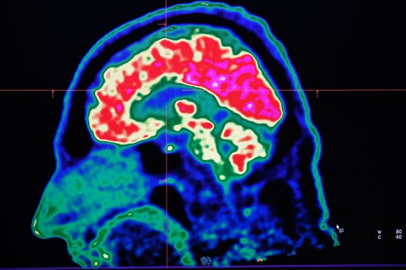
A picture of a human brain taken by a positron emission tomography scanner, also called PET scan, is seen on a screen on January 9, 2019, at the Regional and University Hospital Center of Brest in France.
“Stop faking!” Imagine hearing those words moments after your doctor diagnosed you with, say, a stroke or a brain tumor. That sounds absurd but for many people diagnosed with a condition called functional neurological disorder (FND), this is exactly what happens.
Although the disorder is not well known to many people, FND is actually one of the most common conditions that neurologists like myself encounter. In it, abnormal brain functioning causes symptoms to appear. FND comes in many forms, with symptoms that can include seizures, feelings of weakness and movement disorders. People may lose consciousness or their ability to move or walk. Or they may experience abnormal tremors or tics. It can be highly disabling and just as costly as structural neurological conditions such as amyotrophic lateral sclerosis (ALS), also known as Lou Gehrig’s disease, multiple sclerosis and Parkinson’s disease.
Although men can develop FND, young to middle-aged women receive this diagnosis most frequently. And during the first two years of the COVID pandemic, FND briefly made international headlines when functional tic-like behaviors spread with social media usage, particularly among adolescent girls.
So why would a doctor or other medical professional accuse someone who has lost control of their limbs or has experienced a seizure of faking their symptoms? Unfortunately, many such professionals have a poor or outdated understanding of FND, despite the frequency with which they encounter it. Because nothing is structurally wrong with the brain—there’s no injury noted on clinical testing, for instance—they may write your symptoms off as “all in your head” or dismiss them as psychological. That response, recent research shows, can harm a patient who is already suffering. Fortunately, there is another path forward, rooted in sensitivity, respect and new evidence-based approaches.
Historically, FND was called “conversion disorder.” The term came from the belief that traumatic stress “converted” into functional neurological symptoms via psychological mechanisms. This is no longer how we understand FND. Stress and traumacan play a part. In fact, some researchers believe the unique global stressors our society faced during the COVID pandemic increased some people’s susceptibility to the condition. But not every person with FND has experienced a traumatic event.
Instead recent advances in brain imaging suggest that FND is caused by abnormalities in the functioning of brain networks. Some experts use the analogy that the brain’s hardware (or structure) is unchanged, but the software (or processing) is malfunctioning. For example, studies suggest that, in FND, the neurological networks—electrical and chemical signaling pathways between groups of neurons or larger brain regions—that affect our reactions to traumatic stress, emotional regulation, sensorimotor functioning, attentional processing, body awareness and self-agency are not functioning together, as is typically expected. These networks include limbic system structures, such as the amygdala, which are important in our brain’s processing of emotions and stress.
Neuroimaging underscores that people are not “faking” anything. Scientists have found decreased activity in areas that influence whether a patient’s symptoms feel under their control. There are also abnormalities in the connections between brain areas responsible for interpreting internal physical sensations and motor planning. Simply put, one of the hallmark features of FND is that patients feel their symptoms are involuntary. By contrast, as a research team at the University of Calgary in Alberta explored in a paper published last November, patients with the structural neurological condition Tourette’s syndrome report some degree of control in suppressing their tics.
Clinicians are also finding better ways to diagnose FND. In the past, neurologists considered conversion disorder to be a diagnosis of exclusion, meaning a diagnosis was made after ruling out structural neurological abnormality through examination, radiological imaging, laboratory studies and neurophysiological testing such as electroencephalography (EEG). As a result, many patients with FND felt their doctor had told them what they didn’t have, not what they did have.
But in the past decade neurologists have developed diagnostic criteria to determine which symptoms are linked to functional brain abnormalities. These emphasize characteristic “positive,” or “rule-in,” findings based on a neurologist’s physical examination, which can predict FND as the basis for a patient’s symptoms. A combination of a thorough neurological examination, EEG, brain imaging and laboratory testing can show whether a person’s symptoms are consistent with a structural brain pathology—for instance, a stroke or a brain tumor—or a functional condition such as FND.
Together, these advances in the diagnosis and understanding of FND mean that doctors are in a better position than ever to identify and understand this disorder. Nevertheless, many patients still have the disorienting, distressing experience of being treated with dismissal or disbelief by medical professionals
This reaction has damaging consequences. In January a collaboration of researchers at the University of Sheffield in England, Arizona State University and the Northeast Regional Epilepsy Group laid out case studies and other evidence that clinicians’ unsupportive response to their patients may contribute to a sense of shame in people who are already suffering psychologically from their functional symptoms. In fact, being prone to shame may itself be an additional risk factor for FND.
This connection to shame and stigma takes on an even greater weight when we consider that minoritized groups such as LGBTQ+ community members may be at an increased risk for functional disorders. A person experiencing stressors—such as discrimination, bias and stigma—because of their minoritized identity can internalize feelings of shame when their psychosocial support systems and coping mechanisms are inadequate or overwhelmed. If someone in this situation has FND, receiving treatment from a doctor who lacks empathy or a current understanding of the condition only makes things worse. Telling a patient their condition is “in their head” contributes to medical misinformation and further stigmatizes patients with these disorders.
But this problem can be addressed. Researchers have found that how empathetically a doctor informs their patient about an FND diagnosis influences that patient’s likelihood to accept the diagnosis and successfully complete treatment. And appropriate treatment works. Therapy may combine psychoeducation, medication for any coexisting mental health conditions, psychotherapy and physiotherapy. Outcomes for people who receive sensitive and appropriate care are actually very good.
This year my colleagues and I will publish our observations on the treatment of LGBTQ+ people with FND. Our preliminary findings are promising. Most patients had improvement or complete resolution of their functional symptoms following treatment. In some of our patients, these results can be really important. We have treated patients with functional blindness who then regained the ability to see. And we have watched those in wheelchairs regain the ability to walk. In short, care and compassion can be powerful medicine.
Evidence shows a strong association between stress and heart disease, particularly when it comes to chronic or long-term stress. However, research into the exact nature of the relationship is still ongoing.
Chronic stress can have a cumulative effectTrusted Source on various aspects of human health, including the cardiovascular system. Stress can increase blood pressure and may make a person more likely to develop other risk factors for heart disease.
However, it can be difficult to study this, as the circumstances that lead to chronic stress can also affect health in many other ways.
This article explores the complex relationship between stress and heart disease and gives some tips for reducing the effects of stress on the heart.
Design by MNT; Photography by Hill Street Studios/Getty Images & Artur Debat/Getty Images
Yes, stress affects the heart and cardiovascular system. There are two types of stress: acute and chronic stress.
Acute stress
Acute stress is short-lived and usually occurs because of a single, isolated event. It results from the stress response, which is the body’s way of preparing a person for a perceived threat.
During the stress response:
blood pressure increases
the heart beats faster
breathing becomes faster and more shallow
However, these effects are temporaryTrusted Source. Once the stressful event is over, the heart functions as usual.
Chronic stress
Chronic stress persists regularly over weeks, months, or longer. According to the American Heart Association (AHA)Trusted Source, chronic stress can disrupt the heart’s functioning by:
According to the AHATrusted Source, it is unknown if stress alone can cause heart disease. ResearchTrusted Source suggests it can contribute to the risk when a person already has a predisposition for heart disease.
Acute stress
For most people, acute stress does not have a long-term effect on the heart.
However, researchTrusted Source has shown that acute stress does lead to a temporary increase in the risk of cardiovascular disease (CVD). For example, there is an increased rate of heart attacks in people who experience natural disasters.
Severe stress or intense emotions can also be a factor in takotsubo cardiomyopathy, or “broken heart syndrome.”
Chronic stress
Chronic stress has an association with heart disease but does not necessarily cause it directly. It can raiseTrusted Source blood pressure, which is a significant risk factor for conditions such as stroke, but this does not fully explain why some people get this type of CVD while others with chronic stress do not.
It may be that stress influences the risk in several ways. For example, stress links to behaviorsTrusted Source that can raise the risk of heart disease, such as:
smoking
excessive alcohol use
physical inactivity
an imbalanced diet
This, in itself, could cause the development of heart disease in some cases. However, stress can also result from social and economic circumstances that have additional knock-on effects on heart health.
For example, financial insecurity can raise stress, but it may also be the reason why a person cannot access fresh food or the space or time to exercise.
This is known as a “confounding factor,” as it can obscure the true role of stress in heart disease. As a result, health experts sometimes call stress a “contributory risk” or “underlying determinant” of heart disease.
There are no surefire ways of determining whether stress is directly affecting the heart. People may notice an increase in heart rate and blood pressure when they are stressed, but this does not necessarily indicate that there is long-term damage.
Many factors can influence heart rate and blood pressure, including anxiety, other emotions, physical activity, and more.
That said, people should speak with a doctor if they have concerns. The following symptoms could indicate that they have CVD:
Occasional stress is a part of life, but when it becomes chronic or severe, it can negatively affect health. Wherever possible, it is beneficial to reduce avoidable sources of stress by:
focusing only on the most important tasks
asking for help with things that feel overwhelming
saying “no” to things that are not necessary
delegating tasks to others
For unavoidable stress, it may help to learn relaxation techniques. These help bring heart rate and breathing down, returning the body to a calmer state. Some examples include:
People who think stress might be affecting their heart, or who are experiencing persistent stress, should speak with a doctor.
Sometimes, what a person thinks is stress is actually anxiety. Anxiety is a treatable condition, with the help of talk therapy. There are also medications that can reduce the symptoms.
The symptoms of anxiety can be similar to those of a heart condition. For example, an anxiety attack can cause rapid breathing, a fast heart rate, nausea, and sweating, all of which are also heart attack symptoms.
A doctor can help rule out serious conditions and determine what is causing the symptoms. However, if there is ever any doubt as to whether a person is experiencing a heart attack, they should seek emergency help.
The symptoms of a heart attack can include:
pain, tightness, or ache in the chest
pain spreading into the shoulder, arm, back, neck, or jaw
Stress has an associationTrusted Source with heart disease, but scientists are still learning how one affects the other.
It is clear that stress can lead to behaviors that may contribute to heart disease risk, such as smoking, drinking alcohol, or eating an imbalanced diet. However, this does not entirely explain the connection between stress and CVD.
More research is necessary to understand the role stress plays in heart disease, but reducing and managing stress may help lower the risk.


00011-0&id=gr1.jpg)