A 38-year-old man presented to the pulmonology department with a 6-month history of snoring, witnessed breathing pauses during sleep and daytime sleepiness. He also noted changed sensation in his left hand. He had a history of hypertension, but no drug or alcohol use. On examination, his body mass index was 32.6, he had prickling dysesthesia and a sensation of muscular tension on the left arm and trunk, and symmetric hyperreflexia of the legs. Polysomnography showed an Apnea–Hypopnea Index (AHI) score of 88 and a central AHI of 56. A total AHI lower than 5 is considered normal, and greater than 15 suggests the need for continuous positive airway pressure (CPAP) therapy. Central sleep apnea (CSA) is defined by a central AHI of 5 or more with at least half of the total events being central.1 The patient’s symptoms improved with CPAP treatment.
Given the unexplained CSA and the neurologic findings, we performed brain magnetic resonance imaging, which showed a 2 cm cyst in the medulla oblongata (Figure 1). We performed microsurgical fenestration, and biopsy showed an arachnoid cyst. Fifteen months later, both apnea and dysesthesia had recovered. Polysomnography had improved substantially with a total AHI of 13, of which most events were obstructive. The patient no longer required CPAP.
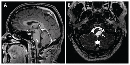
Magnetic resonance imaging scan of the brain of a 38-year-old man with central sleep apnea. The images — gadolinium-enhanced T1-weighted sagittal (A) and T2-weighted axial views (B) — show a 2 cm cyst in the medulla oblongata (arrow) with left dorsolateral compression of the dorsal respiratory group including the solitary tract nucleus (asterisk), which is known to play a role in ventilatory drive.1
The AHI is calculated by adding all apneas and hypopneas and dividing by total sleep time. In central events, there is a reduction of airflow without respiratory effort, whereas obstructive events are accompanied by a high breathing effort.1 Central sleep apnea is uncommon and is associated with heart failure with Cheyne–Stokes breathing, stroke, opioid use, structural cerebral anomalies and treatment of obstructive sleep apnea with CPAP.1 When CSA is unexplained, neuroimaging is indicated to look for a structural cause at the cervicomedullary junction, such as a space-occupying lesion or Chiari malformation, which can be cured with surgery
