ARTEMIS – UCLA’s most advanced humanoid robot – gets ready for action


Robotics lab, already a 5-time RoboCup champ, will bring its newest invention to international competition in July

ARTEMIS humanoid robot kicking a soccer ball
RoMeLa at UCLA ARTEMIS’ major innovation is that its actuators — devices that generate motion from energy — were custom-designed to behave like biological muscles.

Mechanical engineers at the UCLA Samueli School of Engineering have developed a full-sized humanoid robot with first-of-its-kind technology.

Named ARTEMIS, for Advanced Robotic Technology for Enhanced Mobility and Improved Stability, the robot is scheduled to travel in July to Bordeaux, France, where it will take part in the soccer competition of the 2023 RoboCup, an international scientific meeting where robots demonstrate capabilities across a range of categories.

The robot was designed by researchers at the Robotics and Mechanisms Laboratory at UCLA, or RoMeLa, as a general-purpose humanoid robot, with a particular focus on bipedal locomotion over uneven terrain. Standing 4 feet, 8 inches tall and weighing 85 pounds, it’s capable of walking on rough and unstable surfaces, as well as running and jumping. ARTEMIS is able to remain steady even when strongly shoved or otherwise disturbed.

During tests in the lab, ARTEMIS has been clocked walking 2.1 meters per second, which would make it the world’s fastest walking humanoid robot, according to the UCLA researchers. It is also believed to be the first humanoid robot designed in an academic setting that is capable of running, and only the third overall.

The robot’s major innovation is that its actuators — devices that generate motion from energy — were custom-designed to behave like biological muscles. They’re springy and force-controlled, as opposed to the rigid, position-controlled actuators that most robots have.

“That is the key behind its excellent balance while walking on uneven terrain and its ability to run — getting both feet off the ground while in motion,” said Dennis Hong, a UCLA professor of mechanical and aerospace engineering and the director of RoMeLa. “This is a first-of-its-kind robot.”

Another major advance is that ARTEMIS’ actuators are electrically driven, rather than controlled by hydraulics, which uses differences in fluid pressure to drive movement. As a result, it makes less noise and operates more efficiently than robots with hydraulic actuators — and it’s cleaner, because hydraulic systems are notorious for leaking fluids.

ARTEMIS’ ability to respond and adapt to what it senses comes from its system of sensors and actuators. It has custom-designed force sensors on each foot, which help the machine keep its balance as it moves. It also has an orientation unit and cameras in its head to help it perceive its surroundings.

To prepare ARTEMIS for RoboCup, student researchers have been testing the robot on regular walks around the UCLA campus. In the coming weeks, they will fully test the robot’s running and soccer-playing skills at the UCLA Intramural Field. Researchers also will evaluate how well it can traverse uneven terrain and stairs, its capacity for falling and getting back up, and its ability to carry objects. RoMeLa’s Twitter account is regularly sharing information about the robot’s testing results and posting the routes for its campus walks, giving Bruins the chance to catch ARTEMIS in action and chat with researchers.

“We’re very excited to take ARTEMIS out for field testing here at UCLA and we see this as an opportunity to promote science, technology, engineering and mathematics to a much wider audience,” Hong said.

Taoyuanmin Zhu and Min Sung Ahn, both of whom recently earned doctorates in mechanical engineering at UCLA, developed ARTEMIS’ hardware and software systems, respectively.

RoMeLa, which has been making humanoid robots for more than two decades, has had earlier robots win the RoboCup competition five times already; the engineers are hoping ARTEMIS brings home trophy number six.

ARTEMIS’ development was funded in part by 232 donors who contributed more than $118,000 through a UCLA Spark crowdfunding campaign. Additional support came from an Office of Naval Research grant.

ARTEMIS Fun Facts

  • Named in honor of the Greek goddess of the hunt, wild animals, chastity and childbirth, RoMeLa members refer to ARTEMIS as “she.”
  • RoMeLa researchers have joked that ARTEMIS could also stand for “A Robot That Exceeds Messi In Soccer.”
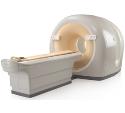
PET Measures Effects of STING-Activating Drugs


A study led by UCLA researchers suggest that PET imaging could provide a widely accessible approach to measure the pharmacodynamic effects of using stimulator of interferon genes, or STING, activating drugs. STING helps regulate immune activation and drugs that activate STING are being tested as a form of cancer immunotherapy and for treating certain infections. Findings from the study published in the Journal of Nuclear Medicine reveal previously unknown functional links between STING signaling and immunometabolism.

Oral and IV administration of STING-activating drugs stimulate potent anti-tumor responses in mouse models of cancer, suggesting this as a possible strategy for human therapy. Until now, however, researchers have not had a way to determine the magnitude, duration and location of immune activation. This research, led by  UCLA Jonsson Comprehensive Cancer Center investigators, investigated whether systemic STING activation induces metabolic alterations in immune cells that can be visualized by PET imaging.

Lab mice were treated with systemic STING agonists and later imaged with PET scans using standard fluorodeoxyglucose (FDG) radiotracing. Immune cells from the spleen were analyzed by RNA sequencing and flow cytometry. The amount of tracer in B and T lymphocytes was measured. In mice bearing prostate or pancreas cancer tumors, the effects of STING agonist treatment on tracer uptake, T-lymphocyte activation marker levels, and tumor growth were evaluated.

Systemic delivery of STING-activating drugs in mice significantly increased PET tracer uptake in the spleen. Analyses of immune cells indicated an increase of tracer in both T and B lymphocytes correlated with the induction of immune cell activation markers. In tumor-bearing mice, STING agonist administration significantly delayed tumor growth and increased radiotracer uptake in secondary lymphoid glands. In addition to identifying previously unknown functional links between STING signaling and immunometabolism, the study suggests that readily available PET scanning could enable a whole-body analysis of the effects of systemic STING-activating therapy in humans. This could potentially provide a diagnostic tool guiding clinical development of this treatment approach.

“A new generation of STING agonists have broadened the available routes of administration to include systemic immune activation and are thus moving quickly in the clinical setting of cancer immunotherapy,” said co-first co-author Hailey Lee, a graduate student in the laboratory of Dr Caius Radu, MD, a professor in the Molecular and Medical Pharmacology and Surgery Departments at UCLA and a member of the UCLA Jonsson Comprehensive Cancer Center.

Dr Radu, the article’s senior author, said, “Given that overstimulation of the immune system could lead to potential toxic side effects for STING agonists, significant questions remain in regard to optimizing the correct dose and treatment schedule for individual patients. Our recent investigation into the identification of a clinically applicable biomarker highlights PET as a readout for measuring STING agonist-induced immune activation in both the preclinical and clinical settings.”

Co-first author Thuc Le, PhD, an adjunct professor of molecular and medical pharmacology at UCLA, added, “Such imaging tools can guide the dosing and scheduling of STING agonists to minimize the risk of immune overstimulation while still retaining therapeutic efficacy. We hope our findings will impact the clinical application of STING agonists.”

Shorter course of radiation therapy effective in treating men with prostate cancer


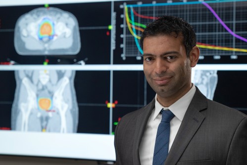
Lead author Dr. Amar Kishan, assistant professor of radiation oncology at the David Geffen School of Medicine at UCLA and researcher at the UCLA Jonsson Comprehensive Cancer Center.

A new UCLA-led study shows that men with low- or intermediate-risk prostate cancer can safely undergo higher doses of radiation over a significantly shorter period of time and still have the same, successful outcomes as from a much longer course of treatment.

This type of radiation, known as stereotactic body radiotherapy, is a form of external beam radiation therapy and reduces the duration of treatment from 45 days to four to five days. The approach has been in use since 2000, but has not yet been widely adopted because of concerns over how safe and effective this approach would be in the long term.

“Most men with low- or intermediate-risk prostate cancer undergo conventional radiation, which requires them to come in daily for treatment and takes an average of nine weeks to complete,” said lead author Dr. Amar Kishan, assistant professor of radiation oncology at the David Geffen School of Medicine at UCLA and researcher at the UCLA Jonsson Comprehensive Cancer Center. “That can be very burdensome on a patient and be a huge interruption in their life. With the improvements being made to modern technology, we’ve found that using stereotactic body radiotherapy, which has a higher dose of radiation, can safely and effectively be done in a much shorter timeframe without additional toxicity or compromising any chance of a cure.”

The UCLA research team analyzed data from 2,142 men with low- or intermediate-risk prostate cancer across multiple institutions who were treated with stereotactic body radiotherapy for prostate cancer between 2000 and 2012.

The men were followed for a median of 6.9 years. Just over half of the men had low-risk disease (53 percent), 32 percent had less aggressive intermediate-risk disease and 12 percent had a more aggressive form of intermediate-risk disease.

The recurrence rate for men with low-risk disease was 4.5 percent, the recurrence rate for the less aggressive intermediate-risk was 8.6 percent, and the recurrence rate for the more aggressive intermediate-risk group was 14.9 percent. Overall, the recurrence rate for intermediate-risk disease was 10.2 percent. These are essentially identical to rates following more conventional forms of radiation, which are about 4 percent to 5 percent for low-risk disease and 10 percent to 15 percent for intermediate-risk disease.

“What is remarkable about this very large study is how favorably stereotactic body radiotherapy compares to all other forms of radiation treatments, both in terms of effectiveness and side effects,” said senior author Dr. Christopher King, professor of radiation oncology and scientist at the UCLA cancer center. “With such long-term follow-up data, we can now offer this approach to patients with full confidence.”

The research team at UCLA had previously found that stereotactic body radiation therapy was more cost effective because of the fewer treatments involved. Other research has also suggested psychological benefits such as less regret about undergoing treatment. The current study now provides long-term data regarding the safety and clinical efficacy of this approach.

Kishan said the data show that the majority of the men followed are free of prostate cancer seven years after treatment. He added that there was no evidence that this therapy caused worse toxicity in the long term. “In fact,” Kishan said, “we not only confirm that this method is both safe and effective, but we provide significant evidence that this could be a viable treatment option for men with low- and intermediate-risk of prostate cancer.”

Pioneering stem cell gene therapy cures infants with bubble baby disease.


Evangelina Vaccaro (far right), who received Dr. Kohn’s treatment for bubble baby disease in 2012, with her family before her first day of school.

FINDINGS
UCLA researchers have developed a stem cell gene therapy cure for babies born with adenosine deaminase-deficient severe combined immunodeficiency, a rare and life-threatening condition that can be fatal within the first year of life if left untreated.

In a phase 2 clinical trial led by Dr. Donald Kohn of the Eli and Edythe Broad Center of Regenerative Medicine and Stem Cell Research at UCLA, all nine babies were cured. A 10th trial participant was a teenager at the time of treatment and showed no signs of immune system recovery. Kohn’s treatment method, a stem cell gene therapy that safely restores immune systems in babies with the immunodeficiency using the child’s own cells, has cured 30 out of 30 babies during the course of several clinical trials.

BACKGROUND
Adenosine deaminase-deficient severe combined immunodeficiency, also known as ADA-SCID or bubble baby disease, is caused by a genetic mutation that results in the lack of the adenosine deaminase enzyme, which is an important component of the immune system. Without the enzyme, immune cells are not able to fight infections. Children with the disease must remain isolated in clean and germ-free environments to avoid exposure to viruses and bacteria; even a minor cold could prove fatal.

Currently, there are two commonly used treatment options for children with ADA-SCID. They can be injected twice a week with the adenosine deaminase enzyme — a lifelong process that is very expensive and often does not return the immune system to optimal levels. Some children can receive a bone marrow transplant from a matched donor, such as a sibling, but bone marrow matches are rare and can result in the recipient’s body rejecting the transplanted cells.

METHOD
The researchers used a strategy that corrects the ADA-SCID mutation by genetically modifying each patient’s own blood-forming stem cells, which can create all blood cell types. In the trial, blood stem cells removed from each child’s bone marrow were corrected in the lab through insertion of the gene responsible for making the adenosine deaminase enzyme. Each child then received a transplant of their own corrected blood stem cells.

The clinical trial ran from 2009 to 2012 and treated 10 children with ADA-SCID and no available matched bone marrow donor. Three children were treated at the National Institutes of Health and seven were treated at UCLA. No children in the trial experienced complications from the treatment. Nine out of ten were babies and they all now have good immune system function and no longer need to be isolated. They are able to live normal lives, play outside, go to school, receive immunizations and, most importantly, heal from common sicknesses such as the cold or an ear infection. The teenager who was not cured continues to receive enzyme therapy.

The fact that the nine babies were cured and the teenager was not indicates that the gene therapy for ADA-SCID works best in the youngest patients, before their bodies lose the ability to restore the immune system

IMPACT
The next step is to seek approval from the Food and Drug Administration for the gene therapy in the hopes that all children with ADA-SCID will be able to benefit from the treatment. Kohn and colleagues have also adapted the stem cell gene therapy approach to treat sickle cell disease and X-linked chronic granulomatous disease, an immunodeficiency disorder commonly referred to as X-linked CGD. Clinical trials providing stem cell gene therapy treatments for both diseases are currently ongoing.

Source:https://stemcell.ucla.edu

UCLA Scientists “Jump-start” the Brain of a Coma Patient.


Article Image

A new ultrasound treatment by UCLA scientists was used to restart brain activity in a 25-year-old man recovering from a coma. Before the treatment, the man showed few signs of consciousness and understanding speech, able to perform very limited movements. Three days after the treatment, the man could fully understand language, communicated by nodding his head and even fist-bumped one of the doctors.

“The changes were remarkable,” said Martin Monti, the study’s lead author and a UCLA associate professor of psychology and neurosurgery.

He elaborated on the technique which used a sonic stimulation device to excite neurons in the thalamus, the brain’s central processing hub. It is the area with a typically diminished performance in post-coma patients.

“It’s almost as if we were jump-starting the neurons back into function,” Monti said. “Until now, the only way to achieve this was a risky surgical procedure known as deep brain stimulation, in which electrodes are implanted directly inside the thalamus. Our approach directly targets the thalamus but is noninvasive.”

The device used is the size of a coffee cup saucer. According to the UCLA press release, it “creates a small sphere of acoustic energy that can be aimed at different regions of the brain to excite brain tissue”.

brain

The researchers targeted the thalamus with low-intensity focused ultrasound pulsation. 

Both the device and the technique, called low-intensity focused ultrasound pulsation, was developed by another UCLA professor Alexander Bystritsky. He is the co-author of the study and founder of Brainsonix, a company that made the device.

The small amount of energy emitted by the device is safe for the patient.

Can this technique be used to help other patients recovering from comas? The researchers are taking a wait and see approach. More studies need to be done to see if they can get consistent results. UCLA researchers will be testing the procedure on several more people in the fall.

“It is possible that we were just very lucky and happened to have stimulated the patient just as he was spontaneously recovering,” cautioned Monti.

Monti hopes that if the technology pans out, their research will lead to the creation of low-cost portable devices that can “wake up” the brain of vegetative or minimally conscious patients.

Modern intensive care medicine has greatly increased the rates of survival after severe brain injury (BI). Nonetheless, a number of patients fail to fully recover from coma, and awaken to a disorder of consciousness (DOC) such as the vegetative state (VS) or the minimally conscious state (MCS) [1]. In these conditions, which can be transient or last indefinitely, patients can lose virtually all autonomy and have almost no treatment options [1,2]. In addition, these conditions place great emotional and financial strain on families, lead to increased burn-out rates among care-takers, impose financial stress on medical structures and public finances due to the costs of prolonged intensive care, and raise difficult legal and ethical questions

UCLA doctors make history ‘jump-starting’ brain of 25-year-old coma patient.


  • Currently, post-coma patients require life-threatening brain surgery
  • A new test by UCLA used new device to pulse ultrasound into the brain
  • The scientists targeted the sensory hub (thalamus) with 10 pulses
  • Within 3 days, the 25-year-old man was conscious, fist-bumped his doctor

A 25-year-old man has become the first coma patient to regain consciousness without life-threatening surgery.

The man, who has not been identified, had his brain jump-started with new ultrasound technology in an experiment by UCLA.

Within days of waking up, he was fully conscious, responding to questions, and even gave his doctor a fist-bump.

It is the first time such an approach has been used to treat severe brain injury.

The procedure marks a significant step in medical understanding that could save and transform millions of lives.

Groundbreaking: Scientists have successfully 'jump-start' a man's brain after a coma using incredibly low-energy ultrasound pulses. The device targeted the thalamus (highlighted here in green), which is the brain's 'sensory hub' controlling waking up, alertness and arousal

Groundbreaking: Scientists have successfully ‘jump-start’ a man’s brain after a coma using incredibly low-energy ultrasound pulses. The device targeted the thalamus (highlighted here in green), which is the brain’s ‘sensory hub’ controlling waking up, alertness and arousal

‘Until now, the only way to achieve [brain function] was a risky surgical procedure known as deep brain stimulation, in which electrodes are implanted directly inside the thalamus,’ said lead author Dr Martin Monti, UCLA professor of neurosurgery.

‘Our approach directly targets the thalamus but is noninvasive.

‘It’s almost as if we were jump-starting the neurons back into function.’

The study, published in the journal Brain Stimulation, focused on the thalamus as that is the part of the brain that is most impaired after a coma.

This is the brain’s sensory hub – an egg-shaped structure relaying signals from different regions and regulating waking, alertness and arousal.

Currently, medications prescribed to coma victims only target the thalamus indirectly.

Before the procedure began the patient showed only minimal signs of consciousness and recognizing speech.

HOW DOES THE NEW PROCEDURE WORK?

Currently, the only way to regain brain function after a coma is through a surgical procedure.

The procedure, dubbed deep brain stimulation, involves implanting electrodes directly inside the thalamus.

The UCLA study used a device about the size of a coffee cup.

It was developed by co-author Professor Alexander Bystritsky in his bio-tech firm Brainsonix.

The researchers placed the device on the man’s head and activated it to send pulses of ultrasound into the thalamus.

They activated it 10 times over 10 minutes, each time for 30 seconds.

By activating the device, they were creating a sphere of acoustic energy that could be aimed at different regions of the brain to stimulate tissue.

The device has incredibly low energy levels.

It emits less energy than a conventional Doppler ultrasound.

He could perform small, limited movements when asked but his reactions were slow.

The treatment involved a device developed by co-author Professor Alexander Bystritsky in his bio-tech firm Brainsonix.

The device – about the size of a saucer – was placed on the side of the man’s head, sending pulses of ultrasound into the thalamus.

This procedure, called low-intensity focused ultrasound pulsation, creates a small sphere of acoustic energy that can be aimed at different regions of the brain to excite brain tissue.

It was repeated 10 times, once a minute for 30 seconds each.

By the day after the treatment, the patient’s responses had improved measurably.

Three days later, the patient had regained full consciousness and full language comprehension.

He could reliably communicate by nodding his head ‘yes’ or shaking his head ‘no’.

He even made a fist-bump gesture to say goodbye to one of his doctors.

‘The changes were remarkable,’ Dr Monti said.

According to the researchers, the device is uniquely safe due to its low energy levels.

It emits less energy than a conventional Doppler ultrasound, thereby minimizing its impact on other delicate parts of the brain.

The researchers plan to test the procedure on more patients this year to develop the treatment.

Ultrasound devices capable of penetrating the human brain are already being tested for other conditions including tremors, chronic pain, and even dementia.


Lens-free microscope can detect cancer at cellular level


A lens-free microscope that can be used to detect the presence of cancer or other cell-level abnormalities with the same accuracy as larger and more expensive optical microscopes, has been developed by researchers. The invention could lead to less expensive and more portable technology for performing common examinations of tissue, blood and other biomedical specimens. It may prove especially useful in remote areas and in cases where large numbers of samples need to be examined quickly.
Tissue sample image created by a new lens-free microscope developed in the UCLA lab of Aydogan Ozcan.
UCLA researchers have developed a lens-free microscope that can be used to detect the presence of cancer or other cell-level abnormalities with the same accuracy as larger and more expensive optical microscopes.
The invention could lead to less expensive and more portable technology for performing common examinations of tissue, blood and other biomedical specimens. It may prove especially useful in remote areas and in cases where large numbers of samples need to be examined quickly.

The microscope is the latest in a series of computational imaging and diagnostic devices developed in the lab of Aydogan Ozcan, the Chancellor’s Professor of Electrical Engineering and Bioengineering at the UCLA Henry Samueli School of Engineering and Applied Science and a Howard Hughes Medical Institute professor. Ozcan’s lab has previously developed custom-designed smartphone attachments and apps that enable quick analysis of food samples for allergens, water samples for heavy metals and bacteria, cell counts in blood samples, and the use of Google Glass to process the results of medical diagnostic tests.

The latest invention is the first lens-free microscope that can be used for high-throughput 3-D tissue imaging — an important need in the study of disease.

“This is a milestone in the work we’ve been doing,” said Ozcan, who also is the associate director of UCLA’s California NanoSystems Institute. “This is the first time tissue samples have been imaged in 3D using a lens-free on-chip microscope.”

The research is the cover article in Science Translational Medicine, which is published by the American Association for the Advancement of Science.

The device works by using a laser or light-emitting-diode to illuminate a tissue or blood sample that has been placed on a slide and inserted into the device. A sensor array on a microchip — the same type of chip that is used in digital cameras, including cellphone cameras — captures and records the pattern of shadows created by the sample.

The device processes these patterns as a series of holograms, forming 3-D images of the specimen and giving medical personnel a virtual depth-of-field view. An algorithm color codes the reconstructed images, making the contrasts in the samples more apparent than they would be in the holograms and making any abnormalities easier to detect.

Ozcan’s team tested the device using Pap smears that indicated cervical cancer, tissue specimens containing cancerous breast cells, and blood samples containing sickle cell anemia. In a blind test, a board-certified pathologist analyzed sets of specimen images that had been created by the lens-free technology and by conventional microscopes. The pathologist’s diagnoses using the lens-free microscopic images proved accurate 99 percent of the time.

Another benefit of the lens-free device is that it produces images that are several hundred times larger in area, or field of view, than those captured by conventional bright-field optical microscopes, which makes it possible to process specimens more quickly.

“While mobile health care has expanded rapidly with the growth of consumer electronics — cellphones in particular — pathology is still, by and large, constrained to advanced clinical laboratory settings,” Ozcan said. “Accompanied by advances in its graphical user interface, this platform could scale up for use in clinical, biomedical, scientific, educational and citizen-science applications, among others.”


Story Source:

The above story is based on materials provided by University of California – Los Angeles. The original article was written by Bill Kisliuk. Note: Materials may be edited for content and length.


Journal Reference:

  1. Aydogan Ozcan et al. Wide-field computational imaging of pathology slides using lens-free on-chip microscopy. Science Translational Medicine, December 2014 DOI: 10.1126/scitranslmed.3009850

Model predicted final menses.


Researchers at UCLA have developed a model to estimate the timing of a woman’s final menstrual period. According to researchers, the model has the potential to help physicians and patients determine when the menopausal transition is complete and estimate bone loss.

“We need a better way to answer women’s questions about when to expect the final menstrual period,” researcher Gail A. Greendale, MD, from UCLA’s David Geffen School of Medicine, said in a press release. “If further research bears out our approach, it could be the first step to developing Web-based calculators and other tools women can use to estimate where they are in the menopause transition and how far away their final period is.”

Greendale and colleagues included 554 women from the Study of Women’s Health Across the Nation (SWAN). They designed the probability of meeting specific landmarks: 2 years before, 1 year before and the final menstrual period (FMP).

“For example, some researchers have proposed that an intervention begun 1 or 2 years before the final menstrual period would greatly decrease future fracture risk by preventing the very rapid bone loss that occurs in the few years before and few years after the final menses,” Greendale said. “But before ideas such as this can be tested, we need to accurately predict where a woman is in her timeline to menopause.”

Therefore, the researchers assessed the probability of being in restricted intervals: 1 to 2 years before FMP, 2 years before FMP and FMP, or 1 year before FMP and FMP. Additionally, the markers that best predicted having crossed each landmark were determined, with the ideal markers defined as the greatest area under the receiver-operator curve (AUC).

Researchers wrote that the final models included the current estradiol and follicle-stimulating hormone (FSH), age, the stage of menopause transition, race/ethnicity and whether serum was collected during the early follicular phase.

Data indicate the AUC of final models predicted the probability of a woman having crossed 2 years before (0.902), 1 year before (0.926) and the FMP (0.945), researchers wrote. If they identified women as having crossed the 2 years before the FMP landmark when predicted probability extended beyond 0.3, the sensitivity was 85% and specificity 77%, they added.

Despite limitations, Greendale and colleagues conclude that the clinical practice implementation of their model is conceivable. However, further studies are warranted to determine validation of these findings.

Source: http://www.healio.com