Three Distinct Brain Circuits in the Thalamus Contribute to Parkinson’s Symptoms


Summary: Neurons in the parafascicular thalamus project to three different parts of the basal ganglia. Targeting these circuits could be a new target for treating motor dysfunction and depression associated with Parkinson’s disease.

Source: MIT

Parkinson’s disease is best-known as a disorder of movement. Patients often experience tremors, loss of balance, and difficulty initiating movement. The disease also has lesser-known symptoms that are nonmotor, including depression.

In a study of a small region of the thalamus, MIT neuroscientists have now identified three distinct circuits that influence the development of both motor and nonmotor symptoms of Parkinson’s. Furthermore, they found that by manipulating these circuits, they could reverse Parkinson’s symptoms in mice.

The findings suggest that those circuits could be good targets for new drugs that could help combat many of the symptoms of Parkinson’s disease, the researchers say.

“We know that the thalamus is important in Parkinson’s disease, but a key question is how can you put together a circuit that that can explain many different things happening in Parkinson’s disease. Understanding different symptoms at a circuit level can help guide us in the development of better therapeutics,” says Guoping Feng, the James W. and Patricia T. Poitras Professor in Brain and Cognitive Sciences at MIT, a member of the Broad Institute of Harvard and MIT, and the associate director of the McGovern Institute for Brain Research at MIT.

Feng is the senior author of the study, which appears today in Nature. Ying Zhang, a J. Douglas Tan Postdoctoral Fellow at the McGovern Institute, and Dheeraj Roy, a NIH K99 Awardee and a McGovern Fellow at the Broad Institute, are the lead authors of the paper.

Tracing circuits

The thalamus consists of several different regions that perform a variety of functions. Many of these, including the parafascicular (PF) thalamus, help to control movement. Degeneration of these structures is often seen in patients with Parkinson’s disease, which is thought to contribute to their motor symptoms.

In this study, the MIT team set out to try to trace how the PF thalamus is connected to other brain regions, in hopes of learning more about its functions.

They found that neurons of the PF thalamus project to three different parts of the basal ganglia, a cluster of structures involved in motor control and other functions: the caudate putamen (CPu), the subthalamic nucleus (STN), and the nucleus accumbens (NAc).

“We started with showing these different circuits, and we demonstrated that they’re mostly nonoverlapping, which strongly suggests that they have distinct functions,” Roy says.

Further studies revealed those functions. The circuit that projects to the CPu appears to be involved in general locomotion, and functions to dampen movement. When the researchers inhibited this circuit, mice spent more time moving around the cage they were in.

The circuit that extends into the STN, on the other hand, is important for motor learning — the ability to learn a new motor skill through practice. The researchers found that this circuit is necessary for a task in which the mice learn to balance on a rod that spins with increasing speed.

Lastly, the researchers found that, unlike the others, the circuit that connects the PF thalamus to the NAc is not involved in motor activity. Instead, it appears to be linked to motivation. Inhibiting this circuit generates depression-like behaviors in healthy mice, and they will no longer seek a reward such as sugar water.

Druggable targets

Once the researchers established the functions of these three circuits, they decided to explore how they might be affected in Parkinson’s disease. To do that, they used a mouse model of Parkinson’s, in which dopamine-producing neurons in the midbrain are lost.

They found that in this Parkinson’s model, the connection between the PF thalamus and the CPu was enhanced, and that this led to a decrease in overall movement. Additionally, the connections from the PF thalamus to the STN were weakened, which made it more difficult for the mice to learn the accelerating rod task.

Lastly, the researchers showed that in the Parkinson’s model, connections from the PF thalamus to the NAc were also interrupted, and that this led to depression-like symptoms in the mice, including loss of motivation.

Using chemogenetics or optogenetics, which allows them to control neuronal activity with a drug or light, the researchers found that they could manipulate each of these three circuits and in doing so, reverse each set of Parkinson’s symptoms.

Then, they decided to look for molecular targets that might be “druggable,” and found that each of the three PF thalamus regions have cells that express different types of cholinergic receptors, which are activated by the neurotransmitter acetylcholine.

By blocking or activating those receptors, depending on the circuit, they were also able to reverse the Parkinson’s symptoms.
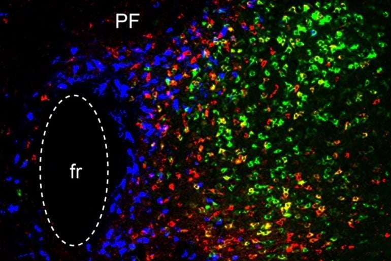
This shows neurons in the thalamus
In this image of the parafascicular (PF) thalamus, the blue cells participate in reward processing/depression, the red cells are critical for motor learning, and the green cells are important for general locomotion. The ‘fr’ stands for a fiber bundle. Credit: Ying Zhang and Dheeraj Roy

“We found three distinct cholinergic receptors that can be expressed in these three different PF circuits, and if we use antagonists or agonists to modulate these three different PF populations, we can rescue movement, motor learning, and also depression-like behavior in PD mice,” Zhang says.

Parkinson’s patients are usually treated with L-dopa, a precursor of dopamine. While this drug helps patients regain motor control, it doesn’t help with motor learning or any nonmotor symptoms, and over time, patients become resistant to it.

The researchers hope that the circuits they characterized in this study could be targets for new Parkinson’s therapies. The types of neurons that they identified in the circuits of the mouse brain are also found in the nonhuman primate brain, and the researchers are now using RNA sequencing to find genes that are expressed specifically in those cells.

“RNA-sequencing technology will allow us to do a much more detailed molecular analysis in a cell-type specific way,” Feng says. “There may be better druggable targets in these cells, and once you know the specific cell types you want to modulate, you can identify all kinds of potential targets in them.”

Funding: The research was funded, in part, by the K. Lisa Yang and Hock E. Tan Center for Molecular Therapeutics in Neuroscience at MIT, the Stanley Center for Psychiatric Research at the Broad Institute, the James and Patricia Poitras Center for Psychiatric Disorders Research at MIT, the National Institutes of Health BRAIN Initiative, and the National Institute of Mental Health.


Abstract

Targeting thalamic circuits rescues motor and mood deficits in PD mice

Although bradykinesia, tremor and rigidity are the hallmark motor defects in patients with Parkinson’s disease (PD), patients also experience motor learning impairments and non-motor symptoms such as depression.

https://imasdk.googleapis.com/js/core/bridge3.517.2_en.html#goog_750152574

The neural circuit basis for these different symptoms of PD are not well understood. Although current treatments are effective for locomotion deficits in PD, therapeutic strategies targeting motor learning deficits and non-motor symptoms are lacking.

Here we found that distinct parafascicular (PF) thalamic subpopulations project to caudate putamen (CPu), subthalamic nucleus (STN) and nucleus accumbens (NAc). Whereas PF→CPu and PF→STN circuits are critical for locomotion and motor learning, respectively, inhibition of the PF→NAc circuit induced a depression-like state.

Whereas chemogenetically manipulating CPu-projecting PF neurons led to a long-term restoration of locomotion, optogenetic long-term potentiation (LTP) at PF→STN synapses restored motor learning behaviour in an acute mouse model of PD.

Furthermore, activation of NAc-projecting PF neurons rescued depression-like phenotypes.

Further, we identified nicotinic acetylcholine receptors capable of modulating PF circuits to rescue different PD phenotypes.

Thus, targeting PF thalamic circuits may be an effective strategy for treating motor and non-motor deficits in PD.

UCLA doctors make history ‘jump-starting’ brain of 25-year-old coma patient.


  • Currently, post-coma patients require life-threatening brain surgery
  • A new test by UCLA used new device to pulse ultrasound into the brain
  • The scientists targeted the sensory hub (thalamus) with 10 pulses
  • Within 3 days, the 25-year-old man was conscious, fist-bumped his doctor

A 25-year-old man has become the first coma patient to regain consciousness without life-threatening surgery.

The man, who has not been identified, had his brain jump-started with new ultrasound technology in an experiment by UCLA.

Within days of waking up, he was fully conscious, responding to questions, and even gave his doctor a fist-bump.

It is the first time such an approach has been used to treat severe brain injury.

The procedure marks a significant step in medical understanding that could save and transform millions of lives.

Groundbreaking: Scientists have successfully 'jump-start' a man's brain after a coma using incredibly low-energy ultrasound pulses. The device targeted the thalamus (highlighted here in green), which is the brain's 'sensory hub' controlling waking up, alertness and arousal

Groundbreaking: Scientists have successfully ‘jump-start’ a man’s brain after a coma using incredibly low-energy ultrasound pulses. The device targeted the thalamus (highlighted here in green), which is the brain’s ‘sensory hub’ controlling waking up, alertness and arousal

‘Until now, the only way to achieve [brain function] was a risky surgical procedure known as deep brain stimulation, in which electrodes are implanted directly inside the thalamus,’ said lead author Dr Martin Monti, UCLA professor of neurosurgery.

‘Our approach directly targets the thalamus but is noninvasive.

‘It’s almost as if we were jump-starting the neurons back into function.’

The study, published in the journal Brain Stimulation, focused on the thalamus as that is the part of the brain that is most impaired after a coma.

This is the brain’s sensory hub – an egg-shaped structure relaying signals from different regions and regulating waking, alertness and arousal.

Currently, medications prescribed to coma victims only target the thalamus indirectly.

Before the procedure began the patient showed only minimal signs of consciousness and recognizing speech.

HOW DOES THE NEW PROCEDURE WORK?

Currently, the only way to regain brain function after a coma is through a surgical procedure.

The procedure, dubbed deep brain stimulation, involves implanting electrodes directly inside the thalamus.

The UCLA study used a device about the size of a coffee cup.

It was developed by co-author Professor Alexander Bystritsky in his bio-tech firm Brainsonix.

The researchers placed the device on the man’s head and activated it to send pulses of ultrasound into the thalamus.

They activated it 10 times over 10 minutes, each time for 30 seconds.

By activating the device, they were creating a sphere of acoustic energy that could be aimed at different regions of the brain to stimulate tissue.

The device has incredibly low energy levels.

It emits less energy than a conventional Doppler ultrasound.

He could perform small, limited movements when asked but his reactions were slow.

The treatment involved a device developed by co-author Professor Alexander Bystritsky in his bio-tech firm Brainsonix.

The device – about the size of a saucer – was placed on the side of the man’s head, sending pulses of ultrasound into the thalamus.

This procedure, called low-intensity focused ultrasound pulsation, creates a small sphere of acoustic energy that can be aimed at different regions of the brain to excite brain tissue.

It was repeated 10 times, once a minute for 30 seconds each.

By the day after the treatment, the patient’s responses had improved measurably.

Three days later, the patient had regained full consciousness and full language comprehension.

He could reliably communicate by nodding his head ‘yes’ or shaking his head ‘no’.

He even made a fist-bump gesture to say goodbye to one of his doctors.

‘The changes were remarkable,’ Dr Monti said.

According to the researchers, the device is uniquely safe due to its low energy levels.

It emits less energy than a conventional Doppler ultrasound, thereby minimizing its impact on other delicate parts of the brain.

The researchers plan to test the procedure on more patients this year to develop the treatment.

Ultrasound devices capable of penetrating the human brain are already being tested for other conditions including tremors, chronic pain, and even dementia.


Dyslexia caused by faulty signal processing in brain.


Dyslexia, a learning disability can result when the medial geniculate nucleus in the thalamus of the brain does not process speech sounds correctly, a new study has claimed.

Researchers from Max Planck Institute for Human Cognitive and Brain Sciences in Leipzig found that many difficulties associated with dyslexia can potentially be traced back to a malfunction of the medial geniculate nucleus in the thalamus.

The thalamus is a large, dual lobed mass of grey matter buried under the cerebral cortex. It is involved in sensory perception and regulation of motor functions.

There are three proposed cognitive subtypes of dyslexia: auditory, visual and attentional.

People who suffer from dyslexia have difficulties with identifying speech sounds in spoken language. For example, while most children are able to recognise whether two words rhyme even before they go to school, dyslexic children often cannot do this until late primary school age.

The research found that dyslexic adults have a malfunction in a structure that transfers auditory information from the ear to the cortex, the medial geniculate body in the auditory thalamus does not process speech sounds correctly.

“This malfunction at a low level of language processing could percolate through the entire system. This explains why the symptoms of dyslexia are so varied,” Begona Diaz from the Institute said.

The researchers conducted two experiments in which several volunteers had to perform various speech comprehension tasks.

When affected individuals performed tasks that required the recognition of speech sounds, as compared to recognise the voices that pronounced the same speech, magnetic resonance tomography (MRT) recordings showed abnormal responses in the area around the medial geniculate body.

In contrast, no differences were apparent between controls and dyslexic participants if the tasks involved only listening to the speech sounds without having to perform a specific task.

“The problem, therefore, has nothing to do with sensory processing itself, but with the processing involved in speech recognition,” Diaz said.

Source: the Hindu.

Keywords: dyslexiafaulty signalbrain function

Gamma Knife surgery for basal ganglia and thalamic arteriovenous malformations.


Gamma Knife surgery (GKS) has emerged as the treatment of choice for small- to medium-sized cerebral arteriovenous malformations (AVMs) in deep locations. The present study aims to investigate the outcomes of GKS for AVMs in the basal ganglia and thalamus.

Methods

Between 1989 and 2007, 85 patients with AVMs in the basal ganglia and 97 in the thalamus underwent GKS and were followed up for more than 2 years. The nidus volumes ranged from 0.1 to 29.4 cm3 (mean 3.4 cm3). The mean margin dose at the initial GKS was 21.3 Gy (range 10–28 Gy). Thirty-six patients underwent repeat GKS for residual AVMs at a median 4 years after initial GKS. The mean margin dose at repeat GKS was 21.1 Gy (range 7.5–27 Gy).

Results

Following a single GKS, total obliteration of the nidus was confirmed on angiograms in 91 patients (50%). In 12 patients (6.6%) a subtotal obliteration was achieved. No flow voids were observed on MR imaging in 14 patients (7.7%). Following single or repeat GKS, total obliteration was angiographically confirmed in 106 patients (58.2%) and subtotal obliteration in 8 patients (4.4%). No flow voids on MR imaging were observed in 18 patients (9.9%). The overall obliteration rates following one or multiple GKSs based on MR imaging or angiography was 68%. A small nidus volume, high margin dose, low number of isocenters, and no history of embolization were significantly associated with an increased rate of obliteration. Twenty-one patients experienced 25 episodes of hemorrhage in 850 risk-years following GKS, yielding an annual hemorrhage rate of 2.9%. Four patients died in this series: 2 due to complications of hemorrhage and 2 due to unrelated diseases. Permanent neurological deficits caused by radiation were noted in 9 patients (4.9%).

Conclusions

Gamma Knife surgery offers a reasonable chance of obliterating basal ganglia and thalamic AVMs and does so with a low risk of complications. It is an optimal treatment option in patients for whom the anticipated risk of microsurgery is too high.

Source: Journal of Neurosurgery