Mapping the Brain: The Largest Neuron Projectome Unveiled.


Summary: Researchers unveiled the most extensive single-neuron projectome database to date, featuring over 10,000 mouse hippocampal neurons.

The study provides an unprecedented view of the spatial connectivity patterns at the mesoscopic level, crucial for understanding learning, memory, and emotional processing in the hippocampus. By employing machine learning algorithms for categorizing axonal trajectories and integrating spatial transcriptome data, researchers identified 43 distinct projectome cell types, revealing intricate projection patterns and soma locations’ correspondence to projection targets.

This work, accessible via the Digital Brain CEBSIT portal, lays the structural foundation for advancing our knowledge of hippocampal functions and their molecular underpinnings.

Key Facts:

  1. The study reconstructed whole-brain axonal morphology of over 10,000 mouse hippocampal neurons, creating the world’s most extensive single-neuron projectome database.
  2. Researchers used machine learning to analyze morphological similarities among neurons, identifying 43 distinct projectome cell types.
  3. The integration of projectome cell types with spatial transcriptome data revealed potential molecular and circuit targets for hippocampal functions, all accessible through a dedicated online platform.

Source: Chinese Academy of Science

A study published in Science on Feb. 1 reported a comprehensive database of single-neuron projectomes consisting of over 10,000 mouse hippocampal neurons, thus revealing the spatial connectivity patterns of mouse hippocampal neurons at the mesoscopic level.   

The study was conducted by teams from the Center for Excellence in Brain Science and Intelligence Technology (CEBSIT), the Institute of Neuroscience of the Chinese Academy of Sciences (CAS), the HUST-Suzhou Institute for Brainsmatics, Hainan University, the Kunming Institute of Zoology of CAS, Lingang Laboratory, and the Shanghai Center for Brain Science and Brain-Inspired Technology.  
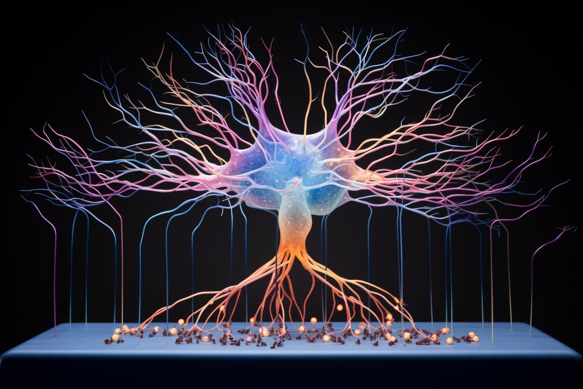
This shows a connectome brain map.
Hippocampal neurons project widely to the brain-wide targets; thus, it is critical to investigate the projection patterns of hippocampal neurons at the single-neuron level. Credit: Neuroscience News

The hippocampus serves as an essential brain region for learning and memory as well as various brain functions such as spatial cognition and emotional processing. It is one of the most extensively studied brain regions.

Hippocampal neurons project widely to the brain-wide targets; thus, it is critical to investigate the projection patterns of hippocampal neurons at the single-neuron level.   

This study reconstructed the whole-brain axonal morphology of over 10,000 neurons in the mouse hippocampus at a single-cell resolution with the neuronal cell bodies covering all subregions and multiple locations along different hippocampal axes, making this the most extensive single-neuron projectome database in the world.   

This study took an innovative approach to categorize axonal trajectories with machine learning algorithms, thus allowing for a more efficient analysis of the morphological similarities among 341 projection patterns for mouse hippocampal neurons and ultimately identifying 43 distinct projectome cell types. It also incorporated the spatial transcriptome of mouse CA1 areas.  

Based on these analyses, the study was able to elucidate the axonal projection pathways of hippocampal neurons along the anterior-posterior axis and reveal new projection patterns of hippocampal neurons. It also outlined the correspondence between hippocampal neuron soma locations and projection targets, and revealed basic organization principles of bilateral projections.

Furthermore, correlation analysis of projectome cell types and spatial transcriptome data identified spatial correspondence between various genes and projectome subtypes, providing potential molecular and circuit targets for hippocampal functions.  

Taken together, this study provides a structural basis for future studies of hippocampal functions and deciphers the potential correspondences between their soma locations, gene expression, and circuitry functions.  

The database for the hippocampal single-neuron projectomes, along with the database on the hippocampal longitudinal axis and spatial transcriptomes, are now publicly accessible through the Digital Brain CEBSIT portal (https://mouse.digital-brain.cn/hipp).

To facilitate broader usage of the databases, a team from the Computing and Data Center of CEBSIT has developed a website to integrate data visualization, user interface, online analysis, and data downloads.


Abstract

Whole-brain spatial organization of hippocampal single-neuron projectomes

INTRODUCTION

In the brain circuitry, a single neuron could broadcast output signals to other neurons located in nearby or distant areas. Therefore, understanding the spatial organization of axon projections at the single-cell level is crucial for elucidating the neural circuitry underlying various brain functions. As a brain structure essential for learning, memory, cognition, stress responses and emotional behaviors, it is known that the hippocampus (HIP) is widely connected with various brain areas, including the cortex, thalamus, hypothalamus, olfactory areas, and amygdala. However, it remains unclear how single HIP neurons project to brain-wide target areas and how a single-neuron projectome can be specified by the soma location within the HIP.

RATIONALE

To reconstruct single-neuron projectomes of the mouse HIP, we combined sparse-labeling methods with fluorescence micro-optical sectioning tomography and reconstructed the soma, axon arbors, and dendrites of individual neurons. We used a wide range of analytic tools for the projectome analysis and created a state-of-art web interface to visualize the projectome data.

RESULTS

We have created an open and comprehensive database with 10,100 single-neuron projectomes throughout the HIP, which allows interactive query, visualization, and analysis of reconstructed brain-wide projectomes. We classified HIP neurons into 341 projection patterns, and then 43 projectome subtypes, based on their axon morphology and brain-wide target areas.

Our study revealed previously unknown axon projection patterns, target-dependent soma distribution within HIP subdomains, a general rule for bihemispheric projections, axon orientation rules for mossy fibers and Schaffer collaterals, and topographic correlation between axon arbors and soma location along HIP axes.

CONCLUSION

Single-neuron projectome analyses have provided unprecedented information on axon projection patterns at the single-cell resolution and elucidated the organizational principles of whole-brain connectivity of HIP neurons. Such knowledge could be further combined with gene expression data to define HIP neuron subtypes and serve as the structural basis for understanding their diverse and coordinated functions.
Mapping the Brain: The Largest Neuron Projectome Unveiled


Summary: Researchers unveiled the most extensive single-neuron projectome database to date, featuring over 10,000 mouse hippocampal neurons.

The study provides an unprecedented view of the spatial connectivity patterns at the mesoscopic level, crucial for understanding learning, memory, and emotional processing in the hippocampus. By employing machine learning algorithms for categorizing axonal trajectories and integrating spatial transcriptome data, researchers identified 43 distinct projectome cell types, revealing intricate projection patterns and soma locations’ correspondence to projection targets.

This work, accessible via the Digital Brain CEBSIT portal, lays the structural foundation for advancing our knowledge of hippocampal functions and their molecular underpinnings.

Key Facts:

  1. The study reconstructed whole-brain axonal morphology of over 10,000 mouse hippocampal neurons, creating the world’s most extensive single-neuron projectome database.
  2. Researchers used machine learning to analyze morphological similarities among neurons, identifying 43 distinct projectome cell types.
  3. The integration of projectome cell types with spatial transcriptome data revealed potential molecular and circuit targets for hippocampal functions, all accessible through a dedicated online platform.

Source: Chinese Academy of Science

A study published in Science on Feb. 1 reported a comprehensive database of single-neuron projectomes consisting of over 10,000 mouse hippocampal neurons, thus revealing the spatial connectivity patterns of mouse hippocampal neurons at the mesoscopic level.   

The study was conducted by teams from the Center for Excellence in Brain Science and Intelligence Technology (CEBSIT), the Institute of Neuroscience of the Chinese Academy of Sciences (CAS), the HUST-Suzhou Institute for Brainsmatics, Hainan University, the Kunming Institute of Zoology of CAS, Lingang Laboratory, and the Shanghai Center for Brain Science and Brain-Inspired Technology.  

This shows a connectome brain map.
Hippocampal neurons project widely to the brain-wide targets; thus, it is critical to investigate the projection patterns of hippocampal neurons at the single-neuron level.

The hippocampus serves as an essential brain region for learning and memory as well as various brain functions such as spatial cognition and emotional processing. It is one of the most extensively studied brain regions.

Hippocampal neurons project widely to the brain-wide targets; thus, it is critical to investigate the projection patterns of hippocampal neurons at the single-neuron level.   

This study reconstructed the whole-brain axonal morphology of over 10,000 neurons in the mouse hippocampus at a single-cell resolution with the neuronal cell bodies covering all subregions and multiple locations along different hippocampal axes, making this the most extensive single-neuron projectome database in the world.   

This study took an innovative approach to categorize axonal trajectories with machine learning algorithms, thus allowing for a more efficient analysis of the morphological similarities among 341 projection patterns for mouse hippocampal neurons and ultimately identifying 43 distinct projectome cell types. It also incorporated the spatial transcriptome of mouse CA1 areas.  

Based on these analyses, the study was able to elucidate the axonal projection pathways of hippocampal neurons along the anterior-posterior axis and reveal new projection patterns of hippocampal neurons. It also outlined the correspondence between hippocampal neuron soma locations and projection targets, and revealed basic organization principles of bilateral projections.

Furthermore, correlation analysis of projectome cell types and spatial transcriptome data identified spatial correspondence between various genes and projectome subtypes, providing potential molecular and circuit targets for hippocampal functions.  

Taken together, this study provides a structural basis for future studies of hippocampal functions and deciphers the potential correspondences between their soma locations, gene expression, and circuitry functions.  

The database for the hippocampal single-neuron projectomes, along with the database on the hippocampal longitudinal axis and spatial transcriptomes, are now publicly accessible through the Digital Brain CEBSIT portal (https://mouse.digital-brain.cn/hipp).

To facilitate broader usage of the databases, a team from the Computing and Data Center of CEBSIT has developed a website to integrate data visualization, user interface, online analysis, and data downloads.


Abstract

Whole-brain spatial organization of hippocampal single-neuron projectomes

INTRODUCTION

In the brain circuitry, a single neuron could broadcast output signals to other neurons located in nearby or distant areas. Therefore, understanding the spatial organization of axon projections at the single-cell level is crucial for elucidating the neural circuitry underlying various brain functions. As a brain structure essential for learning, memory, cognition, stress responses and emotional behaviors, it is known that the hippocampus (HIP) is widely connected with various brain areas, including the cortex, thalamus, hypothalamus, olfactory areas, and amygdala. However, it remains unclear how single HIP neurons project to brain-wide target areas and how a single-neuron projectome can be specified by the soma location within the HIP.

RATIONALE

To reconstruct single-neuron projectomes of the mouse HIP, we combined sparse-labeling methods with fluorescence micro-optical sectioning tomography and reconstructed the soma, axon arbors, and dendrites of individual neurons. We used a wide range of analytic tools for the projectome analysis and created a state-of-art web interface to visualize the projectome data.

RESULTS

We have created an open and comprehensive database with 10,100 single-neuron projectomes throughout the HIP, which allows interactive query, visualization, and analysis of reconstructed brain-wide projectomes. We classified HIP neurons into 341 projection patterns, and then 43 projectome subtypes, based on their axon morphology and brain-wide target areas.

Our study revealed previously unknown axon projection patterns, target-dependent soma distribution within HIP subdomains, a general rule for bihemispheric projections, axon orientation rules for mossy fibers and Schaffer collaterals, and topographic correlation between axon arbors and soma location along HIP axes.

CONCLUSION

Single-neuron projectome analyses have provided unprecedented information on axon projection patterns at the single-cell resolution and elucidated the organizational principles of whole-brain connectivity of HIP neurons. Such knowledge could be further combined with gene expression data to define HIP neuron subtypes and serve as the structural basis for understanding their diverse and coordinated functions.

Gabapentin May Boost Functional Recovery After a Stroke.


https://neurosciencenews.com/gabapentin-stroke-20643/

Stimulating Brain Circuits Promotes Neuron Growth in Adulthood, Improving Cognition and Mood


Summary: Researchers used optogenetics techniques to stimulate specific brain areas to increase neurogenesis and the production of neural stem cells to improve memory, cognition, and emotional processing in animal models.

Source: UNC Health Care

We humans lose mental acuity, an unfortunate side effect of aging. And for individuals with neurodegenerative conditions such as Alzheimer’s and Parkinson’s, the loss of cognitive function often accompanied by mood disorders such as anxiety is a harrowing experience. One way to push back against cognitive decline and anxiety would be to spur the creation of new neurons.

For the first time, University of North Carolina School of Medicine scientists have targeted a specific kind of neuron in mice to increase the production of neural stem cells and spur on the creation of new adult neurons to affect behavior.

Targeting these cells, as reported in the journal Nature Neuroscience, modulated memory retrieval and altered anxiety-like behaviors in mice. Essentially, the UNC scientists boosted the electrical activity between cells in the hypothalamus and the hippocampus to create new neurons – an important process called neurogenesis.

“Targeting the hypothalamic neurons to enhance adult hippocampal neurogenesis will not only benefit brain functions,” said senior author Juan Song, PhD, associate professor of pharmacology, “but also holds the potential to treat cognitive and affective deficits associated with various brain disorders.”

Most neurons we carry for life were created before we were born and get organized during early childhood. But such neurogenesis continues into adulthood and throughout life. In fact, one of the reasons for cognitive decline and anxiety, and even diseases such as Alzheimer’s, is the suspension of neurogenesis. 

Song, a member of the UNC Neuroscience Center, has been studying the detailed interplay between brain cells that keep neurogenesis chugging along. She knew that adult hippocampal neurogenesis plays a critical role in memory and emotion processing, and that neural circuit activity – think ‘electrical activity’ – regulates this process in a constantly changing manner.

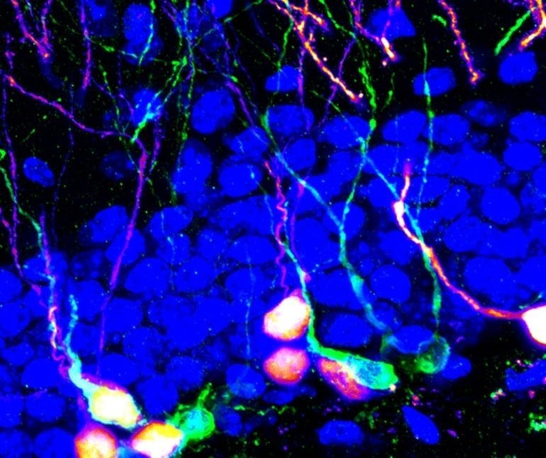
This shows new born neurons
New adult-born neurons that contribute to memory and emotion regulation. Credit: Song Lab, UNC-CH

What no one knew is whether this neural circuit activity could be manipulated to spur neurogenesis to such a degree that the effect would be seen as a changed behavior, such as better memory or less anxiety.

To see the effect of modulating neural activity, the Song lab conducted experiments led by co-first authors Ya-Dong Li, PhD, and Yan-Jia Luo, PhD, both postdoctoral fellows. They used optogenetics – essentially a method using light to trigger neuronal activity – in a small brain structure called supramammillary nucleus (SuM). The SuM is located inside the hypothalamus region of the brain; it helps manage things from cognition to locomotion and sleep/wakefulness.

When Song’s researchers chronically stimulated the SuM neurons, they discovered a robust promotion of neurogenesis at multiple stages. They observed increased production of neural stem cells and the creation of new adult-born neurons with enhanced properties. Optogenetic stimulation of these new neurons then altered memory and anxiety-like behaviors.

“We also show that the SuM neurons are highly responsive when the mice experienced new things in their environment,” Song said. “In fact, in a new environment, mice require these cells for neurogenesis.”

Impaired adult hippocampal neurogenesis correlates with many pathological states, such as aging, neurodegenerative diseases, and mental disorders.

“Therefore,” Song added, “targeting the hypothalamic neurons to enhance adult hippocampal neurogenesis will not only benefit brain functions but also holds the potential to treat cognitive and affective deficits associated with various brain disorders.”

Double-Edged


Amyloid beta protein protects brain from herpes infection by entrapping viral particles

amyloid beta plaques

Amyloid beta plaques (brown), a hallmark of Alzheimer’s disease, in the cerebral cortex.

A new study by Harvard Medical School researchers at Massachusetts General Hospital reveals how amyloid beta, the protein deposited into plaques in the brains of patients with Alzheimer’s disease, protects the brain from the effects of herpes viruses.

Along with another study appearing in the same July 11 issue of Neuron, which found elevated levels of three types of herpesvirus in the brains of patients with Alzheimer’s disease, the HMS team’s results support a potential role for viral infection in accelerating amyloid beta deposition and Alzheimer’s progression.


“There have been multiple epidemiological studies suggesting people with herpes infections are at higher risk for Alzheimer’s disease, along with the most recent findings from Icahn School of Medicine at Mt. Sinai that are being published with our study,” said corresponding author Rudolph Tanzi, the HMS Joseph P. and Rose F. Kennedy Professor of Child Neurology and Mental Retardation at Mass General.

“Our findings reveal a simple and direct mechanism by which herpes infections trigger the deposition of brain amyloid as a defense response in the brain,” Tanzi said. “In this way, we have merged the infection hypothesis and amyloid hypothesis into one ‘antimicrobial response hypothesis’ of Alzheimer’s disease.”

Previous studies led by Tanzi and co-corresponding author Robert Moir, HMS assistant professor of neurology at Mass General, found evidence indicating that amyloid beta, which has long been thought to be useless “metabolic garbage,” was an antimicrobial protein of the body’s innate immune system. Amyloid beta appears capable of protecting animal models and cultured human brain cells from dangerous infections.

Brain infection with herpes simplex, the virus that causes cold sores, is known to increase with aging, leading to almost universal presence of that and other herpes strains in the brain by adulthood. The HMS team set out to find whether amyloid beta could protect against herpes infection and, if so, the mechanism by which such protection takes place.

After first finding that transgenic mice engineered to express human amyloid beta survive significantly longer after injections of herpes simplex into their brains than do nontransgenic mice, the researchers found that amyloid beta inhibited infection of cultured human brain cells with herpes simplex and two other herpes strains by binding to proteins on the viral membranes and clumping into fibrils that entrap the virus and prevent it from entering cells.

Amyloid beta plaques

Further experiments with the transgenic mice revealed that introduction of herpes simplex into the brains of 5- to 6-week-old animals induced rapid development of amyloid beta plaques, which usually appear only when the animals are 10 to 12 weeks old.

“Our findings show that amyloid entrapment of herpesviruses provides immediate, effective protection from infection,” Moir said. “But it’s possible that chronic infection with pathogens like herpes that remain present throughout life could lead to sustained and damaging activation of the amyloid-based immune response, triggering the brain inflammation that drives a cascade of pathologies leading to the onset of Alzheimer’s disease.”

“A key insight is that it’s not direct killing of brain cells by herpes that causes Alzheimer’s, rather, it’s the immune response to the virus that leads to brain-damaging neuroinflammation,” Moir said.

“Our data and the Mt. Sinai findings suggest that an antimicrobial protection model utilizing both anti-herpes and anti-amyloid drugs could be effective against early Alzheimer’s disease,” he added. “Later on, when neuroinflammation has begun, greater benefit may come from targeting inflammatory molecules. However, it remains unclear whether infection is the disease’s root cause. After all, Alzheimer’s is a highly heterogeneous disease, so multiple factors may be involved in its development.”

“We are currently conducting what we call the ‘brain microbiome project,’ to characterize the population of microbes normally found in the brain,” said Tanzi, who is director of the Genetics and Aging Research Unit in the MassGeneral Institute for Neurodegenerative Disease. “The brain used to be considered sterile, but it turns out to have a resident population of microbes, some of which may be needed for normal brain health.”

“Our preliminary findings suggest that the brain microbiome is severely disturbed in Alzheimer’s disease and that bad players, including herpes viruses, seem to take advantage of the situation, leading to trouble for the patient,” Tanzi said. “We are exploring whether Alzheimer’s pathogenesis parallels the disrupted microbiome models seen in conditions like inflammatory bowel disease, and the data generated to date are both surprising and fascinating.”

Scientists Tease Out How the Brain Processes Sensory Experiences


Building a World

Our raw sensory experiences — what we see, hear, feel, taste, and smell — make up our construction the world around us. But how? How does this continuous stream of raw data translate into a seamless understanding of our existence?

Two recent studies from researchers at the International School for Advanced Studies explore how a brain region known as posterior parietal cortex (PPC) influences perception. They showed that, at least in rats, this region contributes to the merging of different sensory information as well as the formation of memories of recent sensory experiences.

One of the two studies, which was published on Jan. 30 in the journal Neuron, reveals how signals that arrive through different channels (from different senses) integrate in this brain region. In this study, researchers wanted to know how we recognize objects without all of its sensory properties. In other words, they wondered how, once we’ve experienced something like an apple, we’re able to know what it is by sight alone (without smelling, tasting, or feeling it).

They explored this by measuring neural activity in the PPCs of trained rats as they interacted with objects. The researchers found that, while neurons varied in how they encoded objects, the neural response was the same for touch, vision, and audition.

“This means that the message of the neurons was the object itself, not the sensory modality through which the object was explored,” Mathew Diamond, senior investigator, said in a press release.

Exploring Senses

In the second paper, published Friday, Jan. 9, in the journal Nature, researchers zeroed in on the exact neural circuit in the PPC that can sometime cause our expectations to actually taint our memories. They examined how recent sensory memories are both formed and kept by training rats to compare the volume of two separated sounds of different volumes — testing them over and over again.

By observing the rats’ PPCs, the researchers found that, as the rodents waited for the second sound, the memory of the latest sound they heard shifted towards the average of all the previous sounds from their previous tests. The results confirmed that PPC can cause memory to slide towards the expected value.

How does the human brain make sense of sensory stimuli like sound? Image Credit: geralt / pixabay
How does the brain make sense of sensory stimuli like sound?

These results still have to be replicated in human brains before we can apply the findings to ourselves. But, the deeper we explore into how and why the brain functions as it does, even in model animals like rats, the more insights we can gain to better we understand the human species.

For decades upon decades, scientists have wondered how the raw sensory data that barrages our brains every day shapes our perception of the world. These studies suggest that the PPC takes part in two critical processes: the integration of sensory signals and the storage a retrieval of stimulus memory. They also indicate that three senses — seeing, hearing, and touch feeling — are integrated in the PPC.

If the brain processes observed in rats are similar in humans, then this new understanding could one day have an impact on technology. The neurological basis of our sensory experiences could play a huge role in developing wearable technologies. It could even support growing research into Brain-Computer Interfaces.

Scientists Just Identified The Physical Source of Anxiety in The Brain


And they can control it with light.


We’re not wired to feel safe all the time, but maybe one day we could be.

A new study investigating the neurological basis of anxiety in the brain has identified ‘anxiety cells’ located in the hippocampus – which not only regulate anxious behaviour but can be controlled by a beam of light.

 The findings, so far demonstrated in experiments with lab mice, could offer a ray of hope for the millions of people worldwide who experience anxiety disorders (including almost one in five adults in the US), by leading to new drugs that silence these anxiety-controlling neurons.

“We wanted to understand where the emotional information that goes into the feeling of anxiety is encoded within the brain,” says one of the researchers, neuroscientist Mazen Kheirbek from the University of California, San Francisco.

To find out, the team used a technique called calcium imaging, inserting miniature microscopes into the brains of lab mice to record the activity of cells in the hippocampus as the animals made their way around their enclosures.

874 anxiety neurons brain light 2Anxiety cells (Hen Lab/Columbia University)

These weren’t just any ordinary cages, either.

The team had built special mazes where some paths led to open spaces and elevated platforms – exposed environments known to induce anxiety in mice, due to increased vulnerability to predators.

Away from the safety of walls, something went off in the mice’s heads – with the researchers observing cells in a part of the hippocampus called ventral CA1 (vCA1) firing up, and the more anxious the mice behaved, the greater the neuron activity became.

“We call these anxiety cells because they only fire when the animals are in places that are innately frightening to them,” explains senior researcher Rene Hen from Columbia University.

The output of these cells was traced to the hypothalamus, a region of the brain that – among other things – regulates the hormones that controls emotions.

Because this same regulation process operates in people, too – not just lab mice exposed to anxiety-inducing labyrinths – the researchers hypothesise that the anxiety neurons themselves could be a part of human biology, too.

“Now that we’ve found these cells in the hippocampus, it opens up new areas for exploring treatment ideas that we didn’t know existed before,” says one of the team, Jessica Jimenez from Columbia University’s Vagelos College of Physicians & Surgeons.

Even more exciting is that we’ve already figured out a way of controlling these anxiety cells – in mice at least – to the extent it actually changes the animals’ observable behaviour.

Using a technique called optogenetics to shine a beam of light onto the cells in the vCA1 region, the researchers were able to effectively silence the anxiety cells and prompt confident, anxiety-free activity in the mice.

“If we turn down this activity, will the animals become less anxious?” Kheirbek told NPR.

“What we found was that they did become less anxious. They actually tended to want to explore the open arms of the maze even more.”

This control switch didn’t just work one way.

By changing the light settings, the researchers were also able to enhance the activity of the anxiety cells, making the animals quiver even when safely ensconced in enclosed, walled surroundings – not that the team necessarily thinks vCA1 is the only brain region involved here.

“These cells are probably just one part of an extended circuit by which the animal learns about anxiety-related information,” Kheirbek told NPR, highlighting other neural cells justify additional study too.

In any case, the next steps will be to find out whether the same control switch is what regulates human anxiety – and based on what we know about the brain similarities with mice, it seems plausible.

If that pans out, these results could open a big new research lead into ways to treat various anxiety conditions.

And that’s something we should all be grateful for.

“We have a target,” Kheirbek explained to The Mercury News. “A very early way to think about new drugs.”

The findings are reported in Neuron.

A Neuron’s Hardy Bunch


How brain cells are able to keep up the chatter .

A normal mouse neuron with intact docking stations (in green). Docking stations, critical parts of a neuron’s communication machinery, house neurotransmitter-packed bubbles (in red) that stand ready to launch when a trigger arrives. A new HMS study reveals that even when these docking stations are dismantled, neurons retain some of their ability to communicate with each other.

Neuroscientists have long known that brain cells communicate with each other through the release of tiny bubbles packed with neurotransmitters—a fleet of vessels docked along neuronal ends ready to launch when a trigger arrives.

Now, a study conducted in mice by neurobiologists at Harvard Medical School reveals that dismantling the docking stations that house these signal-carrying vessels does not fully disrupt signal transmission between cells.

The team’s experiments, described Aug. 17 in the journal Neuron, suggest the presence of mechanisms that help maintain partial communication despite serious structural aberrations.

“Our results not only address one of the most fundamental questions about neuronal activity and the way cells in the brain communicate with each other but uncover a few surprises too,” said Pascal Kaeser, senior author on the study and assistant professor of neurobiology at HMS.

“Our findings point to a fascinating underlying resilience in the nervous system.”

Ultrafast signal transmission between neurons is vital for normal neurologic and cognitive function. In the brain, cell-to-cell communication occurs at the junction that connects two neurons—a structure known as a synapse.

At any given moment, neurotransmitter-carrying vesicles are on standby at designated docking stations, called active zones, each awaiting a trigger to release its load across the synaptic cleft and deliver it to the next neuron.

Signal strength and speed are determined by the number of vesicles ready and capable of releasing their cargo to the next neuron.

Neuroscientists have thus far surmised that destroying the docking stations that house neurotransmitter-loaded bubbles would cause all cell-to-cell communication to cease. The HMS team’s findings suggest otherwise.

“Neurons appear to retain some residual communication even with a key piece of their communication apparatus missing.” – Shan Shan Wang

To examine the relationship between docking stations and signal transmission, researchers analyzed brain cells from mice genetically altered to lack two key building proteins, the absence of which led to the dismantling of the entire docking station.

When researchers measured signal strength in neurons with missing docking stations, they observed that those cells emitted much weaker signals when demand to transmit information was low. However, when stronger triggers were present, these cells transmitted remarkably robust signals, the researchers noticed.

“We would have guessed that signal transmission would cease altogether but it didn’t,” said Shan Shan Wang, a neuroscience graduate student in Kaeser’s lab and a co-first author of the study. “Neurons appear to retain some residual communication even with a key piece of their communication apparatus missing.”

Elimination of one active zone building block, a protein called RIM, led to a three-quarter reduction in the pool of vesicles ready for release. Disruption of another key structural protein, ELKS, resulted in one-third fewer ready-to-deploy vesicles. When both proteins were missing, however, the total reduction in the number of releasable vesicles was far less than expected. More than 40 percent of a neuron’s vesicles remained in a “ready to launch” state even with the entire docking station broken down and vesicles failing to dock.

The finding suggests that not all launch-ready vesicles need to be docked in the active zone when a trigger arrives. Neurons, the researchers say, appear to form a remote critical reserve of vesicles that can be quickly marshaled in times of high demand.

“In the absence of a docking sites, we observed that vesicles could be quickly recruited from afar when the need arises,” said Richard Held, an HMS graduate student in neuroscience and co-first author on the paper.

The team cautions that any clinical implications remain far off, but say that their observations may help explain how defects in genes responsible for making neuronal docking stations may be implicated in a range of neurodevelopmental disorders.

Scale of gene mutations in human neurons examined: Single neuron in adult human brain may carry more than 1,000 genetic mutations.


A single neuron in a normal adult brain likely has more than a thousand genetic mutations that are not present in the cells that surround it, according to new research. The majority of these mutations appear to arise while genes are in active use, after brain development is complete.

Active neuron illustration (stock image). Researchers have found that every neuron’s genome was unique. Each had more than 1,000 point mutations (mutations that alter a single letter of the genetic code), and only a few mutations appeared in more than one cell.

A single neuron in a normal adult brain likely has more than a thousand genetic mutations that are not present in the cells that surround it, according to new research from Howard Hughes Medical Institute (HHMI) scientists. The majority of these mutations appear to arise while genes are in active use, after brain development is complete.

“We found that the genes that the brain uses most of all are the genes that are most fragile and most likely to be mutated,” says Christopher Walsh, an HHMI investigator at Boston Children’s Hospital who led the research. Walsh, Peter Park, a computational biologist at Harvard Medical School, and their colleagues reported their findings in the October 2, 2015, issue of the journalScience.

It’s not yet clear how these naturally occurring mutations impact the function of a normal brain, or to what extent they contribute to disease. But by tracing the distribution of mutations among cells, Walsh and his colleagues are already learning new information about how the human brain develops. “The genome of a single neuron is like an archeological record of that cell,” Walsh says. “We can read its lineage in the pattern of shared mutations. We now know that if we examined enough cells in enough brains, we could deconstruct the whole pattern of development of the human brain.”

Cells of many shapes, sizes, and function are intimately intertwined inside the brain, and scientists have wondered for centuries how that diversity is generated. Scientists are further interested in genome variability between neurons due to evidence from Walsh’s lab and others that mutations that affect only a small fraction of cells in the brain can cause serious neurological disease. Until recently, however, scientists who wanted to explore that diversity were stymied by the scant amount of DNA inside neurons: Although researchers could isolate the genetic material from an individual neuron, there was simply not enough DNA to sequence, so cell-to-cell comparisons were impossible.

Walsh’s team undertook its current study thanks to technology that has become available in the last few years for amplifying the full genomes of individual cells. With plenty of DNA now available, the scientists could fully sequence an individual neuron’s genome and scour it for places where that cell’s genetic code differed from that of other cells.

The scientists isolated and sequenced the genomes of 36 neurons from healthy brains donated by three adults after their deaths. For comparison, the scientists also sequenced DNA that they isolated from cells in each individual’s heart. That effort yielded mountains of data, and Walsh’s group teamed up with Park and Semin Lee, a postdoctoral fellow in Park’s group, to make sense of it all.

What they found was that every neuron’s genome was unique. Each had more than 1,000 point mutations (mutations that alter a single letter of the genetic code), and only a few mutations appeared in more than one cell. What’s more, the nature of the variation was not quite what the scientists had expected.

“We expected these mutations to look like cancer mutations,” Walsh says, explaining that cancer mutations tend to arise when DNA is imperfectly copied in preparation for cell division, “but in fact they have a unique signature all their own. The mutations that occur in the brain mostly seem to occur when the cells are expressing their genes.”

Neurons don’t divide, and most of the time their DNA is tightly bundled and protected from damage. When a cell needs to turn on a gene, it opens up the DNA, exposing the gene so that it can be copied into RNA, the first step in protein production. Based on the types and locations of the mutations they found in the neurons, the scientists concluded that most DNA damage had occurred during this unwinding and copying process.

While most of the mutations in the neurons were unique, a small percentage did turn up in more than one cell. That signaled that those mutations had originated when future brain cells were still dividing, a process that is complete before birth. Those early mutations were passed on as cells divided and migrated, and the scientists were able to use them to reconstruct a partial history of the brain’s development.

“We knew that cells that shared a certain mutation were related, so we could look at how different cells in the adult were related to each other during development,” explains Mollie Woodworth, a postdoctoral researcher in Walsh’s lab. Their mapping revealed that closely relatedly cells could wind up quite distant from one another in the adult brain. A single patch of brain tissue might contain cells from five different lineages that diverged before the developing brain had even separated from other tissues in the fetus. “We could identify mutations that happened really early, before the brain existed, and we found that cells that had those mutations were nestled next to cells that had totally different mutations,” Woodworth says. In fact, the scientists found, a particular neuron might be more closely related to a cell in the heart than to a neighboring neuron.

The scientists say intermingling cells with different developmental origins might protect the brain from the effects of early-arising, potentially harmful mutations. Although most of the mutations the scientists catalogued were harmless, they did encounter mutations that disrupted genes that, when impaired throughout the brain, can cause disease. “By having very mixed populations, cells that are next to each other and responsible for a similar task are not very closely related to each other, so they’re not likely to share the same deleterious mutation,” says Michael Lodato, who is also a postdoctoral researcher in Walsh’s lab. That could reduce the risk of a particular mutation interfering with a localized brain function, he explains.

Still, the scientists say, this abundance of mutations could influence the function of a normal brain. “To what extent do these clonal mutations normally shape the development of the brain, in a negative way or a positive way?” says Walsh. “To what extent do we have a patch of brain that doesn’t work quite right, but not so much that we would call it a disease? That’s a big open question.”

Scientists identify brain molecule that triggers schizophrenia-like behaviors, brain changes


Scientists at The Scripps Research Institute (TSRI) have identified a molecule in the brain that triggers schizophrenia-like behaviors, brain changes and global gene expression in an animal model. The research gives scientists new tools for someday preventing or treating psychiatric disorders such as schizophrenia, bipolar disorder and autism.

“This new model speaks to how schizophrenia could arise before birth and identifies possible novel drug targets,” said Jerold Chun, a professor and member of the Dorris Neuroscience Center at TSRI who was senior author of the new study.

The findings were published April 7, 2014, in the journal Translational Psychiatry.

What Causes Schizophrenia?

According to the World Health Organization, more than 21 million people worldwide suffer from schizophrenia, a severe psychiatric disorder that can cause delusions and hallucinations and lead to increased risk of suicide.

Although psychiatric disorders have a genetic component, it is known that environmental factors also contribute to disease risk. There is an especially strong link between psychiatric disorders and complications during gestation or birth, such as prenatal bleeding, low oxygen or malnutrition of the mother during pregnancy.

In the new study, the researchers studied one particular known risk factor: bleeding in the brain, called fetal cerebral hemorrhage, which can occur in utero and in premature babies and can be detected via ultrasound.

In particular, the researchers wanted to examine the role of a lipid called lysophosphatidic acid (LPA), which is produced during hemorrhaging. Previous studies had linked increased LPA signaling to alterations in architecture of the fetal brain and the initiation of hydrocephalus (an accumulation of brain fluid that distorts the brain). Both types of events can also increase the risk of psychiatric disorders.

“LPA may be the common factor,” said Beth Thomas, an associate professor at TSRI and co-author of the new study.

Mouse Models Show Symptoms

To test this theory, the research team designed an experiment to see if increased LPA signaling led to schizophrenia-like symptoms in animal models.

Hope Mirendil, an alumna of the TSRI graduate program and first author of the new study, spearheaded the effort to develop the first-ever animal model of fetal cerebral hemorrhage. In a clever experimental paradigm, fetal mice received an injection of a non-reactive saline solution, blood serum (which naturally contains LPA in addition to other molecules) or pure LPA.

The real litmus test to show if these symptoms were specific to psychiatric disorders, according to Mirendil, was “prepulse inhibition test,” which measures the “startle” response to loud noises. Most mice—and humans—startle when they hear a loud noise. However, if a softer noise (known as a prepulse) is played before the loud tone, mice and humans are “primed” and startle less at the second, louder noise. Yet mice and humans with symptoms of schizophrenia startle just as much at loud noises even with a prepulse, perhaps because they lack the ability to filter sensory information.

Indeed, the female mice injected with serum or LPA alone startled regardless of whether a prepulse was placed before the loud tone.

Next, the researchers analyzed brain changes, revealing schizophrenia-like changes in neurotransmitter-expressing cells. Global gene expression studies found that the LPA-treated mice shared many similar molecular markers as those found in humans with schizophrenia. To further test the role of LPA, the researchers used a molecule to block only LPA signaling in the brain.

This treatment prevented schizophrenia-like symptoms.

Implications for Human Health

This research provides new insights, but also new questions, into the developmental origins of psychiatric disorders.

For example, the researchers only saw symptoms in female mice. Could schizophrenia be triggered by different factors in men and women as well?

“Hopefully this animal model can be further explored to tease out potential differences in the pathological triggers that lead to disease symptoms in males versus females,” said Thomas.

In addition to Chun, Thomas and Mirendil, authors of the study, “LPA signaling initiates schizophrenia-like brain and behavioral changes in a mouse model of prenatal brain hemorrhage,” were Candy De Loera of TSRI; and Kinya Okada and Yuji Inomata of the Mitsubishi Tanabe Pharma Corporation.