What Is Pollution Doing to Our Brains? ‘Exposomics’ Reveals Links to Many Disease.


The new science of “exposomics” shows how air pollution contributes to Alzheimer’s, Parkinson’s, bipolar disorder and other brain diseases

Backview of school girls in white pants and backpacks walking through smog on a sandy road.
Indian schoolgirls walk to school after days off due to heavy smog in Amritsar. 

By 1992, burgeoning population, choking traffic, and explosive industrial growth in Mexico City had caused the United Nations to label it the most polluted urban area in the world. The problem was intensified because the high-altitude metropolis sat in a valley trapping that atmospheric filth in a perpetual toxic haze. Over the next few years, the impact could be seen not just in the blanket of smog overhead but in the city’s dogs, who had become so disoriented that some of them could no longer recognize their human families. In a series of elegant studies, the neuropathologist Lilian Calderón-Garcidueñas compared the brains of canines and children from “Makesicko City,” as the capital had been dubbed, to those from less polluted areas. What she found was terrifying: Exposure to air pollution in childhood decreases brain volume and heightens risk of several dreaded brain diseases, including Parkinson’s and Alzheimer’s, as an adult.

Calderón-Garcidueñas, today head of the Environmental Neuroprevention Laboratory at the University of Montana, points out that the damaged brains she documented through neuroimaging in young dogs and humans aren’t just significant in later years; they play out in impaired memory and lower intelligence scores throughout life. Other studies have found that air pollution exposure later in childhood alters neural circuitry throughout the brain, potentially affecting executive function, including abilities like decision-making and focus, and raising the risk of psychiatric disorders.

The stakes for all of us are enormous. In places like China, India, and the rest of the global south, air pollution, both indoor and outdoor, has steadily soared over the course of decades. According to the United Nations Foundation, “nearly half of the world’s population breathes toxic air each day, including more than 90 percent of children.” Some 2.3 billion people worldwide rely on solid fuels and open fires for cooking, the Foundation adds, making the problem far worse. The World Health Organization calculates about 3 million premature deaths, mostly in women and children, result from air pollution created by such cooking each year.

In the United States, meanwhile, average air pollution levels have decreased significantly since the passage of the Clean Air Act in 1970. But the key word is average. Millions of Americans are still breathing outdoor air loaded with inflammation-triggering ozone and fine particulate matter. These particles, known as PM2.5 (particles less than 2.5 micrometers in diameter), can affect the lungs and heart and are strongly associated with brain damage. Wildfires—like the ones that raged across Canada this past summer—are a major contributor of PM2.5. A recent study showed that pesticides, paints, cleaners, and other personal care products are another major—and under-recognized—source of PM2.5 and can raise the risk for numerous health problems, including brain-damaging strokes.

Untangling the relationship between air pollution and the brain is complex. In the modern industrial world, we are all exposed to literally thousands of contaminants. And not every person exposed to a given pollutant will develop the same set of symptoms, impairments, or diseases—in part because of their genes, and in part because each exposure may occur at a different point in development or impact a different area of the body or brain. What’s more, social disparities are at play: Poorer populations almost always live closer to factories, toxins, and pollutants.

The effort to figure it out and intervene has sparked a new field of study: exposomics, the science of environmental exposures and their effects on health, disease, and development. Exposomics draws on enormous datasets about the distribution of environmental toxins, genetic and cellular responses, and human behavioral patterns. There is a huge amount of information to parse, so researchers in the field are turning to another emerging science, artificial intelligence, to make sense of it all.

“Anything from our external environment—the air we breathe, food we eat, the water we drink, the emotional stress that we face every day—all of that gets translated into our biology,” says Rosalind Wright, professor of pediatrics and co-director of the Institute for Exposomic Research at the Icahn School of Medicine at Mount Sinai in New York. “All these things plus genes themselves explain the patterns of risk we see.” When an exposure is constant and cumulative, or when it overwhelms our ability to adapt, or “when you’re a fetus in utero, when you’re an infant or in early childhood or in a critical period of growth,” it can have a particularly powerful effect on lifelong cognitive clarity and brain health.

Related Stories

Neuroscientist Megan Herting at the University of Southern California (USC) has been studying the impact of air pollution on the developing brain. “Over the past few years, we have found that higher levels of PM2.5 exposure are linked to a number of differences in the shape, neural architecture, and functional organization of the developing brain, including altered patterns of cortical thickness and differences in the microstructure of gray and white matter,” she says. On the basis of neuroimaging of exposed youngsters, Herting and fellow researchers suspect the widespread differences in brain structure and function linked with air pollution may be early biomarkers for cognitive and emotional problems emerging later in life.

That suspicion gains support from an international meta-analysis (a study of other studies) published in 2023 that correlated exposure to air pollution during critical periods of brain development in childhood and adolescence to risk of depression and suicidal behavior. The imaging parts of the studies showed changes in brain structure, including neurocircuitry potentially involved in movement disorders like Parkinson’s, and white matter of the prefrontal lobes, responsible for executive decision-making, attention, and self-control.

In a 2023 study, Herting and colleagues tracked children transitioning into adolescence, when brains are in a sensitive period of development and thus especially vulnerable to long-term damage from toxins. Among brain regions developing during this period is the prefrontal cortex, which helps with cognitive control, self-regulation, decision-making, attention, and problem-solving, Herting says. “Your emotional reward systems are also still being refined,” she adds.

Looking at scan data from more than 9,000 youngsters exposed to air pollution between ages 9 and 10 and following them over the next couple of years, the researchers found changes in connectivity between brain regions, with some regions having fewer connections and others having more connections than normal. Herting explains that these structural and functional connections allow us to function in our daily lives, but how or even whether the changes in circuitry have an impact, researchers do not yet know.

The specific pollutants involved in the atypical brain circuits appear to be nitrogen dioxide, ozone, and PM2.5—the small particles that worry many researchers the most. Herting explains: Limits set on fine particulate matter are stricter in the United States than in most other countries but still inadequate. The U.S. Environmental Protection Agency currently limits annual average levels of the pollutant to 12 micrograms per cubic meter and permits daily spikes of up to 35 micrograms per cubic meter. Health organizations, on the other hand, have called for the agency to lower levels to 8 micrograms and 25 micrograms per cubic meter, respectively. Thus, even though it may be “safe” by EPA standards, “air quality across America is contributing to changes in brain networks during critical periods of childhood,” Herting says. And that may augur “increased risk for cognitive and emotional problems later in life.” She plans to follow her group of young people into adulthood, when advances in science and the passage of time should reveal more about the effect of air pollution exposure during adolescence.

Other research shows that air pollution increases risk of psychiatric disorder as years go by. In work based on large datasets in the United States and Denmark, University of Chicago computational biologist Andrey Rzhetsky and colleagues found that bad air quality was associated with increased rates of bipolar disorder and depression in both countries, especially when exposure occurs early in life. Rzhetsky and his team used two major sources: in Denmark, the National Health Registry, which contains health data on every citizen from cradle to grave; and in the United States, insurance claims with medical history plus details such as county of residence, age, sex, and importantly, linkages to family—specifics that helped reveal genetic predisposition to develop a psychiatric condition during the first 10 years of life.

“It’s possible that the same environment will cause disease in one person but not in another because of predisposing genetic variants that are different in different people,” Rzhetsky says. “The different genetic predisposition, that’s one part of the puzzle. Another part is varying environment.”

Indeed, these complex diseases are spreading much faster than genetics alone seems to explain. “We definitely don’t know for sure which pollutant is causal. We can’t really pinpoint a smoking gun,” Rzhetsky says. But one pesky culprit continues to prove statistically significant: “It looks like PM2.5 is one of those strong signals.” To figure it out specifically, we’ll need much more data, and exposomics will play a vital role.

Sign Up for Our Daily Newsletter

Email AddressBy giving us your email, you are agreeing to receive the Today In Science newsletter and to our Terms of Use and Privacy Policy.Sign Up

Thank you for signing up!

Check out our other newsletters

“This is a wake-up call,” Frances Jensen told her fellow physicians at the American Neurological Society’s symposium on Neurologic Dark Matter in October 2022. The meeting was an exploration of the exposome –the sum of external factors that a person is exposed to during a lifetime— driving neurodegenerative disease. It was focused in no small part on air pollution. Jensen, a University of Pennsylvania neurologist and president of the American Neurological Association, argued that researchers need to pay more attention to contaminants because the sharp rise in the number of Parkinson’s diagnoses cannot be explained by the aging population alone. “Environmental exposures are lurking in the background, and they’re rising,” she said.

Parkinson’s disease is already the second-most common neurodegenerative disease after Alzheimer’s. Symptoms, which can include uncontrolled movements, difficulty with balance, and memory problems, generally develop in people age 60 and older, but they can occur, though rarely, in people as young as 20. Could something in the air explain the increasing worldwide prevalence of Parkinson’s? Researchers have not identified one specific cause, but they know Parkinson’s symptoms result from degeneration of nerve cells in the substantia nigra, the part of the brain that produces dopamine and other signal-transmitting chemicals necessary for movement and coordination.

A host of air pollution suspects are now thought to play a role in the loss of dopamine-producing cells, according to Emory University environmental health scientist W. Michael Caudle, who uses mass spectrometry to identify chemicals in our bodies. One suspect he’s looking at are lipopolysaccharides, compounds often found in air pollution and bacterial toxins. Although lipopolysaccharides cannot directly enter the brain, they inflame the liver. The liver then releases inflammatory molecules into the bloodstream, which interact with blood vessels in the blood-barrier. “Then the inflammatory response in the brain leads to loss of dopamine neurons, like that seen in Parkinson’s disease,” Caudle says.

More evidence comes from neuroepidemiologist Brittany Krzyzanowski, based at the Barrow Neurological Institute in Phoenix. Krzyzanowski had an “aha!” moment when she saw a map highlighting the high risk of Parkinson’s disease in the Mississippi–Ohio River Valley, including areas of Tennessee and Kentucky. At first she wondered whether the Parkinson’s hotspot was due to pesticide use in the region. But then it hit her: The area also had a network of high-density roads, suggesting that air pollution could be involved. “The pollution in these areas may contain more combustion particles from traffic and heavy metals from manufacturing, which have been linked to cell death in the part of the brain involved in Parkinson’s disease,” she said.

In a study published in Neurology in October 2023, Krzyzanowski and colleagues, using sophisticated geospatial analytic techniques, went on to show that those with median levels of air pollution have a 56 percent greater risk of developing Parkinson’s disease compared to those living in regions with the lowest level of air pollution. Along with the Mississippi-Ohio River Valley, other hotspots included central North Dakota, parts of Texas, Kansas, eastern Michigan, and the tip of Florida. People living in the western half of the U.S. are at a reduced risk of developing Parkinson’s disease compared with the rest of the nation.

As to the hotspot in the Mississippi-Ohio River Valley, Parkinson’s there is 25% higher than in areas with the lowest air particulate matter. Aside from that, Krzyzanowski and her research team noted something especially odd: Frequency of the disease rose with the level of pollution, but then it plateaued even as air pollution continued to soar. One reason could be that other air pollution-linked diseases, including Alzheimer’s, are masking the emergence of Parkinson’s; another reason could be an unusual form of PM2.5. “Regional differences in Parkinson’s disease might reflect regional differences in the composition of the particulate matter, and some areas may have particulate matter containing more toxic components compared to other areas,” Krzyzanowsk says. Tapping the tenets of exposomics, she expects to explore these issues in the months and years ahead.

The hunt is on for the connections between environmental factors and Alzheimer’s as well. USC neurogerontologist Caleb Finch has spent years studying dementia, especially Alzheimer’s disease, which affects more than six million Americans. As with Parkinson’s, Alzheimer’s numbers are rising in the United State and much of the world. Degenerative changes in neurons become increasingly frequent after the age of 60, yet half of the people who make it to 100 will not get dementia. Many factors could explain those discrepancies. Air pollution may be an important one, Finch says.

Researchers like Finch and his USC colleague Jiu-Chiuan Chen are joining forces to explore the connections between environmental neurotoxins and decline in brain health. It’s a challenging project, since air pollution levels and specific pollutants vary on fine scales and can change from hour to hour in many areas of the globe. On the basis of brain scans of hundreds of people over a range of geographic areas, this much we know: “People living in areas of high levels of air pollution and who have been studied on three continents showed accelerated arterial disease, heart attacks, and strokes, and faster cognitive decline,” Finch says.

Not everyone reacts the same way when exposed to pollutants, of course. Greatest risk for Alzheimer’s seems to hit people who have a genetic variant known as apolipoprotein E (APOE4), which is involved in making proteins that help carry cholesterol and other types of fat in the bloodstream. About 25 percent of people have one copy of that gene, and 2 to 3 percent carry two copies. But inheriting the gene alone doesn’t determine a person’s Alzheimer’s risk. Environmental exposures count too.

A recent study by Chen, Finch, and colleagues published in the Journal of Alzheimer’s Disease looked at associations between air pollution exposure and early signs of Alzheimer’s in 1,100 men, all around age 56 when the study began. By age 68, test subjects with high PM2.5 exposures had the worst scores in verbal fluency. People exposed to high levels of nitrogen dioxide (NO2) air pollution were also linked to worsened episodic memory. The men who had APOE4 genes had the worst scores in executive function. The evidence indicates that the process by which air pollution interacts with genetic risk to cause Alzheimer’s in later life may begin in the middle years, at least for men.

A separate USC study of more than 2,000 women found that when air quality improved, cognitive decline in older women slowed. When exposure to pollutants like PM2.5 and NO2 dropped by a few micrograms per cubic foot a year over the course of six years, the women in the study tested as being a year or so younger than their real age. This suggests that when exposure air pollution is lowered, dementia risk can go down.

In parallel, an international study by the Lancet Commission concluded that the risk of dementia, including Alzheimer’s, can be lowered by modifying or avoiding 12 risk factors: hypertension, hearing impairment, smoking, obesity, depression, low social contact, low level of education, physical inactivity, diabetes, excessive alcohol consumption, traumatic brain injury—and air pollution. Together, the 12 modifiable risk factors account for around 40 percent of worldwide dementias, which theoretically could be prevented or delayed.

In light of all this, Finch and Duke University social scientist Alexander Kulminski have proposed the “Alzheimer’s disease exposome” to assess environmental factors that interact with genes to cause dementia. Where medicines have failed, exposomics just might help. Studies of Swedish twins show that half of individual differences in Alzheimer’s risk may be environmental, and thus modifiable; and while vast sums of research funding have been poured into the genetic roots of the disease, it could be that altering the exposome would provide a better preventive than all the ongoing drug trials to date. Environmental toxins broadly disrupt cell repair and protective mechanisms in the brain, the researchers point out. And factors like obesity and stress contribute to chronic inflammation, which likely damages neurons’ ability to function and communicate. The research framework of the Alzheimer’s disease exposome offers a comprehensive, systematic approach to the environmental underpinnings of Alzheimer’s risk over individuals’ lifespans—from the time they are pre-fertilized gametes to life as a fetus in the womb to childhood and beyond.

For three decades, Rosalind Wright at Mount Sinai has wanted to trace critical problems in neurodevelopment and neurodegeneration to pollutants—from highway emissions to heavy metals to specific household chemicals and a host of other factors—but the mass of data has been overwhelming. With the advent of artificial intelligence (AI) and sophisticated neuroimaging technology, high-precision research using vast genomic databanks is finally possible. “I knew we needed to ask these kinds of questions, but I didn’t have the tools to do it. Now we do and it’s very exciting,” Wright says.

Using machine learning—an AI approach to data analysis—Wright looks at giant datasets that include the precise location of an individual’s residence as well as the myriad of pollutants he or she encounters. “It’s no different fundamentally from other statistical models we use,” she says. “It’s just that this one has been developed to be able to take in bigger and bigger data, more and more types of exposures.” The resulting data breakdown should tell us which factors drive which types of risk for which people. That information will help people know where they should target their efforts to reduce exposures to risky pollutants, and ultimately how to lower risk of impairment and disease, brain or otherwise.

The tools used by Wright and her colleagues are being trained on diseases like Alzheimer’s. If you put genes and the environment together, “you start to see who might be at higher risk and also what underlying mechanisms might be driving it in different ways in different populations,” Wright says. The exposome could also explains more subtle cognitive effects of pollution that may emerge over long periods, such as harms to attention, intelligence, and performance.

To address environmental brain risks, it’s important to know which pollutants are present—another target of exposomic research. In the United States, the EPA has placed stationary environmental monitors all over our major cities, conducting daily measurements of small particulates from traffic and industry, along with secondary chemicals that emerge as a result. There are also thousands of satellites all over the globe calibrating heat waves that can alter how the pollutants react with each other.

Pioneers like Wright are just starting to chart the terrain of environmental exposures that affect the brain. “As we measure more and more of the exposome, we may be able to tailor prevention and intervention strategies. New weapons include a silicone bracelet that we have in the laboratory. You wear it and it will tell us what pollutants you are exposed to,” Wright says. She also is exploring more ways to collect data on the toxins people have already encountered: “With a single strand of hair, we can tell you what you’ve been exposed to. Hair grows about a centimeter a month, so if we get a hair from a pregnant woman and she has nine centimeters of hair, we can go back a full nine months, over the entire life of the fetus. Or we can create a life-long exposome history when a child loses a tooth at age six.”

“We’re designed to be pretty resilient,” Wright adds. The problem comes when the exposures are chronic and accumulative and overwhelm our ability to adapt. We’re not going to fix everything, “but if I know more about myself than before, that empowers me to think, ‘I’m optimizing the balance, and I’m intervening as best I can.’ ”

Exercise Reduces Stress in the Brain.


Summary: Physical activity significantly reduces the risk of cardiovascular disease by diminishing stress-related brain signaling. The study, which analyzed data from over 50,000 participants, showed that individuals who adhered to physical activity guidelines had a 23% lower risk of developing heart disease.

Those with stress-related conditions like depression benefited the most, experiencing even greater cardiovascular improvements. This connection is largely attributed to physical activity’s ability to enhance the function of the prefrontal cortex, which helps regulate stress responses in the brain.

Key Facts:

  1. The study involved a comprehensive analysis of medical records, physical activity surveys, and brain imaging from 50,359 participants, showing a clear link between physical activity and reduced cardiovascular risk.
  2. Participants engaging in recommended levels of physical activity displayed reduced activity in brain regions associated with stress, which contributed to lowering their risk of heart disease.
  3. The benefits of physical activity were particularly pronounced in individuals with stress-related conditions such as depression, suggesting that exercise might be especially beneficial for this group.

Source: Mass General

New research indicates that physical activity lowers cardiovascular disease risk in part by reducing stress-related signaling in the brain.

In the study, which was led by investigators at Massachusetts General Hospital (MGH), a founding member of the Mass General Brigham healthcare system and published in the Journal of the American College of Cardiology, people with stress-related conditions such as depression experienced the most cardiovascular benefits from physical activity.

This shows a man on a tread mill.
Individuals with higher levels of physical activity also tended to have lower stress-related brain activity.

To assess the mechanisms underlying the psychological and cardiovascular disease benefits of physical activity, Ahmed Tawakol, MD, an investigator and cardiologist in the Cardiovascular Imaging Research Center at Massachusetts General Hospital, and his colleagues analyzed medical records and other information of 50,359 participants from the Mass General Brigham Biobank who completed a physical activity survey.

A subset of 774 participants also underwent brain imaging tests and measurements of stress-related brain activity.

Over a median follow-up of 10 years, 12.9% of participants developed cardiovascular disease. Participants who met physical activity recommendations had a 23% lower risk of developing cardiovascular disease compared with those not meeting these recommendations.

Individuals with higher levels of physical activity also tended to have lower stress-related brain activity. Notably, reductions in stress-associated brain activity were driven by gains in function in the prefrontal cortex, a part of the brain involved in executive function (i.e., decision making, impulse control) and is known to restrain stress centers of the brain. Analyses accounted for other lifestyle variables and risk factors for coronary disease.

Moreover, reductions in stress-related brain signaling partially accounted for physical activity’s cardiovascular benefit.

As an extension of this finding, the researchers found in a cohort of 50,359 participants that the cardiovascular benefit of exercise was substantially greater among participants who would be expected to have higher stress-related brain activity, such as those with pre-existing depression.

“Physical activity was roughly twice as effective in lowering cardiovascular disease risk among those with depression. Effects on the brain’s stress-related activity may explain this novel observation,” says Tawakol, who is the senior author of the study.

“Prospective studies are needed to identify potential mediators and to prove causality. In the meantime, clinicians could convey to patients that physical activity may have important brain effects, which may impart greater cardiovascular benefits among individuals with stress-related syndromes such as depression.”


Abstract

Effect of Stress-Related Neural Pathways on the Cardiovascular Benefit of Physical Activity

Background

The mechanisms underlying the psychological and cardiovascular disease (CVD) benefits of physical activity (PA) are not fully understood.

Objectives

This study tested whether PA: 1) attenuates stress-related neural activity, which is known to potentiate CVD and for its role in anxiety/depression; 2) decreases CVD in part through this neural effect; and 3) has a greater impact on CVD risk among individuals with depression.

Methods

Participants from the Mass General Brigham Biobank who completed a PA survey were studied. A subset underwent 18F-fluorodeoxyglucose positron emission tomography/computed tomographic imaging. Stress-related neural activity was measured as the ratio of resting amygdalar-to-cortical activity (AmygAC). CVD events were ascertained from electronic health records.

Results

A total of 50,359 adults were included (median age 60 years [Q1-Q3: 45-70 years]; 40.1% male). Greater PA was associated with both lower AmygAC (standardized β: −0.245; 95% CI: −0.444 to −0.046; P = 0.016) and CVD events (HR: 0.802; 95% CI: 0.719-0.896; P < 0.001) in multivariable models. AmygAC reductions partially mediated PA’s CVD benefit (OR: 0.96; 95% CI: 0.92-0.99; P < 0.05). Moreover, PA’s benefit on incident CVD events was greater among those with (vs without) preexisting depression (HR: 0.860; 95% CI: 0.810-0.915; vs HR: 0.929; 95% CI: 0.910-0.949; P interaction = 0.011). Additionally, PA above guideline recommendations further reduced CVD events, but only among those with preexisting depression (P interaction = 0.023).

Conclusions

PA appears to reduce CVD risk in part by acting through the brain’s stress-related activity; this may explain the novel observation that PA reduces CVD risk to a greater extent among individuals with depression.

Stress-Induced Immune Cell Metalloproteinase Alters Brain


neuropsychiatric stress

According to new research from the Icahn School of Medicine at Mount Sinai, stress promotes immune cell interactions with the brain to control social behavior through a circulating myeloid cell-specific proteinase.

In the serum of humans with major depressive disorder (MDD) and stress-susceptible mice subjected to chronic stress, the researchers identified increases in a specific matrix metalloproteinase (MMP). In these mice, increases in this MMP lead to alterations in the extracellular matrix (ECM) and neurophysiology of the nucleus accumbens—a key part of the brain’s reward circuit, which is involved in pleasure, reinforcement learning, and processing motivation and reward—as well as altered social behavior.

Depleting this MMP in these mice stopped the social avoidance behavior caused by stress and stopped changes in the nucleus accumbens neurophysiology and ECM. This mechanism, by which peripheral immune factors can affect central nervous system function and behavior in the context of stress, could constitute novel therapeutic targets for stress-related neuropsychiatric disorders.

The research article, “Circulating myeloid-derived MMP8 in stress susceptibility and depression,” was published in Nature.

The body takes the score

Psychosocial stress profoundly affects the body, including the immune system and the brain. A number of studies have shown or suggested that changes in behavior are caused by peripheral immune factors like cytokines or different types of cells. These studies have shown or suggested mechanisms that directly affect neurons, such as the binding of cytokines to receptors found on neurons. Although a large number of preclinical and clinical studies have linked peripheral immune system alterations to stress-related disorders such as MDD, the underlying mechanisms are not well understood.

MMPs in circulation have been associated with numerous inflammatory processes and disorders, such as cancer and myocardial infarction. Several studies have shown that MMPs in the central nervous system change parts of the ECM to affect synaptic remodeling and transmission. However, little is known about how MMPs from the peripheral immune system affect psychosocial stress.

Flurin Cathomas, PhD, and his colleagues showed a unique way that stress affects the brain and how it controls social behavior. The immune cells secrete MMPs into the bloodstream, changing the ECM and, ultimately, brain function. The researchers showed that the expression of a circulating myeloid cell-specific proteinase, matrix metalloproteinase 8 (MMP8), is increased in the serum of humans with MDD as well as in stress-susceptible mice following chronic social defeat stress (CSDS).

In mice, this increase leads to alterations in extracellular space, neurophysiological changes in the nucleus accumbens, and altered social behavior. In stress-susceptible mice, circulating monocytes and monocytes that traffic to the brain showed increased Mmp8 expression following chronic social defeat stress. Circulating MMP8 directly infiltrates the NAc parenchyma and controls the ultrastructure of the extracellular space. Depleting MMP8 prevented stress-induced social avoidance behavior and alterations in NAc neurophysiology and extracellular space.

Collectively, these data establish a mechanism by which peripheral immune factors can affect central nervous system function and behavior in the context of stress. Targeting specific peripheral immune cell-derived matrix metalloproteinases could constitute novel therapeutic targets for stress-related neuropsychiatric disorders. These results give us important information about the growing role of neuroimmune mechanisms in neuropsychiatric disorders and provide new peripheral targets for advanced biomarkers and treatment options. A question that needs to be addressed in future studies is the extent to which region-specific monocyte trafficking enables local delivery of secreted factors such as MMP8. Further research is needed to disentangle the neuroimmune mechanisms of stress-induced social versus nonsocial behavioral alterations.

Leaky Blood Vessels in the Brain Linked to Brain Fog in Long COVID Patients.


Researchers at Trinity College Dublin and FutureNeuro discovered brain fog in Long COVID patients is caused in part by leaky blood vessels in the brain. They published their study, “Blood–brain barrier disruption and sustained systemic inflammation in individuals with long COVID-associated cognitive impairment” in Nature Neuroscience.

“Vascular disruption has been implicated in coronavirus disease 2019 (COVID-19) pathogenesis and may predispose to the neurological sequelae associated with long COVID, yet it is unclear how blood–brain barrier (BBB) function is affected in these conditions,” write the scientists.

“Here we show that BBB disruption is evident during acute infection and in patients with long COVID with cognitive impairment, commonly referred to as brain fog. Using dynamic contrast-enhanced magnetic resonance imaging, we show BBB disruption in patients with long COVID-associated brain fog. Transcriptomic analysis of peripheral blood mononuclear cells revealed dysregulation of the coagulation system and a dampened adaptive immune response in individuals with brain fog.

“Accordingly, peripheral blood mononuclear cells showed increased adhesion to human brain endothelial cells in vitro, while exposure of brain endothelial cells to serum from patients with long COVID induced expression of inflammatory markers. Together, our data suggest that sustained systemic inflammation and persistent localized BBB dysfunction is a key feature of long COVID-associated brain fog.”

Novel technique to study Long COVID

“For the first time, we have been able to show that leaky blood vessels in the human brain, in tandem with a hyperactive immune system may be the key drivers of brain fog associated with Long COVID. This is critically important, as understanding the underlying cause of these conditions will allow us to develop targeted therapies for patients in the future,” said Matthew Campbell, PhD, professor in genetics and head of genetics at Trinity, and PI at FutureNeuro.

The team used a new form of MRI scan, dynamic contrast-enhanced magnetic resonance imaging, to identify the changes to neural vasculature. The data showed that there is reduced integrity of the blood vessels in Long COVID patients with brain fog and other cognitive impairments (memory loss, difficulty focusing, and thinking), compared with Long COVID patients without this suite of symptoms, but they do exhibit general fatigue, shortness of breath, and joint pain. For those who suffer from Long COVID symptoms for more than 12 weeks after a bout of COVID, it’s a challenge to find answers or relief. Researchers and clinicians struggle to identify and treat patients with Long COVID, who can account for up to 10% of patients who contract the SARS-CoV-2 virus.

This team also aimed to examine how COVID’s impact on the blood-brain barrier affects different categories of Long COVID symptoms.

“We investigated the functioning of the blood-brain barrier [and] established the barrier is not functioning normally in these patients,” noted Colin Doherty, PhD, professor of neurology and head of the school of medicine at Trinity, and PI at FutureNeuro in a video interview about the study’s implications. “Now we know there is a definite pathological basis for long COVID. Not only that, we have at least a range of possible treatments now to try to repair the barrier. And so, the next phase of these studies will be really exciting for the potential of a cure in the distance.”

Connecting the dots

This is by far not the first study to explore the root cause of neuropathy in Long COVID patients. Many recent reports have focused on the impacts of the disease on the immune system. Just two weeks ago, another team explored a similar phenomenon of neurological impacts triggered by viral infections. This group identified viral-induced neuropathy caused by Zika virus infection activating a lasting immune response. The common and often lasting neurological symptom of loss of smell was studied two years ago by examining inflammation markers in nasal biopsies, finding increases in those markers, and in number of T cells.

The current study opened a different door looking at the impact of viral infection on the vasculature integrity. The group wanted to examine the effect on the brain if there were disruption in the blood-brain barrier coupled with increased inflammation. This study illuminates an area of neurological research that scientists are beginning to explore. Recent work has concluded that viral infections are likely triggers for several neurological conditions, including Long COVID, multiple sclerosis (MS), and others. This study suggests that leaky blood vessels and a disruption of the blood-brain barrier may be factors in the development of these conditions.

“Our findings have now set the stage for further studies examining the molecular events that lead to post-viral fatigue and brain fog. Without doubt, similar mechanisms are at play across many disparate types of viral infection and we are now tantalizingly close to understanding how and why they cause neurological dysfunction in patients,” added postdoctoral researcher Chris Greene, PhD.

Mapping Love and Sex in the Brain


Summary: Researchers developed the first comprehensive brain map showing activity in prairie voles during mating and bonding, uncovering 68 brain regions involved in forming enduring monogamous relationships. This study challenges previous assumptions that male and female brains operate differently during these processes, revealing nearly identical patterns of brain activity in both sexes.

Surprisingly, the most significant predictor of bonding-related brain activity was found to be male ejaculation, suggesting a profound emotional state that facilitates pair bonding. This groundbreaking research not only offers insights into the neurobiological basis of monogamy but also hints at potential parallels in human relationship formation and maintenance.

Key Facts:

  1. Comprehensive Brain Activity Mapping: The study identified 68 distinct brain regions involved in the stages of mating, bonding, and the development of stable relationships in prairie voles.
  2. Gender Similarities in Brain Patterns: Contrary to previous beliefs, the research found nearly identical patterns of brain activity in both male and female voles during bonding processes.
  3. Emotional State Tied to Male Ejaculation: The strongest predictor of bonding-related brain activity was male ejaculation, indicating its significant role in facilitating pair bonding and potentially suggesting orgasm-like responses in both sexes.

Source: UT Austin

How does sex relate to lasting love?

To answer that question, scientists have long studied a small Midwestern rodent called the prairie vole, one of the few mammals known to form long-term, monogamous relationships.

A team of researchers including Steven Phelps at The University of Texas at Austin has created the first brain-wide map of regions that are active in prairie voles during mating and pair bonding.

The researchers found that bonding voles experience a storm of brain activity distributed across 68 distinct brain regions that make up seven brain-wide circuits. The brain activity correlates with three stages of behavior: mating, bonding and the emergence of a stable, enduring bond.

Most of these brain regions the researchers identified were not previously associated with bonding, so the map reveals new places to look in the human brain to understand how we form and maintain close relationships.

Earlier studies concluded that male and female brains often use fundamentally different mechanisms to produce the same behaviors, such as mating and nurturing offspring. But in this study, bonding males and females had nearly identical patterns of brain activity.

“That was a surprise,” said Phelps, a professor of integrative biology at UT Austin and senior author of the new study in the journal eLife.

“Sex hormones like testosterone, estrogen and progesterone are important for sexual, aggressive and parental behaviors, so the prevailing hypothesis was that brain activity during mating and bonding would also be different between the sexes.”

Compared with humans, prairie voles have whirlwind courtships. Within half an hour of being together, a male and female begin to have sex, and they will do so repeatedly, often many times an hour.

Within a day, their amorousness will lead the pair to form a bond that can last a lifetime. Bonded pairs will groom each other, console each other when stressed, defend their shared territory and rear their young together.

The researchers were able to pinpoint with high resolution which brain cells were active in vole brains at various points over the course of the process that leads to and includes bonding.

This is the first time such a method has been applied to prairie voles. By studying more than 200 prairie voles across multiple times during mating and bonding, the researchers produced an unprecedented and foundational data set.

The strongest predictor of activity across the 68 brain regions that the researchers identified surprised them. It was male ejaculation, suggesting the experience elicits a profound emotional state—and not only in the affected males. Females, too, had more bonding-related brain activity with males who reached that milestone.

“The brain and behavior data suggest that both sexes may be having orgasm-like responses, and these ‘orgasms’ coordinate the formation of a bond,” Phelps said. “If true, it would imply that orgasms can serve as a means to promote connection, as has long been suggested in humans.”

Phelps cautioned that it’s impossible to know whether a female prairie vole is having an orgasm simply by watching its sexual behavior, though previous research has found that some female animals such as monkeys have these physiological responses.

Saffron, Sunshine Spice to Keep Your Brain Healthy


Saffron, Sunshine Spice to Keep Your Brain Healthy

The exotic spice saffron, long-honored and known for being the most expensive spice in the world because of the labor involved in harvesting it, also is known for more. Saffron has potent powers for supporting and promoting brain health and boosting mood. 

What Is Saffron?

Saffron is harvested by hand from the saffron crocus or Crocus sativus flower. The term “saffron” refers to the thread-like structures, or stigmas, of the flowers. Harvesting these threads is challenging, which is why one pound of saffron can cost between $500 and $5,000

This spice is a rich source of various antioxidants, including crocetin, crocin, kaempferol, and safranal. These compounds have been credited with a number of health benefits, especially those affecting emotional health and cognitive function. That’s why the nickname “sunshine spice” has been given to saffron—because using it can brighten your spirits.

Saffron and Brain Health

Daniel G. Amen, MD, author of Memory Rescue: Supercharge Your Brain, Reverse Memory Loss, and Remember What Matters Most, has called saffron “nature’s antidepressant.” This vivid crimson spice has been shown to help with a variety of emotional and mental health challenges. 

In a five-study (2 placeboes, 3 antidepressants) review, for example, saffron supplements were significantly more effective than placebo in managing individuals who had mild-to-moderate depression. In the three studies that compared saffron with antidepressants, the spice and the drugs provided similar benefits in reducing depression symptoms.

In a 2018 review, investigators evaluated laboratory and clinical evidence concerning the use of saffron in the treatment of anxiety, depression, and other mental conditions. They noted that “saffron and its active constituents possess antidepressant properties similar to those of current antidepressant medications such as fluoxetine, imipramine, and citalopram, but with fewer reported side effects.” 

Saffron and especially its most active component, crocin, have also been found to provide other benefits for the brain.

  • They help prevent spatial learning impairment and memory problems related to chronic stress, according to an animal study.
  • Saffron was found to be as efficient as imipramine in a double-blind, randomized trial.
  • 30 mg per day of crocin helped enhance the effects of antidepressants (selective serotonin reuptake inhibitors) in patients with mild-to-moderate depression and without significant side effects.
  • In a double-blind, placebo-controlled, randomized study, 56 healthy adults with subclinical low mood, anxiety, and/or stress were given either a saffron extract or a placebo. Those who used the supplement had reduced depression scores and an improvement in social relationships after the eight-week study.
By marco mayer/Shutterstock

Saffron Rice Recipe

Want to enjoy the delicious taste of saffron while also benefiting from its healing powers? Try this recipe.

  • 1/4 tsp high-quality saffron threads
  • 1/4 cup hot water
  • 2 Tbs extra virgin olive oil
  • 3/4 cup minced yellow onion
  • 2 cups organic white basmati rice
  • 3 cups vegetable stock
  • 3/4 tsp salt (if using a low sodium stock, use 1 tsp)

Grind 1/8 teaspoon saffron threads in a spice mortar to a powder. Add the remaining threads to the powder and pour the hot water into the mortar. Soak the saffron for 5 minutes. In the meantime, rinse the rice in a colander and drain. In a large pot, heat the olive oil over medium heat and add the onion. Saute for about 10 minutes. Add the rice to the pot and saute for one minute. Pour the saffron liquid over the rice. Add the vegetable stock and salt. Bring to a boil and stir. Return to a boil for 30 seconds. Cover the pot and reduce heat to simmer for 20 minutes. Turn off the heat and keep the pot covered. Allow the rice to sit and steam for an additional 10 minutes. Fluff rice before serving.

Bottom Line

Saffron has demonstrated that it can help support brain health and manage mental health issues such as stress, depression, and anxiety as well as some of the common antidepressants on the market. More research is needed to determine the optimal dosing of saffron for mental health and brain health.

Why does the left side of the brain control the right side of the body, and vice versa? Why shouldn’t each side of the brain control its respective side of the body?


Our brains are wired bizarrely. The right hemisphere controls a lot but not all functions of the left side of the body and vice versa. There are some new ideas why it’s like this, but this weird arrangement still puzzles us, and there is no agreement yet on why it’s this way. It’s straightforward with hands; the right hemisphere controls the left one and vice versa. With our visual system, some signals are processed in both hemispheres, maybe due to the importance of depth perception. On the other hand, the nerves leading from our olfactory system are not crisscrossed at all.

This arrangement is quite difficult to achieve during our embryonic development making millions of nerves crisscross from one side of our bodies to the other; this is why it is speculated that it must have some essential benefits. Perhaps it’s this way because lenses in our eyes create an image upside down, and crisscrossing maps it to be downside down, but it doesn’t explain why in some blind animals, this crisscrossing also exists. Also contradicting this theory is that humans who wear glasses that reverse the image upside down after some time get normal vision; their brain adapts and corrects the image without rewiring the living tissue.

A new idea involves body mapping of sensory signals into our brain, being more accurate with such crisscrossing than without it. It posits that 3-dimensional space projected into two dimensions of our brain is easier to solve and less prone to errors when the left hemisphere controls the right side and vice versa, as shown in the below image.

Another suggestion is that our distant ancestors, still living in the oceans hundreds of millions of years ago, were under selective pressure to twist the fronts of their bodies, which became locked this way forever. Vertebrate animals, in comparison to invertebrate ones, like insects, have the nervous system running at the back of the body, while in insects, it runs at the abdomen due to this. We share a distant ancestor with invertebrates, and this twist must have happened after our lineages split.

Funny enough, a similar thing might be evolving again in flatfish, as shown above; they live at the bottom of seas and oceans and are born like other fish with their eyes at the top of their heads on both sides, but when they mature, their eyes migrate to a weird position so that they can see what’s above them better. They already have crisscrossed nerves like other vertebrate animals. Perhaps in the distant future, they will evolve to have this crisscrossing double or even reverse if it goes on to the opposite side of the existing one. However, for this to happen, they must start swimming belly up instead of living in the muddy or sandy bottoms of aquatic habitats.

The question was: Why does the left side of the brain control the right side of the body and vice versa? Why shouldn’t each side of the brain control its respective side of the body?

Mapping a 3-dimensional environment into 2-dimensional brain tissue might be easier and less prone to errors with crisscrossing.

In humans and other vertebrate animals, the nervous system runs at the back but in protosomes, like insects, at the abdomen.

In vertebrate animals, the front twisted and remained like this hundreds of millions of years ago.

Identification of direct connections between the dura and the brain


The arachnoid barrier delineates the border between the central nervous system and dura mater. Although the arachnoid barrier creates a partition, communication between the central nervous system and the dura mater is crucial for waste clearance and immune surveillance1,2. How the arachnoid barrier balances separation and communication is poorly understood. Here, using transcriptomic data, we developed transgenic mice to examine specific anatomical structures that function as routes across the arachnoid barrier. Bridging veins create discontinuities where they cross the arachnoid barrier, forming structures that we termed arachnoid cuff exit (ACE) points. The openings that ACE points create allow the exchange of fluids and molecules between the subarachnoid space and the dura, enabling the drainage of cerebrospinal fluid and limited entry of molecules from the dura to the subarachnoid space. In healthy human volunteers, magnetic resonance imaging tracers transit along bridging veins in a similar manner to access the subarachnoid space. Notably, in neuroinflammatory conditions such as experimental autoimmune encephalomyelitis, ACE points also enable cellular trafficking, representing a route for immune cells to directly enter the subarachnoid space from the dura mater. Collectively, our results indicate that ACE points are a critical part of the anatomy of neuroimmune communication in both mice and humans that link the central nervous system with the dura and its immunological diversity and waste clearance systems.

Brain’s Hidden Highway: Neural Pathway Linking Motivation, Addiction and Disease


A groundbreaking study reveals a direct pathway between the brain’s habit-forming basal ganglia and the cerebellum, involved in motor learning. This connection could reshape our understanding of brain functions and inform new treatments for disorders like Parkinson’s. Credit: SciTechDaily.com

Researchers say one brain region, the cerebellum, may hold more influence over these dopamine neurons than realized.

New findings published today (January 25) in the journal Nature Neuroscience have shed light on a mysterious pathway between the reward center of the brain that is key to how we form habits, known as the basal ganglia, and another anatomically distinct region where nearly three-quarters of the brain’s neurons reside and assist in motor learning, known as the cerebellum.

Researchers say the connection between the two regions potentially changes our fundamental view of how the brain processes voluntary movements and conditioned learning, and may lend fresh insight into the neural mechanisms underlying addiction and neurodegenerative diseases like Parkinson’s.

Exploring Uncharted Neural Connections

“We are exploring a direct communication between two major components of our brain’s movement system, which is absent from neuroscience textbooks. These systems are traditionally thought to function independently,” said Farzan Nadim, chair of NJIT’s Department of Biological Sciences, whose research in collaboration with the Khodakhah lab at Albert Einstein College of Medicine is being funded by the National Institutes of Health.

“This pathway is physiologically functional and potentially affects our behaviors every day.”

While both subcortical structures have long been known for their separate roles in coordinating movement through the cerebral cortex, they are also critical to both conditioned and error-correction learning.

The basal ganglia, a group of midbrain nuclei that Nadim describes as the “brain’s go-no-go system” for determining whether we initiate or suppress movement, is also involved in reward-based learning of behavior triggered by the release of dopamine.

“It’s the learning system that promotes motivated behavior, like studying for a good grade. It’s also hijacked in cases of addiction,” said Nadim, co-author of the study. “On the other hand, every behavior that we learn — whether it’s to hit a baseball or play violin — this motor learning is happening in your cerebellum at the back of the brain. It’s your brain’s optimization machine.”

However, the team’s latest research suggests the cerebellum could be involved in both.

Implications for Movement and Cognitive Disorders

In their study, Nadim and collaborators say they have reported the first direct evidence that the two systems are intertwined — showing the cerebellum modulates basal ganglia dopamine levels that influence movement initiation, vigor of movement, and reward processing.

“This connection starts at the cerebellum and goes to neurons in the midbrain that provide dopamine to the basal ganglia, called the substantia nigra pars compacta. …. We have brain recordings showing this signal is strong enough to activate the release of dopamine within the basal ganglia,” explained Nadim. “This circuit may be playing a role in linking the cerebellum to motor and nonmotor dysfunctions.”

The team is seeking to identify exactly where cerebellar projections to the dopamine system originate at the nuclei level, a key step in learning whether the function of this pathway can be manipulated, Nadim said.

However, the team’s findings so far could have research implications for neurodegenerative diseases like Parkinson’s, which is associated with the death of dopamine-producing neurons in the substantia nigra.

“This pathway seems very important to our vigor of movement and speed of cognitive processes. Parkinson’s patients not only suffer from suppression of movement, but apathy in some cases,” said Nadim. “The cerebellum’s location at the back of the brain makes it a much easier target for novel therapeutic techniques, such as non-invasive transmagnetic or direct-current stimulation.

“Since we’ve shown the cerebellum is directly exciting dopamine neurons in the substantia nigra, we might now use mouse models for Parkinson’s to explore such techniques to see if that jumpstarts activity of these neurons and relieves symptoms of the disease.”

The road to Alzheimer’s disease could start with brain changes in the womb


New research suggests that the origins of Alzheimer’s disease may go all the way back to when a patient is still in the womb, where abnormalities in brain development may lay the groundwork for this memory-depriving illness. These findings could potentially lead to a screening program that identifies at-risk individuals at birth.

Although there is currently no cure for Alzheimer’s, increasing attention is being paid to protective lifestyle changes, which include maintaining physical fitness and consuming a diet rich in fish, fruit, and vegetables. Neurodegenerative diseases are generally diagnosed between the ages of 40 and 60. However, it is believed that clinical symptoms emerge several decades after the onset of decline in specific brain cell connections.

A French team of scientists explain that this decline might stem from molecular-scale anomalies present from childhood, or potentially even earlier.

“We were interested in the amyloid precursor protein, or APP, which is highly expressed throughout the development of the nervous system,” says lead author Bassem Hassan of the Paris Brain Institute.

“It is an exciting research target as its fragmentation produces the famous amyloid peptides, whose toxic aggregation is associated with neuronal death observed in Alzheimer’s disease. We, therefore, suspect that APP may play a central role in the early stages of the disease,” Hassan continues in a media release.

Alzheimer's Disease

In many species, APP is involved in various biological processes, such as repairing cerebral lesions, orchestrating cellular response after oxygen deprivation or controlling brain plasticity.

APP is highly expressed during the differentiation and migration of cortical neurons, which are responsible for functions such as speech and swallowing – functions often compromised by dementia. The complex process of formation from stem cells begins in the fetus from five weeks gestation and is almost complete by 28 weeks.

“In humans, neurogenesis lasts particularly long compared with other species,” explains Khadijeh Shabani, a post-doctoral researcher at Paris Brain Institute. “Neural stem cells remain in a progenitor state for an extended period. Only later do they differentiate into glial cells, astrocytes, or oligodendrocytes that will form the architecture of the brain and spinal cord.”

Until now, it remained unclear how this balance between stem cell proliferation and differentiation into various cell types was regulated. Moreover, it was unknown whether the extended duration of human neurogenesis could potentially contribute to neurodegenerative diseases.

Historically, Alzheimer’s drugs have proved unsuccessful due to their prescription at a stage when the disease has already taken hold. However, this study, published in the journal Science Advances, paves the way for the development of medications that target APP in middle age – or even earlier.

The researchers utilized cell sequencing data from fetuses at 10 weeks and 18 weeks gestation to track APP expression during human brain development. They found that the protein was initially expressed in six cell types and later in 16 types.

By using gene editing to produce neural stem cells lacking APP expression and comparing these modified cells with those from fetuses, the researchers gathered important data.

“This comparison provided us with valuable data,” Shabani explains. “We observed that in the absence of APP, neural stem cells produced many more neurons, more rapidly, and were less inclined to proliferate in the progenitor cell state.”

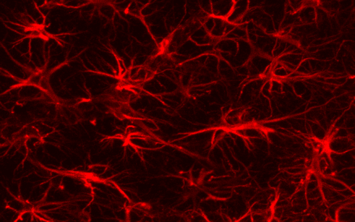
brain astrocytes
Image of brain cell astrocytes

Specifically, APP regulates neurogenesis timing by influencing two finely-tuned genetic mechanisms – one chemical pathway controlling stem cell proliferation and another triggering the production of new neurons.

“In mouse models, neurogenesis is already very fast – too fast for APP deprivation to accelerate it further. We can imagine that the regulatory role of this protein is negligible in mice, while it is essential in the neurodevelopment of our species: to acquire its final form, our brain needs to generate huge quantities of neurons over a very long period, and according to a definite plan. APP-related abnormalities could cause premature neurogenesis and significant cellular stress, the consequences of which would be observable later,” suggests Hassan.

“Moreover, the brain regions in which early signs of Alzheimer’s disease appear also take the longest to mature during childhood and adolescence.”

There could be a direct link between the timing of human neurogenesis and the mechanisms of neurodegeneration. Given that the number of dementia cases worldwide is expected to triple to over 150 million by 2050 due to ageing populations, finding a therapy that targets the root cause is paramount in medical research.

APP appears to play a critical role in this respect, although further studies are necessary to confirm its centrality. Such research could significantly advance our understanding of Alzheimer’s disease and the brain’s development, potentially leading to early identification methods and new treatments.

Medicine, pills on top of brain MRI scans
(© Katsiaryna – stock.adobe.com)

The Paris Brain Institute team’s findings have added a crucial piece to the Alzheimer’s puzzle. This work suggests a paradigm shift in our understanding, positioning the disease’s origins much earlier in life than previously assumed. If their hypothesis holds true, we may be able to detect those at risk at a much earlier stage, offering the potential for targeted preventative measures and therapies.

In a world where Alzheimer’s cases are set to skyrocket, the implications of this research are vast and could significantly alter our approach to tackling this debilitating disease. The promise of a therapy that intervenes at the cause, rather than just managing symptoms, represents a “holy grail” in medical research. With Alzheimer’s’ seeds potentially sown in the womb, this research offers a new understanding that could bring us closer to achieving this goal.

“These disturbances lead to the formation of a brain that functions normally at birth but is particularly vulnerable to certain biological events – such as inflammation, excitotoxicity or somatic mutations – and certain environmental factors such as a poor diet, lack of sleep, infections, etc.,” the study authors conclude.

“Over time, these different stresses could lead to neurodegeneration – a phenomenon specific to the human species and made particularly visible by the increase in life expectancy.”