AI can predict the effectiveness of neoadjuvant chemotherapy in breast cancer patients


Engineers at the University of Waterloo have developed artificial intelligence (AI) technology to predict if women with breast cancer would benefit from chemotherapy prior to surgery.

The new AI algorithm, part of the open-source Cancer-Net initiative led by Dr. Alexander Wong, could help unsuitable candidates avoid the serious side effects of chemotherapy and pave the way for better surgical outcomes for those who are suitable.

“Determining the right treatment for a given breast cancer patient is very difficult right now, and it is crucial to avoid unnecessary side effects from using treatments that are unlikely to have real benefit for that patient,” said Wong, a professor of systems design engineering.

“An AI system that can help predict if a patient is likely to respond well to a given treatment gives doctors the tool needed to prescribe the best personalized treatment for a patient to improve recovery and survival.”

In a project led by Amy Tai, a graduate student with the Vision and Image Processing (VIP) Lab, the AI software was trained with images of breast cancer made with a new magnetic image resonance modality, invented by Wong and his team, called synthetic correlated diffusion imaging (CDI).

With knowledge gleaned from CDI images of old breast cancer cases and information on their outcomes, the AI can predict if pre-operative chemotherapy treatment would benefit new patients based on their CDI images.

Known as neoadjuvant chemotherapy, the pre-surgical treatment can shrink tumors to make surgery possible or easier and reduce the need for major surgery such as mastectomies.

I’m quite optimistic about this technology as deep-learning AI has the potential to see and discover patterns that relate to whether a patient will benefit from a given treatment.”

Dr. Alexander Wong, Director of the VIP Lab and the Canada Research Chair in Artificial Intelligence and Medical Imaging

A paper on the project, Cancer-Net BCa: Breast Cancer Pathologic Complete Response Prediction using Volumetric Deep Radiomic Features from Synthetic Correlated Diffusion Imaging, was recently presented at Med-NeurIPS as part of NeurIPS 2022, a major international conference on AI.

The new AI algorithm and the complete dataset of CDI images of breast cancer have been made publicly available through the Cancer-Net initiative so other researchers can help advance the field.

Critical Advance Announced in Imaging the Living Parkinson’s Brain


ACI-12589 PET images.png

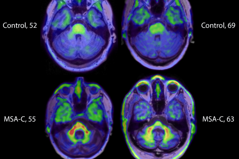
MJFF grantee AC Immune, a Swiss biotechnology company, has shared the first images of alpha-synuclein in the living human brain. Photo credit: AC Immune SA

Today in Barcelona, Spain, at the International Conference on Alzheimer’s & Parkinson’s Diseases (AD/PD), a research team funded by The Michael J. Fox Foundation is announcing important progress in the pursuit of the alpha-synuclein imaging tracer — a top research priority for the Foundation because of its potential to transform drug development.

Misfolding and clumping of the alpha-synuclein protein in brain and body cells are the pathological hallmark of Parkinson’s disease. Scientists believe this toxic dysfunction gives rise to Parkinson’s onset and progression. The ability to visualize alpha-synuclein activity in the living brain would be a game-changer for testing and developing potential new Parkinson’s drugs. (Similar strategies in Alzheimer’s have had a major effect on accelerating drug development for that disease.)

Now AC Immune, a Swiss biotechnology company, has shared the first images of alpha-synuclein in the living human brain. They’ve achieved this major step forward working in individuals living with multiple system atrophy (MSA) — a related parkinsonism that, like Parkinson’s, is characterized by misfolding and clumping of alpha-synuclein in the brain. In its press release, the company said their PET (positron emission tomography) tracer can differentiate people with MSA from control volunteers, people with Parkinson’s disease and people with Lewy body dementia. (Learn more about these conditions related to Parkinson’s.)

If validated, AC Immune’s tracer should be a powerful steppingstone toward the critical tracer tool for alpha-synuclein in Parkinson’s disease patients.

Jamie Eberling, PhD, MJFF Senior Vice President of Research Resources, leads the Foundation’s neuroimaging programs. “Our Foundation has long supported the development of these critical but elusive imaging tools, and we are heartened by this progress toward their widespread application,” she said. “As they have for Alzheimer’s disease, PET tracers would be pivotal in transforming the future of Parkinson’s research and care.”

VIDEO: Dr. Eberling discusses the power of an alpha-synuclein imaging tracer.

The Michael J. Fox Foundation has been working to drive progress in alpha-synuclein imaging for more than a decade. The Ken Griffin Alpha-synuclein Imaging Competition, announced in 2020, awarded AC Immune and two other teams (at Mass General Brigham and Merck) significant funding to accelerate work in this area. Additionally, in 2016, we announced an Alpha-synuclein Imaging Prize to be awarded to the first team ruled by a jury of experts to have achieved successful imaging of alpha-synuclein in the living human brain.

Update (03/21/22): AC Immune presented further data on its ACI-12589 tracer results at the AD/PD conference on Friday, March 18.

“It’s the talk of the meeting,” said Dr. Eberling. “There is work to be done, but we are cautiously optimistic.”

Added Ken Marek, MD, special scientific advisor to MJFF, who was also in Barcelona for the presentation, “This is an important first step. More data is needed, but these findings — made possible by support from The Michael J. Fox Foundation — will help energize the field to deliver additional tracers.” 

The Physics of Imaging


No longer working only behind the scenes, today’s medical physicists are providing clinical guidance to improve patient care.

Physics ADJUSTED

When the radiology department at Cincinnati Children’s Hospital hired a full-time medical physicist about three years ago, the chief of thoracic imaging, Alan S. Brody, MD, didn’t expect the move to significantly impact the department’s operations. Brody, who is also a professor of radiology and pediatrics at the University of Cincinnati College of Medicine, assumed the physicist would work primarily on special projects and calibrate the department’s imaging equipment to meet federal and state regulations. While he supported the hire, Brody wondered whether that was enough work to justify having a medical physicist on staff full time.
But once Keith J. Strauss, MS, FACR, clinical imaging physicist, began working with the department’s radiologists and technologists to optimize dose and enhance image quality beyond the regulatory mandates, Brody soon realized that he had misunderstood what an on-staff medical physicist could do for the department. “I thought a medical physicist did measurements when there was a regulatory need or when someone in the department wanted to better understand radiation dose,” Brody explains. “But our medical physicist has taken an active role in the department and has identified new techniques that have dramatically decreased dose and improved the consistency of our image quality.”
 Brody’s assumptions about the role of medical physicists in radiology are not uncommon. In the past, medical physicists have worked largely behind the scenes — evaluating imaging equipment after hours and issuing reports in much the same way that radiologists issue imaging reports to referring physicians. They traditionally have not collaborated closely with radiologists and other practice members to reduce dose and improve image quality, but that is changing. “As a profession, we are trying to increase our visibility and move from an environment where we are known as the purely technological people to a place where we use our knowledge in a consultative way to help in patient care,” says Robert J. Pizzutiello Jr., MS, FACR, senior vice president of imaging at Landauer Medical Physics, headquartered in Greenwood, Ill., and president and residency program director at Upstate Medical Physics, PC., in Victor, N.Y.

Where Physicists Work

Most medical physicists work in radiation oncology, with only about 25 percent in diagnostic imaging. Imaging medical physicists work either in-house at an institution or as consultants who serve many institutions at once. In-house medical physicists typically work for large hospitals or practices, while consultants tend to serve smaller institutions. “It takes a pretty sizable institution to make it cost-effective to have a medical physicist on staff full time,” says Richard A. Geise, PhD, FACR, a medical physicist who works part-time at Abbott Northwestern Hospital in Minneapolis and as a consultant for several hospitals, clinics, and radiology groups in the Twin Cities area. “So as health care organizations become larger and have more of an umbrella parent organization over multiple hospitals, these facilities might hire a physicist or multiple physicists to support all of the hospitals within their systems.”
Some institutions may wonder whether it’s best to hire an in-house or a consulting medical physicist. Pizzutiello, who founded the consulting group Upstate Medical Physics, notes that each approach has its advantages. “In-hospital physicists have a more intimate knowledge of all the people, processes, and equipment at a single location,” he says. “Consultants, on the other hand, know how different processes and equipment have worked across facilities.” Pizzutiello adds that both in-house and consulting medical physicists can participate in CT protocol optimization. “Our consulting group has been helping clients optimize CT protocols since 2010, and I know many others who have expanded into this more consultative role,” he says. From a radiologist’s perspective, Brody says that either a full-time or consulting medical physicist can make an important difference as long as he or she is available for both regulatory needs and to answer clinical questions.

“We have a high degree of respects for our physicists because this is very detailed physics up to the nuclear reactor level.” — Geoffrey R. Bodeau, MD


Whether in-house or consulting, the most basic job of medical physicists is to ensure that institutions meet the scores of imaging regulations that state and federal governments impose. These include the United States Nuclear Regulatory Commission’s ALARA dose requirements and the Joint Commission’s new mandate requiring hospitals to have medical physicists evaluate the performance of their CT imaging equipment annually (which follows the ACR’s longstanding accreditation requirements). Some medical physicists also engage in clinical care, which can significantly improve image quality and patient safety. “As an in-house medical physicist, I make sure the compliance issues are met, but the majority of my time is spent helping the technologists and the radiologists leverage the strengths of their imaging equipment to get better images and properly manage dose levels,” Strauss says.

What Physicists Do

No matter the setting, medical physicists work with radiologists and technologists to achieve three primary goals: ensure image quality, develop radiation safety procedures, and optimize dose. “I apply physics principles of imaging to come up with a plan to improve the process the department uses to create the images,” Strauss explains. “Depending on the situation, the idea might be that we’re improving image quality and maintaining dose or maybe we’re trying to maintain image quality and reduce dose — or maybe we’re trying to do both.”


While medical physicists sometimes have to assess imaging equipment during off hours to keep from disrupting patient exams, they are becoming more visible by advising radiologists and technologists on quality-improvement techniques. Once they understand how the department is using its imaging equipment, medical physicists have the expertise to develop protocol improvements that others in the department may not consider. “We have a high degree of respect for our physicists because this is very detailed physics up to the nuclear reactor level,” says Geoffrey R. Bodeau, MD, medical director of nuclear medicine at Abbott Northwestern Hospital. “As doctors, we appreciate the fact that they really know this stuff inside and out, and we realize that the work of the hospital couldn’t go on without them.”

At Cincinnati Children’s Hospital, for instance, Strauss developed a completely new way for determining X-ray dose after examining some of the department’s plain films. Brody explains that the department was using age and weight to determine dose. But when Strauss analyzed the techniques the technologists were using, he noticed that the same techniques were used for tall, thin patients as short, stocky patients, which leads to inconsistent dose levels for identical exams for patients of the same age. To more accurately and consistently determine dose, Strauss recommended using body thickness rather than age and weight. “As a result, we’re producing higher quality images and decreasing dose in some cases, which is fantastic,” Brody says.


Physicists’ Additional Roles

Medical physicists also play several other important roles inside of health care institutions. For instance, medical physicists may advise institutions on equipment purchasing decisions. They may also assist with facility design to determine how much lead shielding is necessary to prevent radiation from escaping through the walls of an exam room. “There are all of these calculations that radiologists wouldn’t necessarily think of, but medical physicists have the expertise to help make sure our facilities are safe for patients, radiologists, technologists, and the general public,” Bodeau says. Medical physicists are also trained to assist in emergency situations such as radioactive material spills and terrorist attacks involving nuclear reactor byproducts.

At the ACR, medical physicists are involved in several initiatives, including ACR Select™ (the electronic version of the ACR Appropriateness Criteria®) and the ACR Dose Index Registry™ (a data registry that allows institutions to compare their CT dose levels to regional and national values). They also assist with the ACR’s accreditation program and the technical standards and practice parameters related to radiation issues and safety. “We’re able to look at these programs from the medical physics perspective and contribute to their development and ongoing operation,” Pizzutiello says. “For example, in all of the accreditation programs not only does a radiologist assess the clinical images, but a medical physicist also evaluates the phantom images and physics reports to ensure they meet the accreditation standards.”


Still, medical physicists’ most important role is working directly with radiologists and technologists to improve patient care. Strauss says that radiologists can help medical physicists fulfill this role by encouraging their institutions to hire medical physicists to do more than simply address regulatory requirements. “There are many institutions that want to take care of the compliance issues but aren’t necessarily willing to spend the time, money, and effort needed to optimize things as much as they could,” he says. “Radiologists should advocate for hiring medical physicists as part of their institutions’ overall efforts to improve patient care.”
BRAIN IMAGING SHOWS BRAIN DIFFERENCES IN RISK-TAKING TEENS.


140815102326-large

According to the CDC, unintentional injuries are the leading cause of death for adolescents. Compared to the two leading causes of death for all Americans, heart disease and cancer, a pattern of questionable decision-making in dire situations comes to light in teen mortality. Newresearch from the Center for BrainHealth at The University of Texas at Dallas investigating brain differences associated with risk-taking teens found that connections between certain brain regions are amplified in teens more prone to risk.

“Our brains have an emotional-regulation network that exists to govern emotions and influence decision-making,” explained the study’s lead author, Sam Dewitt. “Antisocial or risk-seeking behavior may be associated with an imbalance in this network.”

The study, published June 30 in Psychiatry Research: Neuroimaging, looked at 36 adolescents ages 12-17; eighteen risk-taking teens were age- and sex-matched to a group of 18 non-risk-taking teens. Participants were screened for risk-taking behaviors, such as drug and alcohol use, sexual promiscuity, and physical violence and underwent functional MRI (fMRI) scans to examine communication between brain regions associated with the emotional-regulation network. Interestingly, the risk-taking group showed significantly lower income compared to the non-risk taking group.

Dewitt paper“Most fMRI scans used to be done in conjunction with a particular visual task. In the past several years, however, it has been shown that performing an fMRI scan of the brain during a ‘mind-wandering’ state is just as valuable,”said Sina Aslan, Ph.D., President of Advance MRI and Adjunct Assistant Professor at the Center for BrainHealth at The University of Texas at Dallas.“In this case, brain regions associated with emotion and reward centers show increased connection even when they are not explicitly engaged.”

The study, conducted by Francesca Filbey, Ph.D., Director of Cognitive Neuroscience Research of Addictive Behaviors at the Center for BrainHealth and her colleagues, shows that risk-taking teens exhibit hyperconnectivity between the amygdala, a center responsible for emotional reactivity, and specific areas of the prefrontal cortex associated with emotion regulation and critical thinking skills. The researchers also found increased activity between areas of the prefrontal cortex and the nucleus accumbens, a center for reward sensitivity that is often implicated in addiction research.

“Our findings are crucial in that they help identify potential brain biomarkers that, when taken into context with behavioral differences, may help identify which adolescents are at risk for dangerous and pathological behaviors in the future,” Dewitt explained.

He also points out that even though the risk-taking group did partake in risky behavior, none met clinical criteria for behavioral or substance use disorders.

By identifying these factors early on, the research team hopes to have a better chance of providing effective cognitive strategies to help risk-seeking adolescents regulate their emotions and avoid risk-taking behavior and substance abuse.

Fast presurgical functional mapping using task-related intracranial high gamma activity.


Abstract

OBJECT

Electrocorticography (ECoG) is a powerful tool for presurgical functional mapping. Power increase in the high gamma band has been observed from ECoG electrodes on the surface of the sensory motor cortex during the execution of body movements. In this study the authors aim to validate the clinical usage of high gamma activity in presurgical mapping by comparing ECoG mapping with traditional direct electrical cortical stimulation (ECS) and functional MRI (fMRI) mapping.

METHODS

Seventeen patients with epilepsy participated in an ECoG motor mapping experiment. The patients executed a 5-minute hand/tongue movement task while the ECoG signal was recorded. All 17 patients also underwent extraoperative ECS mapping to localize the motor cortex. Eight patients also participated in a presurgical fMRI study. The high gamma activity on ECoG was modeled using the general linear model (GLM), and the regions showing significant gamma power increase during the task condition compared with the rest condition were localized. The maps derived from GLM-based ECoG mapping, ECS, and fMRI were then compared.

RESULTS

High gamma activity in the motor cortex can be reliably modulated by motor tasks. Localization of the motor regions achieved with GLM-based ECoG mapping was consistent with the localization determined by ECS. The maps also appeared to be highly localized compared with the fMRI activations. Using the ECS findings as the reference, GLM-based ECoG mapping showed a significantly higher sensitivity than fMRI (66.7% for ECoG, 52.6% for fMRI, p < 0.05), while the specificity was high for both techniques (> 97%). If the current-spreading effect in ECS is accounted for, ECoG mapping may produce maps almost identical to those produced by ECS mapping (100% sensitivity and 99.5% specificity).

CONCLUSIONS

General linear model–based ECoG mapping showed a superior performance compared to traditional ECS and fMRI mapping in terms of efficiency and accuracy. Using this method, motor functions can be reliably mapped in less than 5 minutes.

Source: JNS


n cZ�iiP���nd more medication use after GKS (p < 0.05). Conversely, increase in numbness intensity after GKS was associated with a decrease in pain intensity and pain length (p < 0.05).


CONCLUSIONS

Gamma Knife surgery using a maximum dose of 90 Gy to the trigeminal nerve provides satisfactory long-term pain control, reduces the use of medication, and improves quality of life. Physicians must be aware that higher doses may be associated with an increase in bothersome sensory complications. The benefits and risks of higher dose selection must be carefully discussed with patients, since facial numbness, even if bothersome, may be an acceptable trade-off for patients with severe pain.

Source: JNS


Targeted molecular imaging in oncology.


Improvement of scintigraphic tumor imaging is extensively determined by the development of more tumor specific radiopharmaceuticals. Thus, to improve the differential diagnosis, prognosis, planning and monitoring of cancer treatment, several functional pharmaceuticals have been developed. Application of molecular targets for cancer imaging, therapy and prevention using generator-produced isotopes is the major focus of ongoing research projects. Radionuclide imaging modalities (positron emission tomography, PET; single photon emission computed tomography,

SPECT) are diagnostic cross-sectional imaging techniques that map the location and concentration of radionuclide-labeled radiotracers.

99mTc- and 68Galabeled agents using ethylenedicysteine (EC) as a chelator were synthesized and their potential uses to assess tumor targets were evaluated.

99mTc (t1/2 = 6 hr, 140 keV) is used for SPECT and 68Ga (t1/2 = 68 min, 511 keV) for PET. Molecular targets labeled with Tc-99m and Ga-68 can be utilized for prediction of therapeutic response, monitoring tumor response to treatment and differential diagnosis. Molecular targets for oncological research in (1) cell apoptosis, (2) gene and nucleic acid-based approach, (3) angiogenesis (4) tumor hypoxia,and (5) metabolic imaging are discussed. Numerous imaging ligands in these categories have been developed and evaluated in animals and humans. Molecular targets were imaged and their potential to redirect optimal cancer diagnosis and therapeutics were demonstrated.

Source: http://www.jsnm.org


Rupture of bicornuate uterus.


Summary

A primigravida aged 20 years was referred to Vydehi Institute of Medical Sciences with diagnosis of 30 weeks of period of gestation with eclampsia and failure to respond to induction with misoprostol and she was on Pritchard regimen for the treatment of eclampsia and there was no response to induction of labour and emergency ultrasound was taken and it showed an extrauterine gestation of 30 weeks gestation with fetal demise and free fluid in peritoneum. A tentative diagnosis of secondary abdominal pregnancy with eclampsia was made and she was taken for emergency laprotomy. Intra operative findings showed haemoperitoneum, fetus with placenta and membranes in the peritoneal cavity, there was bicornuate uterus and right horn was ruptured from the fundus to about 8 cm down in the posterior aspect and ruptured part was sutured in two layers. After securing perfect haemostasis, abdomen was closed. This paper illustrates a case report of uterine anomaly with 30 weeks period of gestation and eclampsia and rupture following induction with prostaglandins.

Background

Pregnancy with uterine anomolies is rare in clinical practice and only few cases are reported in literature. Most of the obstetricians do not keep it in mind during induction of labour. In developing countries like India majority of the pregnant women are not booked for early antenatal care due to financial constraints. Most of the uterine anomalies go unnoticed because they are often symptomless. Majority are first recognised during pregnancy and judicious use of prostaglandins should be advocated in unbooked cases.

This paper illustrates a rare case of rupture of bicornuate uterus following induction with prostaglandins and gives us a message that antenatal diagnosis of uterine anomalies is very important to prevent rupture in later pregnancy and uterine anomalies should be kept in mind when there is failure of induction of labour.

Case presentation

Primigravida aged 20 years presented to a private hospital with two episodes of convulsions and she was treated there with magnesium sulphate with Pritchard regimen1 and labour was induced with misoprostol. Four doses of misoprostol 50 microgram were inserted pervaginally 4 hourly, but there was no progress of labour and emergency ultrasound was taken and it showed an extra uterine gestation of 30 weeks gestation with fetal demise and free fluid in peritoneum. The patient was referred to Vydehi Institute of Medical Sciences for further management. Her married life was 2 years and she has not taken any antenatal visits. Her personal and family history was not significant.

At presentation, the patient was pale, drowsy, irritable, with grade 2 pedal oedema, blood pressure was 110/70 mm hg and thready pulse was 104 beats per min. Her respiratory and cardiovascular examinations were with in normal limits. Central nervous system examination patient was drowsy, irritable and deep tendon reflexes were present. The abdomen was very tender and irregularly shaped with easily palpable fetal parts and the fetal heart sounds could not be heard. Vaginal assessment revealed scanty vaginal bleeding and cervical os was 2 cm dilated and soft in consistency. The station of the presenting part could not be made out and there was tenderness in both the fornices.

Investigations

Her haemoglobin was 8.4 g%, blood group was O positive and urine routine proteinurea was present. Her liver function and renal function tests were normal and platelet count was two lakhs. Emergency ultrasound showed an extra uterine gestation of 30 weeks with fetal demise and free fluid in peritoneal cavity.

Differential diagnosis

Primigravida at 30 weeks period of gestation with abdominal pregnancy with fetal demise with eclampsia was presented, this case may be rarely confused for acute abdominal conditions, but presence of gravid uterus helps in diagnosis.

Treatment

A tentative diagnosis of primigravida at 30 weeks period of gestation with abdominal pregnancy with fetal demise with eclampsia was made and patient was taken for emergency laprotomy and magnesium sulphate was continued according to Pritchard’s regimen for the treatment of eclampsia.

Intraoperatively there was haemoperitoneum of around 500 cc, dead fetus lying with intact membranes, baby extracted by breech and the placenta came along with the cord during breech extraction. There was clot of around 50 g in the peritoneal cavity and after removing the blood clot, a bicornuate uterus was seen with left horn of normal size and intact and the right horn showed rupture in the posterior aspect extending from fundus to about 8 cm below (figures 1 and 2). The ruptured horn is sutured in two layers, perfect haemostasis was achieved. There was a thick fibrous band extending between the two horns (figure 3). Peritoneal wash was given. Abdomen was closed in layers.

Post operatively two units of packed cells were transfused in the intra and immediate post operative period. Post operative recovery was uneventful. Magnesium sulphate was discontinued after 24 h and the patient was discharged on the seventh post operative day. Contraceptive advice was given and importance to continue for minimum of 2 years was explained to the patient.

Outcome and follow-up

Patient was followed up for 6 months regularly and postoperative period was uneventful. She resumed her menstrual cycles after 6 months and her menstrual cycles were regular and scan was done after 1 year after surgery which showed bicornuate uterus.

Discussion

Incidence of uterine anomalies is 0.1%–3% in general population, 3.5% in infertile women and about 13% in the women with recurrent pregnancy loss. Among that bicornuate uterus constitutes 1.2%.2 Bicornuate uterus belongs to the class IV according to The American Fertility Society Classification of Mullerian anomalies (1988).3

The uterus is formed during embryogenesis by the fusion of the two paramesonephric ducts (also called Mullerian ducts). This process usually fuses the two Mullerian ducts into a single uterine body. Lack of fusion of these Mullerian ducts can lead to various types of malformations of the female genital tract.4

Uterus bicornis unicollis (bicornuate uterus), which is a common type seen represents an uterine malformation where the uterus is present as a paired organ resulting from the failure of the embryogenetic fusion of part of the Mullerian ducts. As a result there is a double uterus with a single cervix and vagina. Each uterus has a single horn linked to the ipsilateral fallopian tube that faces its ovary. The bicornuate uterus often have an unusually thick strong round ligaments and a thick vesicorectal fold running between them. Implication of uterine malformation relates inversely to the degree of fusion defect and may be associated with renal tract anomalies.5

Incidence of pregnancy in rudimentary horn is 1/40 000 pregnancies. Rupture in such cases occurs because of inability of malformed uterus to expand as a normal uterus.6 The walls of the anomalous uteri tend to become abnormally thin as pregnancies advances. Thickness can be inconsistent over different aspects of the myometrium, and the placenta does not adhere properly.7 The rupture in rudimentary horn is likely to occur in late first trimester or even in second trimester. Rarely pregnancy can go on till late second trimester before rupturing. The haemorrhage occurring because of rupture is massive and can be life-threatening, unless diagnosed and treated promptly. Ravasia et al reported an 8% incidence of uterine rupture in women with congenitally malformed uteri compared with 0.61% in those with normal uteri (p=.013) who were attempting vaginal birth after caesarean.8 Induction with prostaglandin E1 is an important risk factor associated with rupture. Pregnancies implanted in the rudimentary horn of the uterus pose special risk for those women undergoing induction of labour, with a uterine rupture rate of up to 81%, 80% of ruptures occurred before the third trimester, with 67% occurring during the second trimester. The timing of rupture can occur from fifth week onwards. Rupture is usually associated with catastrophe and bleeding is torrential and 90% of maternal deaths have been reported within 10–15 months after rupture.

Rudimentary horn pregnancy should be suspected whenever difficulty is experienced while terminating a pregnancy and every attempt should be made to rule out the same. Uterine abnormalities, though rare can be encountered in pregnancy. There is need to build capacity for making an antenatal diagnosis in order to ensure appropriate management.9

Prostaglandin E1 is a cheap drug and it is stored in room temperature, so it is often used for the induction of labour. There are no specific guidelines for induction of labour with prostaglandin E1, but American Congress of Obstetricians and Gynaecologists (ACOG) recommends use of prostaglandin E1 at lower dose (25 mcg every 3 to 6 h) for the induction of labour as level A recommendation. ACOG said that the data on the safety of high-dose misoprostol (50 mcg every 6 h) were ‘limited or inconsistent’, making its recommendation on high-dose misoprostol an evidence level ‘B’ recommendation.

Learning points

  • ▶ Bicornuate uterus belongs to the class IV according to The American Society for Reproductive Medicine Classification of Mullerian anomalies (1988) and results from failure of fusion of Mullarian ducts in the upper part.
  • ▶ Rupture in such cases occurs because of inability of malformed uterus to expand as a normal uterus. The walls of the anomalous uteri tend to become abnormally thin as pregnancies advances. Thickness can be inconsistent over different aspects of the myometrium, and the placenta does not adhere properly.
  • ▶ Rupture of anomalous uterus pregnancy should be suspected whenever difficulty is experienced while terminating a pregnancy and every attempt should be made to rule out the same particularly in unbooked cases.
  • Competing interests None.
  • Patient consent Obtained.

Footnotes

References

    1. Tukur J

. The use of magnesium sulphate for the treatment of severe preeclampsia and eclampsia. Ann Afr Med 2009;:7680.

[CrossRef][Medline]

    1. Byrne J,
    2. Nussbaum-Blask A,
    3. Taylor WS,
    4. et al

. Prevalence of Müllerian duct anomalies detected at ultrasound. Am J Med Genet 2000;:912.

[CrossRef][Medline][Web of Science]

    1. Elyan A,
    2. Saeed M

. Mullarian duct anamolies: clinical aoncept. ASJOG 2004;:1120.

Search Google Scholar

    1. Nwosu OB,
    2. Ugboaja OJ,
    3. Obi-Nwosu A

. Spontaneous rupture of gravid horn of bicornuate uterus at term – A case report. Niger J Med 2010;:1845.

Search Google Scholar

    1. Bhattachary TK,
    2. Sengupta P

. Rudimentary horn pregnancy. MJAFI 2005;:3778.

Search Google Scholar

    1. Kore S,
    2. Pandole A,
    3. Akolekar R,
    4. et al

. Rupture of left horn of bicornuate uterus at twenty weeks of gestation. J Postgrad Med 2000;:3940.

[Medline]

    1. Ashelby L,
    2. Toll G,
    3. Patel RR,
    4. et al

. Live birth after rupture of a non-communicatinghorn of a bicornuate uterus. BJOG 2005;:15767.

[CrossRef][Medline]

    1. Ravasia DJ,
    2. Brain PH,
    3. Pollard JK

. Incidence of uterine rupture among women with müllerian duct anomalies who attempt vaginal birth after cesarean delivery. Am J Obstet Gynecol 1999;:87781.

[CrossRef][Medline]

    1. Dasari P

. Case report: pre-rupture ultrasound diagnosis of rudimentary horn pregnancy. Biomed Res 2007;:1857.

Search Google Scholar

Source: BMJ

Ultrasound-assisted convection-enhanced delivery to the brain in vivo with a novel transducer cannula assembly.


In convection-enhanced delivery (CED), drugs are infused locally into tissue through a cannula inserted into the brain parenchyma to enhance drug penetration over diffusion strategies. The purpose of this study was to demonstrate the feasibility of ultrasound-assisted CED (UCED) in the rodent brain in vivo using a novel, low-profile transducer cannula assembly (TCA) and portable, pocket-sized ultrasound system.

Methods

Forty Sprague-Dawley rats (350–450 g) were divided into 2 equal groups (Groups 1 and 2). Each group was divided again into 4 subgroups (n = 5 in each). The caudate of each rodent brain was infused with 0.25 wt% Evans blue dye (EBD) in phosphate-buffered saline at 2 different infusion rates of 0.25 μl/minute (Group 1), and 0.5 μl/minute (Group 2). The infusion rates were increased slowly over 10 minutes from 0.05 to 0.25 μl/minute (Group 1) and from 0.1 to 0.5 μl/minute (Group 2). The final flow rate was maintained for 20 minutes. Rodents in the 4 control subgroups were infused using the TCA without ultrasound and without and with microbubbles added to the infusate (CED and CED + MB, respectively). Rodents in the 4 UCED subgroups were infused without and with microbubbles added to the infusate (UCED and UCED + MB) using the TCA with continuous-wave 1.34-MHz low-intensity ultrasound at a total acoustic power of 0.11 ± 0.005 W and peak spatial intensity at the cannula tip of 49.7 mW/cm2. An additional 4 Sprague-Dawley rats (350–450 g) received UCED at 4 different and higher ultrasound intensities at the cannula tip ranging from 62.0 to 155.0 mW/cm2 for 30 minutes. The 3D infusion distribution was reconstructed using MATLAB analysis. Tissue damage and morphological changes to the brain were assessed using H & E.

Results

The application of ultrasound during infusion (UCED and UCED + MB) improved the volumetric distribution of EBD in the brain by a factor of 2.24 to 3.25 when there were no microbubbles in the infusate and by a factor of 1.16 to 1.70 when microbubbles were added to the infusate (p < 0.001). On gross and histological examination, no damage to the brain tissue was found for any acoustic exposure applied to the brain.

Conclusions

The TCA and ultrasound device show promise to improve the distribution of infused compounds during CED. The results suggest further studies are required to optimize infusion and acoustic parameters for small compounds and for larger molecular weight compounds that are representative of promising antitumor agents. In addition, safe levels of ultrasound exposure in chronic experiments must be determined for practical clinical evaluation of UCED. Extension of these experiments to larger animal models is warranted to demonstrate efficacy of this technique.

Source: Journal of neurosurgery.