Brain Scans Reveal How Drinking Turns People Into Raging Assholes


We all have that friend who gets a little out of hand when they start drinking alcohol. Maybe he gets loud, or maybe she starts fights with strangers for looking at her funny. Alcohol seems to induce aggression, changing the brain in a way that makes a drunk person more likely to see minor social cues as threats, but how it does so has always been a bit of biological mystery.

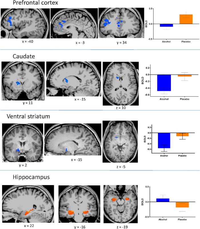
Scientists found that alcohol-induced aggression was correlated to decreased activity in the prefrontal cortex.

But in a paper published in the journal Cognitive, Affective, & Behavioral Neuroscience, a team of researchers led by Thomas Denson, Ph.D., of the University of New South Wales School of Psychology use brain scans to show that alcohol changes activity in certain key parts of the brain related to aggression and emotion.

Using functional magnetic resonance imaging (fMRI), a technique that tracks changes in blood flow in the brain, the team looked at the brains of 50 young men after they consumed either two alcoholic drinks or two non-alcoholic placebo drinks. These volunteers engaged in a task that gauged their level of aggression in the face of provocation, which revealed the parts of the brain that become more active in such situations.

These scans show how alcohol-induced aggression was related to decreased activity in the prefrontal cortex, caudate, and ventral striatum, but increased activity in the hippocampus.

The researchers found that alcohol-induced aggression was correlated with decreased activity in prefrontal cortex, caudate, and ventral striatum, but increased activity in the hippocampus. These parts of the brain all control key factors in aggression: The prefrontal cortex is associated with thoughtful action and social behavior, the caudate is linked to the brain’s reward system and inhibitory control, and the ventral striatum is a part of the reward system that makes you feel good when you do something good. The hippocampus, meanwhile, is associated with emotion and memory.

These results support previous hypotheses that prefrontal cortex dysfunction is associated with alcohol-induced aggression. Taking all these brain areas together, the researchers say their findings suggest that intoxicated people have trouble processing information through their working memory. In short, they suspect that alcohol focuses a person’s attention on the cues that could instigate aggression while taking attention away from their knowledge of social norms that say violence is not acceptable.

Along similar lines, they also suspect that alcohol could make relatively minor cues seem aggressive or violent, which can cause a drunk person to overreact to a minor incident, like someone looking at them funny or accidentally bumping into them at the bar. Denson’s previous research on the angry brain found a lot of overlap in the way the prefrontal cortex behaves when someone is drunk and angry versus when they’re simply ruminating on their anger while sober.

This research proposes some possible brain biomarkers for alcohol-induced aggression, which is a significant public health issue. According to the Centers for Disease Control and Prevention, in the United States, alcohol-related violence — including homicide, child abuse, suicide, and firearm injuries — was responsible for more than 16,000 deaths between 2006 and 2010, the most recent years the agency reported figures.

While the new study doesn’t propose a solution per se, it does build on our body of knowledge around an age-old question: Why do some people become assholes when they get drunk?

Abstract: Alcohol intoxication is implicated in approximately half of all violent crimes. Over the past several decades, numerous theories have been proposed to account for the influence of alcohol on aggression. Nearly all of these theories imply that altered functioning in the prefrontal cortex is a proximal cause. In the present functional magnetic resonance imaging (fMRI) experiment, 50 healthy young men consumed either a low dose of alcohol or a placebo and completed an aggression paradigm against provocative and nonprovocative opponents. Provocation did not affect neural responses. However, relative to sober participants, during acts of aggression, intoxicated participants showed decreased activity in the prefrontal cortex, caudate, and ventral striatum, but heightened activation in the hippocampus. Among intoxicated participants, but not among sober participants, aggressive behavior was positively correlated with activation in the medial and dorsolateral prefrontal cortex. These results support theories that posit a role for prefrontal cortical dysfunction as an important factor in intoxicated aggression.

Completely Blind People Still Able To React To Light.


Photo credit: gun4hire

 1484  77  2 reddit0 googleplus1

Humans need light for a variety of reasons. Beyond allowing us to perceive our environment with sight, light also activates activity in the brain. A recent study has unexpectedly shown that even individuals who are completely blind are influenced by the presence of light. The presence or absence of light controls many bodily functions, including heart rate, attentiveness, mood, and reflexes. The study will be published in an upcoming edition of Journal of Cognitive Neuroscience. The work is a collaboration between a research team at the University of Montreal and the Brigham and Women’s Hospital in Boston.

The experiment was performed by exposing people who are completely blind to a blue light. The light was turned on and off and the participants were asked whether the light was on or off. The participants were shown to have a non-conscious response to the light, despite not being able to see it. There were more positive identifications made than could be explained by chance alone, though the awareness was non-conscious. This light perception comes from ganglion cells in the retina, which are different from the rod and cone cells that process light for sight.

Next, researchers tested if attentiveness was affected by the presence of light. For this activity, participants had to match sounds with lights on or off. Even though the participants could not visualize the light, they showed an increased attentiveness when light was shining into their eyes.

Finally, the test participants completed a brain scan with functional MRI (fMRI) to measure alertness, memory, and cognition recognition while performing tasks of matching sounds. Across the board, the tasks were completed more efficiently when light was present.

Because of these results, the researchers are speculating that light perception is part of the default mode network. This is the name for the brain activity that occurs non-consciously in the background, while other tasks take priority. They speculate that the ability to perceive light even without actively converting it into images is done to continually pay attention to and monitor the environment. If this is correct, it might help explain why cognitive performance is improved in the presence of light.

– See more at: http://www.iflscience.com/brain/completely-blind-people-still-able-react-light#sthash.KvGYh5Ew.dpuf

The Lasting Impacts of Poverty on the Brain.


Poverty shapes people in some hard-wired ways that we’re only now beginning to understand. Back in August, we wrote about some provocative new research that found that poverty imposes a kind of tax on the brain. It sucks up so much mental bandwidth – capacity spent wrestling with financial trade-offs, scarce resources, the gap between bills and income – that the poor have fewer cognitive resources left over to succeed at parenting, education, or work. Experiencing poverty is like knocking 13 points off your IQ as you try to navigate everything else. That’s like living, perpetually, on a missed night of sleep.

That finding offered a glimpse of what poverty does to a person during a moment in time. Picture a mother trying to accomplish a single task (making dinner) while preoccupied with another (paying the rent on time). But scientists also suspect that poverty’s disadvantages – and these moments – accumulate across time. Live in poverty for years, or even generations, and its effects grow more insidious. Live in poverty as a child, and it affects you as an adult, too.

Some new research about the long-term arc of poverty, particularly on the brain, was recently published in theProceedings of the National Academy of Sciences, and these findings offer a useful complement to the earlier study. In this new paper, researchers from the University of Illinois at Chicago, Cornell, the University of Michigan, and the University of Denver followed children from the age of 9 through their early 20s.

The Lasting Impacts of Poverty on the Brain

Those who grew up poor later had impaired brain function as adults—a disadvantage researchers could literally see in the activity of the amygdala and prefrontal cortex on an fMRI scan. Children who were poor at age 9 had greater activity in the amygdala and less activity in the prefrontal cortex at age 24 during an experiment when they were asked to manage their emotions while looking at a series of negative photos. This is significant because the two regions of the brain play a critical role in how we detect threats and manage stress and emotions.

Poor children, in effect, had more problems regulating their emotions as adults (regardless of what their income status was at 24). These same patterns of “dysregulation” in the brain have been observed in people with depression, anxiety disorders, aggression and post-traumatic stress disorders.

Over the course of the longitudinal study – which included 49 rural, white children of varying incomes – these same poor children were also exposed to chronic sources of stress like violence and family turmoil, or crowded and low-quality housing. Those kinds of stressors, the researchers theorize, may help explain the link between income status in childhood and how well the brain functions later on. That theory, they write, is consistent with the idea that “early experiences of poverty become embedded within the organism, setting individuals on lifelong trajectories.”

To add some of these findings together: Poverty taxes the ability of parents to do all kinds of things, including care for their children. And the developmental challenges that children face in a home full of stressed adults may well influence the adults that they, themselves, become.

Scans pinpoint moment anaesthetic puts brain under.


Anaesthetics usually knock you out like a light. But by slowing the process down so that it takes 45 minutes to become totally unresponsive, researchers have discovered a new signature for unconsciousness. The discovery could lead to more personalised methods for administering anaesthetics and cut the risks associated with being given too high or too low a dose. It also sheds new light on what happens to our brain when we go under the knife.

How much do you need? <i>(Image: Wicki58/Getty Images)</i>

Hundreds of thousands of people are anaesthetised every day, yet researchers still don’t fully understand what’s going on in the anaesthetised brain. Nor is there a direct way of measuring when someone is truly unresponsive. Instead, anaesthetists rely on indirect measures such as heart and breathing rate, and monitoring reflexes.

To investigate further, Irene Tracey and her colleagues at Oxford University slowed the anaesthesia process down. Instead of injecting the anaesthetic propofol in one go, which triggers unconsciousness in seconds, the drug was administered gradually so that it took 45 minutes for 16 volunteers to become fully anaesthetised. Their brain activity was monitored throughout using electroencephalography (EEG). The study was then repeated on 12 of these volunteers using functional magnetic resonance imaging (fMRI).

EEG recordings revealed that before the volunteers became completely unresponsive to external stimuli they progressed through a sleep-like state characterised by slow-wave oscillations – a hallmark of normal sleep, in which neurons cycle between activity and inactivity. As the dose of anaesthetic built up, more and more neurons fell into this pattern, until a plateau was reached when no more neurons were recruited, regardless of the dose administered.

Interestingly, the time it took to reach this plateau varied from individual to individual, and seemed to be determined by the number of neurons people possessed – something that decreases as we age.

Meanwhile, fMRI revealed what was happening in different regions of the brain as they lost consciousness. One theory is that an anaesthetic switches off one of the brain’s central relay hubs, the thalamus, meaning it no longer speaks to the cerebral cortex. However, Tracey’s team found that conversations between the cortex and the thalamus continued, even during deep anaesthesia – but there was no propagation of messages to wider regions of the brain.

“The thalamus is actually in a lot of dialogue with the cortex, but because it’s in this lockdown, sensory events that normally come into the cortex and would be routed out to the logical parts of the brain so that you could perceive ‘ouch that hurts’, is not happening,” says Tracey. “To have true perception you’ve got to have all the right bits active and their activity coordinated.”

Importantly, the point at which messages stopped being routed out was the same point at which the slow-wave oscillations reached a plateau. The hope is that this “saturation point” could be used as a measure of when to stop administering drugs, to reduce the risk of side effects such as headaches, dizziness and memory loss.

The next step is to monitor anaesthetised patients while they are undergoing surgery.

Journal reference: Science Translational Medicine, doi.org/ph4

Brain decoding: Reading minds.


By scanning blobs of brain activity, scientists may be able to decode people’s thoughts, their dreams and even their intentions.

Jack Gallant perches on the edge of a swivel chair in his lab at the University of California, Berkeley, fixated on the screen of a computer that is trying to decode someone’s thoughts.

On the left-hand side of the screen is a reel of film clips that Gallant showed to a study participant during a brain scan. And on the right side of the screen, the computer program uses only the details of that scan to guess what the participant was watching at the time.

Anne Hathaway’s face appears in a clip from the film Bride Wars, engaged in heated conversation with Kate Hudson. The algorithm confidently labels them with the words ‘woman’ and ‘talk’, in large type. Another clip appears — an underwater scene from a wildlife documentary. The program struggles, and eventually offers ‘whale’ and ‘swim’ in a small, tentative font.

“This is a manatee, but it doesn’t know what that is,” says Gallant, talking about the program as one might a recalcitrant student. They had trained the program, he explains, by showing it patterns of brain activity elicited by a range of images and film clips. His program had encountered large aquatic mammals before, but never a manatee.

Groups around the world are using techniques like these to try to decode brain scans and decipher what people are seeing, hearing and feeling, as well as what they remember or even dream about.

Media reports have suggested that such techniques bring mind-reading “from the realms of fantasy to fact”, and “could influence the way we do just about everything”. The Economist in London even cautioned its readers to “be afraid”, and speculated on how long it will be until scientists promise telepathy through brain scans.

Although companies are starting to pursue brain decoding for a few applications, such as market research and lie detection, scientists are far more interested in using this process to learn about the brain itself. Gallant’s group and others are trying to find out what underlies those different brain patterns and want to work out the codes and algorithms the brain uses to make sense of the world around it. They hope that these techniques can tell them about the basic principles governing brain organization and how it encodes memories, behaviour and emotion (see ‘Decoding for dummies’).

Applying their techniques beyond the encoding of pictures and movies will require a vast leap in complexity. “I don’t do vision because it’s the most interesting part of the brain,” says Gallant. “I do it because it’s the easiest part of the brain. It’s the part of the brain I have a hope of solving before I’m dead.” But in theory, he says, “you can do basically anything with this”.

Beyond blobology

Brain decoding took off about a decade ago1, when neuroscientists realized that there was a lot of untapped information in the brain scans they were producing using functional magnetic resonance imaging (fMRI). That technique measures brain activity by identifying areas that are being fed oxygenated blood, which light up as coloured blobs in the scans. To analyse activity patterns, the brain is segmented into little boxes called voxels — the three-dimensional equivalent of pixels — and researchers typically look to see which voxels respond most strongly to a stimulus, such as seeing a face. By discarding data from the voxels that respond weakly, they conclude which areas are processing faces.

Decoding techniques interrogate more of the information in the brain scan. Rather than asking which brain regions respond most strongly to faces, they use both strong and weak responses to identify more subtle patterns of activity. Early studies of this sort proved, for example, that objects are encoded not just by one small very active area, but by a much more distributed array.

These recordings are fed into a ‘pattern classifier’, a computer algorithm that learns the patterns associated with each picture or concept. Once the program has seen enough samples, it can start to deduce what the person is looking at or thinking about. This goes beyond mapping blobs in the brain. Further attention to these patterns can take researchers from asking simple ‘where in the brain’ questions to testing hypotheses about the nature of psychological processes — asking questions about the strength and distribution of memories, for example, that have been wrangled over for years. Russell Poldrack, an fMRI specialist at the University of Texas at Austin, says that decoding allows researchers to test existing theories from psychology that predict how people’s brains perform tasks. “There are lots of ways that go beyond blobology,” he says.

In early studies12 scientists were able to show that they could get enough information from these patterns to tell what category of object someone was looking at — scissors, bottles and shoes, for example. “We were quite surprised it worked as well as it did,” says Jim Haxby at Dartmouth College in New Hampshire, who led the first decoding study in 2001.

Soon after, two other teams independently used it to confirm fundamental principles of human brain organization. It was known from studies using electrodes implanted into monkey and cat brains that many visual areas react strongly to the orientation of edges, combining them to build pictures of the world. In the human brain, these edge-loving regions are too small to be seen with conventional fMRI techniques. But by applying decoding methods to fMRI data, John-Dylan Haynes and Geraint Rees, both at the time at University College London, and Yukiyasu Kamitani at ATR Computational Neuroscience Laboratories, in Kyoto, Japan, with Frank Tong, now at Vanderbilt University in Nashville, Tennessee, demonstrated in 2005 that pictures of edges also triggered very specific patterns of activity in humans34. The researchers showed volunteers lines in various orientations — and the different voxel mosaics told the team which orientation the person was looking at.

ILLUSTRATION BY PETER QUINNELL; PHOTO: KEVORK DJANSEZIAN/GETTY

Edges became complex pictures in 2008, when Gallant’s team developed a decoder that could identify which of 120 pictures a subject was viewing — a much bigger challenge than inferring what general category an image belongs to, or deciphering edges. They then went a step further, developing a decoder that could produce primitive-looking movies of what the participant was viewing based on brain activity5.

From around 2006, researchers have been developing decoders for various tasks: for visual imagery, in which participants imagine a scene; for working memory, where they hold a fact or figure in mind; and for intention, often tested as the decision whether to add or subtract two numbers. The last is a harder problem than decoding the visual system says Haynes, now at the Bernstein Centre for Computational Neuroscience in Berlin, “There are so many different intentions — how do we categorize them?” Pictures can be grouped by colour or content, but the rules that govern intentions are not as easy to establish.

Gallant’s lab has preliminary indications of just how difficult it will be. Using a first-person, combat-themed video game called Counterstrike, the researchers tried to see if they could decode an intention to go left or right, chase an enemy or fire a gun. They could just about decode an intention to move around; but everything else in the fMRI data was swamped by the signal from participants’ emotions when they were being fired at or killed in the game. These signals — especially death, says Gallant — overrode any fine-grained information about intention.

The same is true for dreams. Kamitani and his team published their attempts at dream decoding inScience earlier this year6. They let participants fall asleep in the scanner and then woke them periodically, asking them to recall what they had seen. The team tried first to reconstruct the actual visual information in dreams, but eventually resorted to word categories. Their program was able to predict with 60% accuracy what categories of objects, such as cars, text, men or women, featured in people’s dreams.

The subjective nature of dreaming makes it a challenge to extract further information, says Kamitani. “When I think of my dream contents, I have the feeling I’m seeing something,” he says. But dreams may engage more than just the brain’s visual realm, and involve areas for which it’s harder to build reliable models.

Reverse engineering

Decoding relies on the fact that correlations can be established between brain activity and the outside world. And simply identifying these correlations is sufficient if all you want to do, for example, is use a signal from the brain to command a robotic hand . But Gallant and others want to do more; they want to work back to find out how the brain organizes and stores information in the first place — to crack the complex codes the brain uses.

That won’t be easy, says Gallant. Each brain area takes information from a network of others and combines it, possibly changing the way it is represented. Neuroscientists must work out post hocwhat kind of transformations take place at which points. Unlike other engineering projects, the brain was not put together using principles that necessarily make sense to human minds and mathematical models. “We’re not designing the brain — the brain is given to us and we have to figure out how it works,” says Gallant. “We don’t really have any math for modelling these kinds of systems.” Even if there were enough data available about the contents of each brain area, there probably would not be a ready set of equations to describe them, their relationships, and the ways they change over time.

“Media reports have suggested that such techniques bring mind-reading ‘from the realms of fantasy to fact’.”

Computational neuroscientist Nikolaus Kriegeskorte at the MRC Cognition and Brain Sciences Unit in Cambridge, UK, says that even understanding how visual information is encoded is tricky — despite the visual system being the best-understood part of the brain (see Nature 502, 156–158; 2013). “Vision is one of the hard problems of artificial intelligence. We thought it would be easier than playing chess or proving theorems,” he says. But there’s a lot to get to grips with: how bunches of neurons represent something like a face; how that information moves between areas in the visual system; and how the neural code representing a face changes as it does so. Building a model from the bottom up, neuron by neuron, is too complicated — “there’s not enough resources or time to do it this way”, says Kriegeskorte. So his team is comparing existing models of vision to brain data, to see what fits best.

Real world

Devising a decoding model that can generalize across brains, and even for the same brain across time, is a complex problem. Decoders are generally built on individual brains, unless they’re computing something relatively simple such as a binary choice — whether someone was looking at picture A or B. But several groups are now working on building one-size-fits-all models. “Everyone’s brain is a little bit different,” says Haxby, who is leading one such effort. At the moment, he says, “you just can’t line up these patterns of activity well enough”.

Standardization is likely to be necessary for many of the talked-about applications of brain decoding — those that would involve reading someone’s hidden or unconscious thoughts. And although such applications are not yet possible, companies are taking notice. Haynes says that he was recently approached by a representative from the car company Daimler asking whether one could decode hidden consumer preferences of test subjects for market research. In principle it could work, he says, but the current methods cannot work out which of, say, 30 different products someone likes best. Marketers, he says, should stick to what they know for now. “I’m pretty sure that with traditional market research techniques you’re going to be much better off.”

Companies looking to serve law enforcement have also taken notice. No Lie MRI in San Diego, California, for example, is using techniques related to decoding to claim that it can use a brain scan to distinguish a lie from a truth. Law scholar Hank Greely at Stanford University in California, has written in the Oxford Handbook of Neuroethics (Oxford University Press, 2011) that the legal system could benefit from better ways of detecting lies, checking the reliability of memories, or even revealing the biases of jurors and judges. Some ethicists have argued that privacy laws should protect a person’s inner thoughts and desires as private, but Julian Savulescu, a neuroethicist at the University of Oxford, UK, sees no problem in principle with deploying decoding technologies. “People have a fear of it, but if it’s used in the right way it’s enormously liberating.” Brain data, he says, are no different from other types of evidence. “I don’t see why we should privilege people’s thoughts over their words,” he says.

Haynes has been working on a study in which participants tour several virtual-reality houses, and then have their brains scanned while they tour another selection. Preliminary results suggest that the team can identify which houses their subjects had been to before. The implication is that such a technique might reveal whether a suspect had visited the scene of a crime before. The results are not yet published, and Haynes is quick to point out the limitations to using such a technique in law enforcement. What if a person has been in the building, but doesn’t remember? Or what if they visited a week before the crime took place? Suspects may even be able to fool the scanner. “You don’t know how people react with countermeasures,” he says.

Other scientists also dismiss the implication that buried memories could be reliably uncovered through decoding. Apart from anything else, you need a 15-tonne, US$3-million fMRI machine and a person willing to lie very still inside it and actively think secret thoughts. Even then, says Gallant, “just because the information is in someone’s head doesn’t mean it’s accurate”. Right now, psychologists have more reliable, cheaper ways of getting at people’s thoughts. “At the moment, the best way to find out what someone is going to do,” says Haynes, “is to ask them.”

Source: Nature

Witnessing hateful people in pain modulates brain activity in regions associated with physical pain and reward.


How does witnessing a hateful person in pain compare to witnessing a likable person in pain? The current study compared the brain bases for how we perceive likable people in pain with those of viewing hateful people in pain. While social bonds are built through sharing the plight and pain of others in the name of empathy, viewing a hateful person in pain also has many potential ramifications. In this functional Magnetic Resonance Imaging (fMRI) study, Caucasian Jewish male participants viewed videos of (1) disliked, hateful, anti-Semitic individuals, and (2) liked, non-hateful, tolerant individuals in pain. The results showed that, compared with viewing liked people, viewing hateful people in pain elicited increased responses in regions associated with observation of physical pain (the insular cortex, the anterior cingulate cortex, and the somatosensory cortex), reward processing (the striatum), and frontal regions associated with emotion regulation. Functional connectivity analyses revealed connections between seed regions in the left anterior cingulate cortex and right insular cortex with reward regions, the amygdala, and frontal regions associated with emotion regulation. These data indicate that regions of the brain active while viewing someone in pain may be more active in response to the danger or threat posed by witnessing the pain of a hateful individual more so than the desire to empathize with a likable person’s pain.


Study shows maths experts are ‘made, not born’


A new study of the brain of a maths supremo supports Darwin’s belief that intellectual excellence is largely due to “zeal and hard work” rather than inherent ability.

University of Sussex took fMRI scans of champion ‘mental calculatorYusnier Viera during arithmetical tasks that were either familiar or unfamiliar to him and found that his did not behave in an extraordinary or unusual way.

The paper, published this week (23 September 2013) in PloS One, provides scientific evidence that some calculation abilities are a matter of practice. Co-author Dr Natasha Sigala says: “This is a message of hope for all of us. Experts are made, not born.”

Cuban-born Yusnier holds world records for being able to name the days of the week for any dates of the past 400 years, giving his answer in less than a second. This is the kind of ability sometimes found in those with autism, although Yusnier is not on the autistic spectrum. Unlike those with autism or the related condition Asperger’s, he is able to explain exactly how he calculates his answers – and even teaches his system and has written books on the subject.

The study, carried out at the Clinical Imaging Sciences Centre on the University of Sussex campus, suggests that Yusnier has honed his ability to create short cuts to his answers by storing information in the middle part of the brain specialised for long-term (the and surrounding cortex). This type of memory helps us carry out tasks in our area of expertise with speed and efficiency.

Although the left side of his brain was activated during – which is normal for all brains – the scientists observed that something slightly different happened when Yusnier was presented with unfamiliar problems.

The scans showed marked connectivity of the anterior (prefrontal cortex), which are involved in decision making, during the unfamiliar calculations. This supports Yusnier’s report that he was building in an extra step to his mental processes to turn an unfamiliar problem into a familiar one. His answers to the unfamiliar questions had an 80 per cent degree of accuracy (compared with more than 90 per cent for familiar questions) and his responses were slightly slower.

https://i0.wp.com/phys.org/newman/gfx/news/2013/studyshowsma.jpg

Dr Sigala explains: “Although this kind of ability is seen among some people with autism, it is much rarer in those not on that spectrum. Brain scans of those with autism tend to show a variety of activity patterns, and autistic people are not able to explain how they reach their answer.

“With Yusnier, however, it is clear that his expertise is a result of long-term practice – and motivation.”

She adds: “It was beyond the scope of our paper to discuss the debate on deliberate practice vs. innate ability. But our study does not provide evidence for specific innate ability for mental calculations. As put by Charles Darwin to Francis Galton: ‘ […] I have always maintained that, excepting fools, men did not differ much in intellect, only in zeal and hard work; I still think this an eminently important difference.'”

DHA Linked to Intelligence in Children.


Story at-a-glance

  • Low levels of the omega-3 fat DHA were associated with poorer reading, memory and behavioral problems in healthy school-aged children
  • Children who consumed an omega-3 fat supplement as infants scored higher on rule learning, vocabulary and intelligence testing at ages 3-5
  • Previous studies have also found children with attention deficit hyperactivity disorder (ADHD) and related behavior/learning disabilities are more likely to have low omega-3 fat levels, as well as benefit from supplementation
  • I recommend supplementing with animal-based omega-3 fats like krill oil before and during pregnancy, and while breastfeeding (infants receive DHA through breast milk); as soon as your child can safely swallow a capsule, he or she can start taking a high-quality, animal-based omega-3 supplement
  • dha
  • If you want your child to reach his or her maximum intellectual potential, the research is clear that plentiful intake of the omega-3 fat DHA (docosahexaenoic acid) is essential.
  • In the US, most kids get hardly any of this healthful fat, found primarily in seafood, in their diets, and may be missing out on this simple opportunity to boost brain performance.
  • Most recently, two new studies have confirmed that boosting your child’s intake of DHA as an infant and into the school-age years may be a simple way to generate measurable improvements in their brain function.
  • The first study involved children aged 7-9 who had below-average reading scores. In these kids, low levels of DHA and other omega-3 fats were associated with poor reading, memory and behavioral problems.1
  • Previous studies have also found children with attention deficit hyperactivity disorder (ADHD) and related behavior/learning disabilities are more likely to have low omega-3 fat levels that could benefit from supplementation.
  • The new study was unique in that it looked at healthy children without learning disabilities, but with poor reading skills, and still found a link with low omega-3 levels.
  • “These findings require confirmation, but suggest that the benefits from dietary supplementation with Omega-3 LC-PUFA [long-chain polyunsaturated fatty acids] found for ADHD, Dyspraxia, Dyslexia, and related conditions might extend to the general school population,” the researchers concluded.
  • In the second study, a group of infants received either an omega-3 fat supplement or a placebo.2 Tests to evaluate their cognition were given every six months starting at age 18 months and continuing until they were 6 years old.
  • While no changes were noted in the early test done at 18 months, the study found that infants consuming omega-3 fats consistently outscored the placebo group later, between 3 and 5 years old.
  • Specifically, the omega-3 fat group scored higher on rule learning, vocabulary and intelligence testing, which suggests early omega-3 fat supplementation, during the key period when your child’s brain is still developing, may translate directly into greater intelligence in the pre-school and school-aged years. The researchers noted:
  • “ … although the effects of LCPUFAs [omega-3 fats] may not always be evident on standardized developmental tasks at 18 mo[nths], significant effects may emerge later on [for] more specific or fine-grained tasks.”
  • Sixty percent of your brain is made up of fat. DHA alone makes up about 15 percent to 20 percent of your brain’s cerebral cortex. It’s found in relatively high levels in your neurons – the cells of your central nervous system, where it provides structural support.
  • Because your brain is literally built from omega-3 fats, it makes sense that it would play an integral role in brain function (and even may help support healing after a brain injury).
  • Still more research found, for instance, that DHA supplementation might affect functional cortical brain activity in 8-10-year-old boys.3
  • The study included 33 healthy boys who were randomly assigned to receive a daily dose of either 400 milligrams (mg) of DHA, 1,200 mg of DHA, or a placebo, for two months. Researchers then measured the boys’ brain activation patterns, using functional magnetic resonance imaging (fMRI), while the boys were playing video games.
  • In the group receiving the highest daily dose, the DHA levels in the membrane of red blood cells (erythrocytes) increased by a whopping 70 percent. The lower dose group saw an increase of 47 percent, while the placebo group had an 11 percent reduction in DHA levels while performing this type of sustained attention task.
  • The fMRI data indicates that there were significant increases in the activation of the dorsolateral prefrontal cortex part of the brain in the groups receiving supplemental DHA. This is an area of your brain that is associated with working memory.

    They also noticed changes in other parts of the brain, including the occipital cortex (the visual processing center) and the cerebellar cortex (which plays a role in motor control). The researchers noted:

  • “These findings suggest that this imaging paradigm could be useful for elucidating neurobiological mechanisms underlying deficits in cortical activity in psychiatric disorders associated with DHA deficiencies, including ADHD and major depression.”
  • A high-quality, animal-based omega-3 supplement is something that I recommend for virtually everyone, especially if you’re pregnant, as the benefits likely begin in utero. Research has, in fact, linked inadequate intake of omega-3 fats in pregnant women to premature birth and low birth weight, in addition to hyperactivity in children. So not only is this one healthful fat your children should be consuming, but you should likely be consuming as well – and this includes in later life, too.
  • It is a point well worth emphasizing that omega-3 fats are considered essential because your body cannot produce them, and must get them from your daily diet. DHA-rich foods include wild fish, liver, and brain—all of which are no longer consumed in great amounts by most Americans. When your omega-3 intake is inadequate, your nerve cells become stiff and more prone to inflammation as the missing omega-3 fats are substituted with cholesterol and omega-6 instead. Once your nerve cells become rigid and inflamed, proper neurotransmission from cell to cell and within cells become compromised.
  • It’s thought that the unsaturated fatty acid composition of normal brain tissue is age-specific, which could imply that in addition to their importance during brain development, the older you get, the greater your need for animal-based omega-3 fat to prevent mental decline and brain degeneration becomes.4
  • For example, low DHA levels have been linked to memory loss and Alzheimer’s disease, and research suggests degenerative conditions can not only be prevented but also potentially reversed. For example, in one study, 485 elderly volunteers suffering from memory deficits saw significant improvement after taking 900 mg of DHA per day for 24 weeks, compared with controls.5 The point is, consuming omega-3 fats is a lifelong habit you should get into, just as important as drinking plenty of pure water and eating vegetables…
  • While a helpful form of omega-3 (ALA) can be found in flaxseed, chia, hemp, and a few other foods, the most beneficial form of omega-3 — containing the two fatty acids, DHA and EPA, which are essential to brain function — can only be found in fish and krill. While your body can convert ALA into DHA/EPA, it does so at a very low ratio, and only when sufficient enzymes (that many people are deficient in) are present.
  • Unfortunately, nearly all EPA- and DHA-rich fish are now severely contaminated with toxic mercury, which is why I generally don’t recommend consuming fish on a regular basis. About the only exception to this rule is wild-caught Alaskan salmon or very small fish, like sardines. Alaskan salmon is really the ONLY fish I eat regularly, and the only one I feel comfortable recommending as a good source of healthful fats. AVOID farmed salmon, as it contains only about half of the omega-3 levels of wild salmon. Farmed salmon may also contain a range of harmful contaminants, including environmental toxins, synthetic astaxanthin, and dangerous metabolic byproducts and agrichemical residues of genetically engineered organisms from the corn- and soy-based feed they’re given.
  • My latest recommendation for a source of high-quality omega-3 fats is krill oil. The omega-3 in krill is attached to phospholipids that increase its absorption, which means you need less of it, and it won’t cause belching or burping like many other fish oil products. Additionally, it contains naturally occurring astaxanthin, a potent antioxidant—almost 50 times morethan is present in fish oil.
  • This prevents the highly perishable omega-3 fats from oxidizing before you are able to integrate them into your cellular tissue. In laboratory tests, krill oil remained undamaged after being exposed to a steady flow of oxygen for 190 hours. Compare that to fish oil, which went rancid after just one hour. That makes krill oil nearly 200 times more resistant to oxidative damage (i.e. rancidity) compared to fish oil! When purchasing krill oil, you’ll want to read the label and check the amount of astaxanthin it contains. The more the better, but anything above 0.2 mg per gram of krill oil will protect it from rancidity.
  • As for your kids, I recommend supplementing with krill oil before and during pregnancy, and while breastfeeding. Infants receive vital DHA through your breast milk, so if you can continue breastfeeding through the first year, you will give your child a great head start for success in life.
  • Then, as soon as your child can safely swallow a capsule, he or she can start taking a high-quality krill oil supplement. The capsules should be kid-sized – about half the size of a regular capsule – and odor-free, making them easy and palatable for kids to swallow.

·         Low DHA Levels May Impact Reading, Memory and Behavior

·         DHA Supplementation Early in Life Increases Intelligence as Older Children

·         Omega-3s Found to Alter and Boost Brain Function

·         Omega-3 Fats Are Essential During Pregnancy (and Later in Life) Too

·         What’s the Optimal Source of Omega-3 Fats?

·         Tips for Giving Omega-3 Fats to Kids

Source: mercola.com


Are Probiotics the New Prozac?


Story at-a-glance

  • The secret to improving your mental health is in your gut, as unhealthy gut flora can have a detrimental impact your brain health, leading to issues like anxiety and depression
  • A recent proof-of-concept study found that women who regularly ate yogurt containing beneficial bacteria had improved brain function compared to those who did not consume probiotics
  • Research has also shown that certain probiotics can help alleviate anxiety by modulating the vagal pathways within the gut-brain; affecting GABA levels; and lowering the stress-induced hormone corticosterone
  • What you eat can alter the composition of your gut flora. Specifically, eating a high-vegetable, fiber-based diet produces a more beneficial composition of microbiota than a more typical Western diet high in carbs and processed fats
  • Limiting sugar, eating traditionally fermented foods, and taking a probiotic supplement are among the best ways to optimize your gut flora and subsequently support your brain health and normalize your mood.
  • probiotics

While many think of their brain as the organ in charge of their mental health, yourgut may actually play a far more significant role.

The big picture many of us understand is one of a microbial world that we just happen to be living in. Our actions interfere with these microbes, and they in turn respond having more effects to our individual health as well as the entire environment.

There is some truth to the old expression, having ‘dirt for brains’.  The microbes in our soil, on our plants, in our stomachs are all a result of our actions.  Antibiotics, herbicides, vaccines, and pesticides, and the tens of thousands of synthetic chemicals we’ve created all have impacts and result in reactions from these microbes.

Mounting research indicates that problems in your gut can directly impact your mental health, leading to issues like anxiety and depression.

The gut-brain connection is well-recognized as a basic tenet of physiology and medicine, so this isn’t all that surprising, even though it’s often overlooked. There’s also a wealth of evidence showing intestinal involvement in a variety of neurological diseases.

With this in mind, it should also be crystal clear that nourishing your gut flora is extremely important, because in a very real sense you have two brains, one inside your skull and one in your gut, and each needs its own vital nourishment. A recent article1 titled “Are Probiotics the New Prozac?” reviews some of the most recent supporting evidence.

Probiotics Alter Brain Function, Study Finds

The featured proof-of-concept study, conducted by researchers at UCLA, found that probiotics (beneficial bacteria) actually altered participants’ brain function. The study2 enlisted 36 women between the ages of 18 and 55 who were divided into three groups:

  • The treatment group ate yogurt containing several probiotics thought to have a beneficial impact on intestinal health, twice a day for one month
  • Another group ate a “sham” product that looked and tasted like the yogurt but contained no probiotics
  • Control group ate no product at all

Before and after the four-week study, participants underwent functional magnetic resonance imaging (fMRI) scans, both while in a state of rest, and in response to an “emotion-recognition task.”

For the latter, the women were shown a series of pictures of people with angry or frightened faces, which they had to match to other faces showing the same emotions.

“This task, designed to measure the engagement of affective and cognitive brain regions in response to a visual stimulus, was chosen because previous research in animals had linked changes in gut flora to changes in affective behaviors,” the researchers explained.

Compared to the controls, the women who consumed probiotic yogurt had decreased activity in two brain regions that control central processing of emotion and sensation:

  • The insular cortex (insula), which plays a role in functions typically linked to emotion (including perception, motor control, self-awareness, cognitive functioning, and interpersonal experience) and the regulation of your body’s homeostasis, and
  • The somatosensory cortex, which plays a role in your body’s ability to interpret a wide variety of sensations

During the resting brain scan, the treatment group also showed greater connectivity between a region known as the ‘periaqueductal grey’ and areas of the prefrontal cortex associated with cognition. In contrast, the control group showed greater connectivity of the periaqueductal grey to emotion- and sensation-related regions.

The fact that this study showed any improvement at all is remarkable, considering they used commercial yogurt preparations that are notoriously unhealthy; loaded with artificial sweeteners, colors, flavorings, and sugar. Most importantly, the vast majority of commercial yogurts have clinically insignificant levels of beneficial bacteria. Clearly, you would be far better off making your own yogurt from raw milk—especially if you’re seeking to address depression through dietary interventions.

Yes, Your Diet Affects Your Mood and Mental Health

According to lead author Dr. Kirsten Tillisch:34

“Time and time again, we hear from patients that they never felt depressed or anxious until they started experiencing problems with their gut. Our study shows that the gut–brain connection is a two-way street… ‘When we consider the implications of this work, the old sayings ‘you are what you eat’ and ‘gut feelings’ take on new meaning.’”

The implications are particularly significant in our current era of rampant depression and emotional “malaise.” And as stated in the featured article, the drug treatments available today are no better than they were 50 years ago. Clearly, we need a new approach, and diet is an obvious place to start.

Previous studies have confirmed that what you eat can alter the composition of your gut flora. Specifically, eating a high-vegetable, fiber-based diet produces a profoundly different composition of microbiota than a more typical Western diet high in carbs and processed fats.

The featured research tells us that the composition of your gut flora not only affects your physical health, but also has a significant impact on your brain function and mental state. Previous research has also shown that certain probiotics can help alleviate anxiety:

  • The Journal of Neurogastroenterology and Motility5 reported the probiotic known as Bifidobacterium longum NCC3001 normalized anxiety-like behavior in mice with infectious colitis by modulating the vagal pathways within the gut-brain.
  • Other research6 found that the probiotic Lactobacillus rhamnosus had a marked effect on GABA levels—an inhibitory neurotransmitter that is significantly involved in regulating many physiological and psychological processes—in certain brain regions and lowered the stress-induced hormone corticosterone, resulting in reduced anxiety- and depression-related behavior. It is likely other lactobacillus species also provide this benefit, but this was the only one that was tested.

It’s important to realize that you have neurons both in your brain and your gut — including neurons that produce neurotransmitters like serotonin. In fact, the greatest concentration of serotonin, which is involved in mood control, depression and aggression, is found in your intestines, not your brain! Perhaps this is one reason why antidepressants, which raise serotonin levels in yourbrain, are often ineffective in treating depression, whereas proper dietary changes often help…

Your Gut Bacteria Are Vulnerable to Your Diet and Lifestyle

Processed, refined foods in general will destroy healthy microflora and feed bad bacteria and yeast, so limiting or eliminating these from your diet should be at the top of your list. Following my recently revised nutrition plan is a simple way to automatically reduce your intake of sugar from all sources. Processed foods wreak havoc on your gut in a number of different ways:

  • First, they are typically loaded with sugar, and avoiding sugar (particularly fructose) is in my view, based on the evidence, a critical aspect of preventing and/or treating depression. Not only will sugar compromise your beneficial gut bacteria by providing the preferred fuel for pathogenic bacteria, it also contributes to chronic inflammation throughout your body, including your brain.
  • Many contain artificial sweeteners and other synthetic additives that can wreak havoc with brain health. In fact, depression and panic attacks are two of the reported side effects of aspartame. Preliminary findings presented at the 65th annual meeting of the American Academy of Neurology also report that drinking sweetened beverages―whether they’re sweetened with sugar or artificial sweeteners—is associated with an increased risk of depression.7
  • Processed foods are also typically loaded with refined grains, which turn into sugar in your body. Wheat in particular has also been implicated in psychiatric problems, from depression to schizophrenia, due to Wheat Germ Agglutinin (WGA), which has neurotoxic activity.
  • The majority of processed foods also contain genetically engineered (GE) ingredients (primarily corn and soy), which have been shown to be particularly detrimental to beneficial bacteria. There are several mechanisms of harm at work here. For example:
    • Eating genetically engineered Bt corn may turn your intestinal flora into a sort of “living pesticide factory,” essentially manufacturing Bt-toxin from within your digestive system on a continuing basis
    • Beneficial gut bacteria are very sensitive to residual glyphosate (the active ingredient in Roundup). Due to mounting resistance, GE Roundup Ready crops are being drenched with increasing amounts of this toxic herbicide. Studies have already confirmed that glyphosate alters and destroys beneficial gut flora in animals, as evidenced by the increasing instances of lethal botulism in cattle
    • Recent research also reveals that your gut bacteria are a key component of glyphosate’s mechanism of harm, as your gut microbes have the identical pathway used by glyphosate to kill weeds!

Your gut bacteria are also very sensitive to and can be harmed by:

Antibiotics, unless absolutely necessary (and when you do, make sure to reseed your gut with fermented foods and/or a probiotics supplement) Conventionally-raised meats and other animal products, as CAFO animals are routinely fed low-dose antibiotics, plusgenetically engineered grains, which have also been implicated in the destruction of gut flora
Chlorinated and/or fluoridated water Antibacterial soap

How to Reseed Your Gut Flora

Considering the fact that an estimated 80 percent of your immune system is located in your gut, reseeding your gut with healthy bacteria is important for the prevention of virtually ALL disease, both physical and mental. The first step is to clean up your diet and lifestyle by avoiding the items listed above. Then, to actively reseed your gut with beneficial bacteria, you’ll want to:

  • Radically reduce your sugar intake. I’m being repetitive here, to drive home the point that you can take the best fermented foods and/or probiotic supplements, but if you fail to reduce your sugar intake you will sabotage your efforts to rebuild your gut flora. This would be similar to driving your car with one foot on the accelerator and one on the brake simultaneously. Simply not a good strategy at all. When you consume sugar at the level of the typical American you are virtually guaranteed to have a preponderance of pathogenic bacteria, yeast and fungi, no matter what supplements you are taking.
  • Eat traditionally fermented, unpasteurized foods: Fermented foods are the best route to optimal digestive health, as long as you eat the traditionally made, unpasteurized versions. Some of the beneficial bacteria found in fermented foods are also excellent chelators of heavy metals and pesticides, which will also have a beneficial health effect by reducing your toxic load. Healthy choices include:
    • Fermented vegetables
    • Lassi (an Indian yoghurt drink, traditionally enjoyed before dinner)
    • Fermented milk, such as kefir
    • Natto (fermented soy)

Ideally, you want to eat a variety of fermented foods to maximize the variety of bacteria you’re consuming. Fermented vegetables, which are one of my new passions, are an excellent way to supply beneficial bacteria back into our gut. And, unlike some other fermented foods, they tend to be palatable, if not downright delicious, to most people.

As an added bonus, they can also be a great source of vitamin K2 if you ferment your own using the proper starter culture. We tested samples of high-quality fermented organic vegetables made with our specific starter culture, and a typical serving (about two to three ounces) contained not only 10 trillion beneficial bacteria, it also had 500 mcg of vitamin K2, which we now know is a vital co-nutrient to both vitamin D and calcium. Most high-quality probiotics supplements will only supply you with a fraction of the beneficial bacteria found in such homemade fermented veggies, so it’s your most economical route to optimal gut health as well.

  • Take a high-quality probiotic supplement. Although I’m not a major proponent of taking many supplements (as I believe the majority of your nutrients need to come from food), probiotics are an exception if you don’t eat fermented foods on a regular basis.

Nurture Your Gut for Optimal Health and Mental Well-Being

Foods have an immense impact on your body and your brain, and eating whole foods as described in my nutrition plan is the best way to support your mental and physical health.

Mounting research indicates the bacterial colonies residing in your gut may in fact play key roles in the development of brain, behavioral and emotional problems—from depression to ADHD, autism and more serious mental illness like schizophrenia. Certainly, when you consider the fact that the gut-brain connection is recognized as a basic tenet of physiology and medicine, and that there’s no shortage of evidence of gastrointestinal involvement in a variety of neurological diseases, it’s easy to see how the balance of gut bacteria can play a significant role in your psychology and behavior.

With this in mind, it should also be crystal clear that nourishing your gut flora is extremely important, from cradle to grave, because in a very real sense you have two brains, one inside your skull and one in your gut, and each needs its own vital nourishment.

Cultured foods like raw milk yogurt and kefir, some cheeses, and fermented vegetables are good sources of natural, healthy bacteria. So my strong recommendation would be to make cultured or fermented foods a regular part of your diet; this can be your primary strategy to optimize your body’s good bacteria.

If you do not eat fermented foods on a regular basis, taking a high-quality probiotic supplement is definitely recommended. A probiotic supplement can be incredibly useful to help maintain a well-functioning digestive system when you stray from your healthy diet and consume excess grains or sugar, or if you have to take antibiotics.

Source: mercola.com

Brain Imaging Study Confirms Addictive Nature of Processed Carbs.


Story at-a-glance

  • Using brain imaging, researchers confirm that highly processed carbohydrates stimulate brain regions involved in reward and cravings, promoting excess hunger
  • Previous research has demonstrated that refined sugar is more addictive than cocaine, giving you pleasure by triggering an innate process in your brain via dopamine and opioid signals
  • Food manufacturers have gotten savvy to the addictive nature of certain foods and tastes, including saltiness and sweetness, and have turned addictive taste into a science in and of itself
  • Refined carbohydrates like breakfast cereals, bagels, waffles, pretzels, and most other processed foods quickly break down to sugar, increasing your insulin levels, which eventually leads to insulin resistance.
  • pretzel
  • A staggering two-thirds of Americans are now overweight, and one in four are either diabetic or pre-diabetic.
  • Carb-rich processed foods are a primary driver of these statistics, and while many blame Americans’ overindulgence of processed junk foods on lack of self-control, scientists are now starting to reveal the truly addictive nature of such foods.
  • Most recently, researchers at the Boston Children’s Hospital concluded that highly processed carbohydrates stimulate brain regions involved in reward and cravings, promoting excess hunger.1 As reported by Science Daily:2
  • “These findings suggest that limiting these ‘high-glycemic index’ foods could help obese individuals avoid overeating.”
  • While I don’t agree with the concept of high glycemic foods, it is important that they are at least thinking in the right direction. Also, the timing is ironic, considering the fact that the American Medical Association (AMA) recently declared obesity adisease, treatable with a variety of conventional methods, from drugs to novel anti-obesity vaccines…
  • The featured research is on the mark, and shows just how foolhardy the AMA’s financially-driven decision really is. Drugs and vaccines are clearly not going to doanything to address the underlying problem of addictive junk food.
  • The study, published in the American Journal of Clinical Nutrition3 examined the effects of high-glycemic foods on brain activity, using functional magnetic resonance imaging (fMRI). One dozen overweight or obese men between the ages of 18 and 35 each consumed one high-glycemic and one low-glycemic meal. The fMRI was done four hours after each test meal. According to the researchers:
  • “Compared with an isocaloric low-GI meal, a high-glycemic index meal decreased plasma glucose, increased hunger, and selectively stimulated brain regions associated with reward and craving in the late postprandial period, which is a time with special significance to eating behavior at the next meal.”
  • The study demonstrates what many people experience: After eating a high-glycemic meal, i.e. rapidly digesting carbohydrates, their blood sugar initially spiked, followed by a sharp crash a few hours later. The fMRI confirmed that this crash in blood glucose intensely activated a brain region involved in addictive behaviors, known as the nucleus accumbens.
  • Dr. Robert Lustig, Professor of Pediatrics in the Division of Endocrinology at the University of California, a pioneer in decoding sugar metabolism, weighed in on the featured research in an article by NPR:4
  • “As Dr. Robert Lustig… points out, this research can’t tell us if there’s a cause and effect relationship between eating certain foods and triggering brain responses, or if those responses lead to overeating and obesity.
  • ‘[The study] doesn’t tell you if this is the reason they got obese,’ says Lustig, ‘or if this is what happens once you’re already obese.’ Nonetheless… he thinks this study offers another bit of evidence that ‘this phenomenon is real.’”
  • Previously, Dr. Lustig has explained the addictive nature of sugar as follows:
  • “The brain’s pleasure center, called the nucleus accumbens, is essential for our survival as a species… Turn off pleasure, and you turn off the will to live… But long-term stimulation of the pleasure center drives the process of addiction… When you consume any substance of abuse, including sugar, the nucleus accumbens receives a dopamine signal, from which you experience pleasure. And so you consume more.
  • The problem is that with prolonged exposure, the signal attenuates, gets weaker. So you have to consume more to get the same effect — tolerance. And if you pull back on the substance, you go into withdrawal. Tolerance and withdrawal constitute addiction. And make no mistake, sugar is addictive.”
  • Previous research has demonstrated that refined sugar is more addictive than cocaine, giving you pleasure by triggering an innate process in your brain via dopamine and opioid signals. Your brain essentially becomes addicted to stimulating the release of its own opioids.
  • Researchers have speculated that the sweet receptors located on your tongue, which evolved in ancestral times when the diet was very low in sugar, have not adapted to the seemingly unlimited access to a cheap and omnipresent sugar supply in the modern diet.

    Therefore, the abnormally high stimulation of these receptors by our sugar-rich diets generates excessive reward signals in your brain, which have the potential to override normal self-control mechanisms, thus leading to addiction.

  • But it doesn’t end there. Food manufacturers have gotten savvy to the addictive nature of certain foods and tastes, including saltiness and sweetness, and have turned addictive taste into a science in and of itself.
  • In a recent New York Times article,5 Michael Moss, author of Salt Sugar Fat, dished the dirt on the processed food industry, revealing that there’s a conscious effort on behalf of food manufacturers to get you hooked on foods that are convenient and inexpensive to make.

    I recommend reading his article in its entirety, as it offers a series of case studies that shed light on the extraordinary science and marketing tactics that make junk food so hard to resist.

  • Sugar, salt and fat are the top three substances making processed foods so addictive. In a Time Magazine interview6discussing his book, Moss says:
  • “One of the things that really surprised me was how concerted and targeted the effort is by food companies to hit the magical formulation. Take sugar for example. The optimum amount of sugar in a product became known as the ‘bliss point.’ Food inventors and scientists spend a huge amount of time formulating the perfect amount of sugar that will send us over the moon, and send products flying off the shelves. It is the process they’ve engineered that struck me as really stunning.”
  • It’s important to realize that added sugar (typically in the form of high fructose corn syrup) is not confined to junky snack foods. For example, most of Prego’s spaghetti sauces have one common feature, and that is sugar—it’s the second largest ingredient, right after tomatoes. A half-cup of Prego Traditional contains the equivalent of more than two teaspoons of sugar.
  • Another guiding principle for the processed food industry is known as “sensory-specific satiety.” Moss describes this as “the tendency for big, distinct flavors to overwhelm your brain, which responds by depressing your desire to have more.” The greatest successes, whether beverages or foods, owe their “craveability” to complex formulas that pique your taste buds just enough, without overwhelming them, thereby overriding your brain’s inclination to say “enough.”
  • Novel biotech flavor companies like Senomyx also play an important role.
  • Senomyx specializes in helping companies find new flavors that allow them to use less salt and sugar in their foods. But does that really make the food healthier? This is a questionable assertion at best, seeing how these “flavor enhancers” are created using secret, patented processes. They also do not need to be listed on the food label, which leaves you completely in the dark. As of now, they simply fall under the generic category of artificial and/or natural flavors, and they don’t even need to be tested for safety, as they’re used in minute amounts.

·         Brain Imaging Shows Food Addiction Is Real

·         The Extraordinary Science of Addictive Junk Food

·         Novel Flavor-Enhancers May Also Contribute to Food Addiction

How to Combat Food Addiction and Regain Your Health

To protect your health, I advise spending 90 percent of your food budget on whole foods, and only 10 percent on processed foods. It’s important to realize that refined carbohydrates like breakfast cereals, bagels, waffles, pretzels, and most other processed foods quickly break down to sugar, increase your insulin levels, and cause insulin resistance, which is the number one underlying factor of nearly every chronic disease and condition known to man, including weight gain.

By taking the advice offered in the featured study and cutting out these high-glycemic foods you can retrain your body to burn fat instead of sugar. However, it’s important to replace these foods with healthy fats, not protein—a fact not addressed in this research. I believe most people may need between 50-70 percent of their daily calories in the form of healthful fats, which include:

Olives and olive oil Coconuts and coconut oil Butter made from raw, organic grass-fed milk
Organic raw nuts, especially macadamia nuts, which are low in protein and omega-6 fat Organic pastured eggs and pastured meats Avocados


I’ve detailed a step-by-step guide to this type of healthy eating program in my comprehensive nutrition plan, and I urge you to consult this guide if you are trying to lose weight. A growing body of evidence also suggests that intermittent fasting is particularly effective if you’re struggling with excess weight as it provokes the natural secretion of human growth hormone (HGH), a fat-burning hormone. It also increases resting energy expenditure while decreasing insulin levels, which allows stored fat to be burned for fuel. Together, these and other factors will turn you into an effective fat-burning machine.

Best of all, once you transition to fat burning mode your cravings for sugar and carbs will virtually disappear, as if by magic… While you’re making the adjustment, you could try an energy psychology technique called Turbo Tapping, which has helped many sugar addicts kick their sweet habit. Other tricks to help you overcome your sugar cravings include:

  • Exercise: Anyone who exercises intensely on a regular basis will know that significant amounts of cardiovascular exercise is one of the best “cures” for food cravings. It always amazes me how my appetite, especially for sweets, dramatically decreases after a good workout. I believe the mechanism is related to the dramatic reduction in insulin levels that occurs after exercise.
  • Organic black coffee: Coffee is a potent opioid receptor antagonist, and contains compounds such as cafestrol — found plentifully in both caffeinated and decaffeinated coffee — which can bind to your opioid receptors, occupy them and essentially block your addiction to other opioid-releasing foods.7 This may profoundly reduce the addictive power of other substances, such as sugar.

Source: mercola.com