‘Mini-brains’ reveal genetic mutation linked to dementia and other diseases


Lab-made “mini-brains” are helping scientists makes a breakthrough involving a gene implicated in various neurodegenerative diseases linked to old age. These include frontotemporal dementia (FTD), amyotrophic lateral sclerosis (ALS), and Parkinson’s disease. Scientists in the United Kingdom say this work may pave the way for new detection methods and treatments to address these awful diseases before symptoms ever appear.

Scientists at the University of Bath explain that in its healthy state, the gene in question (called Angiogenin or ANG) actually plays a very important role in the speed at which undifferentiated stem cells develop into specialized nerve cells. The mutated version of the gene is where things go awry.

In its mutated form, ANG causes stem cells to persist in their original state far longer than they should. In lab experiments, this slowing down of the differentiation process resulted in striking neurodevelopmental defects across nerve cells upon reaching their adult form.

“This suggests nerve-cell degeneration may be primed by defects occurring during early development,” says Dr. Vasanta Subramanian, who led the research from the Department of Life Sciences, in a university release.

During an earlier study, the same Bath research group discovered that ANG, in its healthy form, works to protect nerve cells against damage, degeneration, and any impairment of function. Conversely, the mutated form of the gene makes nerve cells more susceptible to stress (a natural occurrence as cells age and experience wear and tear), eventually leading to premature cell death.

“This new discovery adds to our understanding of Angiogenin and its importance in protecting us from diseases associated with aging,” Dr. Subramanian adds.

Elderly, older hands
pexels.com

To conduct this latest study, the research team chose to focus on a family affected by both frontotemporal dementia and ALS. Genetic testing showed that certain family members had mutations in Angiogenin while others did not.

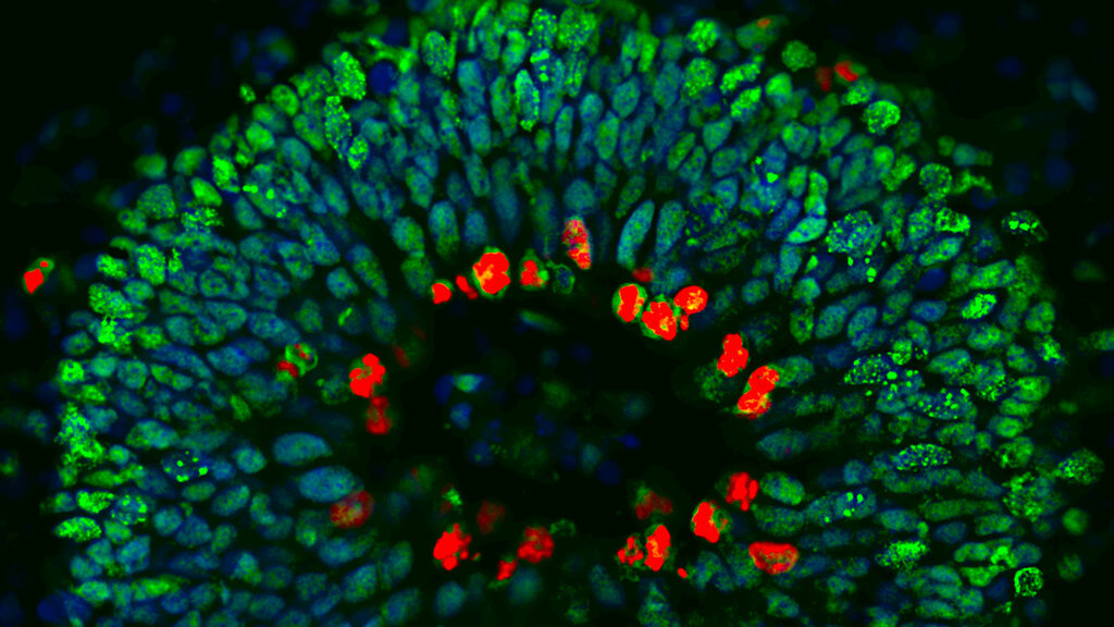
Scientists grew “mini-brains” in a lab for all the family members. A mini-brain is a tiny 3D structure grown using clusters of human stem cells. They serve as realistic models for scientists to study the step-by-step development of disease, and also serve as ideal structures to screen drugs. Study authors noted striking neurodevelopmental defects in the mini-brains of family members carrying the ANG mutation.

“This seems to indicate that subtle development defects play a role in disease susceptibility or onset,” Dr. Subramanian adds. “I envisage a time when we will be identifying people who are susceptible to these diseases, screening them for genetic mutations and offering early-intervention gene therapy to fix the defects.”

human brain organoid
Section through a human brain organoid showing stem cells containing protective antibodies (stained green and red). The cells’ nuclei are stained blue. Credit: Ross Ferguson and Vasanta Subramanian

In conclusion, Dr. Subramanian stresses the need for more research in order to further clarify the mechanisms by which ANG protects cells and better understand its function in stem cells. This study received funding from the National Centre for the Replacement, Refinement and Reduction of Animals in Research (NC3Rs), BRACE, and the Wellcome Trust VIP award.

“We applaud Dr Subramanian’s innovative research, which could make a big difference in tackling frontotemporal dementia. Better understanding of the Angiogenin gene and its link to FTD could support treatment to slow down or stop the disease in the future. This type of dementia tends to have an early onset between the ages of 45-65 years, and often has a devastating impact during middle age. We are hopeful that this BRACE funded research may play a key role in one day reducing the impact of the condition,” says BRACE CEO Chris Williams.

“Research into the brain and neurological disorders relies in large part on animal models and it is fantastic to see Vasanta’s mini-brain ‘organoids’ delivering new insights into neurodegenerative diseases. It is testament to the utility of these models that they are still being applied to new research questions, almost 15 years after we awarded Vasanta the initial funding to develop human cell-based alternatives to the use of animals in ALS (the most common form of motor neuron disease) research,” comments Dr. Jessica Eddy, NC3Rs Regional Program Manager.

What keeps love alive? Scientists discover ‘chemical imprint of desire’


Experts have uncovered a biological signature of desire that causes us to lust over certain people more than others. Study authors observed that this imprint fades if there is no contact with the person for a while and that time can indeed heal heartbreak.

“As humans, our entire social world is basically defined by different degrees of selective desire to interact with different people, whether it’s your romantic partner or your close friends,” says Associate Professor Zoe Donaldson from the University of Colorado Boulder, in a media release. “This research suggests that certain people leave a unique chemical imprint on our brain that drives us to maintain these bonds over time.”

The study utilized neuroimaging technology on prairie voles, chosen for their propensity to form monogamous pair bonds, a trait shared by only three to five percent of mammals. In scenarios where a vole attempted to reach its partner in another room, researchers noticed that dopamine levels in the brain spiked, illuminating the sensor.

woman hugging man
Researchers say some people create a distinctive chemical mark in our brains that drives us to maintain connections over time. (Credit: Photo by Tim Mossholder On Unsplash)

Researchers describe the reunion as lighting up “like a rave,” with continued activity as the voles snuggled and sniffed each other. However, when faced with a stranger, the sensor dimmed significantly.

“This suggests that not only is dopamine really important for motivating us to seek out our partner, but there’s actually more dopamine coursing through our reward center when we are with our partner than when we are with a stranger,” explains study first author and graduate student Anne Pierce.

Interestingly, after a four-week separation, equivalent to an eternity in the rodent world, the dopamine surge nearly vanished upon reunion.

“We think of this as sort of a reset within the brain that allows the animal to now go on and potentially form a new bond,” Dr. Donaldson concludes.

“The hope is that by understanding what healthy bonds look like within the brain, we can begin to identify new therapies to help the many people with mental illnesses that affect their social world.”

Hungry at night? Study proclaims cottage cheese the ideal before-bed snack


 If you’ve been on the lookout for the perfect late night snack, look no further. Researchers from Florida State University say that cottage cheese before bed has a positive effect on the metabolism and overall health, helps promote muscle recovery, and doesn’t result in any body fat gains.

If cottage cheese isn’t exactly your snack of choice, the study’s authors say any helping of 30 grams of protein about a half hour before turning in for the night should do the trick.

For the study, a group of active young women in their early 20s were asked to eat cottage cheese 30-60 minutes before going to bed. Researchers specifically wanted to see what effect the cottage cheese would have on the participants’ metabolisms and muscle recovery process.

This study is especially noteworthy because it is among the first ever to have subjects consume a whole food product before bed, as opposed to a dietary supplement such as a protein shake.

“Until now, we presumed that whole foods would act similarly to the data on supplemental protein, but we had no real evidence,” comments Michael Ormsbee, Associate Professor of Nutrition, Food and Exercise Sciences at FSU, in a release. “This is important because it adds to the body of literature that indicates that whole foods work just as well as protein supplementation, and it gives people options for presleep nutrition that go beyond powders and shaker bottles.”

According to study co-author Samantha Leyh, a research dietitian with the U.S. Air Force, these findings will serve as a helpful jumping off point for future research investigating the impact of whole food consumption on precise metabolic responses.

“While protein supplements absolutely have their place, it is important to begin pooling data for foods and understanding the role they can play in these situations,” Leyh says. “Like the additive and synergistic effects of vitamins and minerals when consumed in whole food form such as fruits or veggies, perhaps whole food sources may follow suit. While we can’t generalize for all whole foods as we have only utilized cottage cheese, this research will hopefully open the door to future studies doing just that.”

Moving forward, the research team plan to conduct additional research on other potential late night snacks, in an effort to determine the optimal food choices one can make before bed in order to promote muscle regeneration and overall improved health.

“There is much more to uncover in this area of study,” Ormsbee concludes.

Humanized mice reveal arsenic may raise diabetes risk only for males


Chronic exposure to arsenic, often through contaminated groundwater, has been associated with Type 2 diabetes in humans, and there are new clues that males may be more susceptible to the disease when exposed.

A new study – using lab mice genetically modified with a human gene to shed light on the potential link – revealed that while the male mice exposed to arsenic in drinking water developed diabetes, the female mice did not.

These results would not have been possible without using a mouse model engineered to express a human enzyme for metabolizing arsenic, since normal mice process arsenic much more efficiently than humans and require very high levels of exposure before they become diabetic.

In the paper, the researchers also identified a transcription factor called Klf11, which might be the master regulator of the difference in how the livers of males and females respond to arsenic.

“Our paper lays the foundation for future investigations into the mechanism of how arsenic exposure leads to diabetes, why there are striking male-female differences, and potential therapeutic strategies,” said Praveen Sethupathy ’03, professor of physiological genomics and chair of the Department of Biomedical Sciences at the College of Veterinary Medicine, and the study’s senior author.

“It highlights how important it is to use an appropriate genetic model [in this case, humanized mice], because none of these results were seen in the wild-type mice,” said Jenna Todero, a doctoral student in Sethupathy’s lab.

Todero is the first author of “Molecular and Metabolic Analysis of Arsenic-exposed Humanized Arsenic +3 Methyl Transferase (AS3MT) Mice,” which published Dec. 27 in the journal Environmental Health Perspectives.

Arsenic is the top priority substance for study, according to the Agency for Toxic Substances and Disease Registry. Human exposure to arsenic, which is common in the environment, often comes through contaminated groundwater. The safe exposure limit is 10 parts per billion, according to the World Health Organization and the Environmental Protection Agency.

Endemic levels of arsenic above safe limits in both Bangladesh and Mexico led to studies that showed an association between higher levels of arsenic exposure and Type 2 diabetes. Though these studies had very small sample sizes, they offered clues for further research.

The current study was performed on regular lab mice – known as wild-type mice – as a control and humanized mice that were engineered to express a human form of an enzyme, AS3MT, which is found across species, and is important for metabolizing arsenic in the body. Efficiency of this enzyme differs across species, but mice are far faster than humans at metabolizing arsenic.

All the mice were exposed for a month to doses of arsenic in drinking water that were nonlethal but sufficient to potentially promote Type 2 diabetes. The researchers then examined liver and white adipose tissues that are implicated in diabetes. In the humanized male mice alone, they found increased expression in genes related to insulin resistance. Also, in both liver and white adipose tissues of the humanized male mice, they identified a biomarker called miR-34a, which is highly associated with insulin resistance in Type 2 diabetes and other metabolic diseases.

“This would suggest miR-34a is potentially a way to screen individuals who live in areas that have endemic arsenic levels,” Todero said. “If you have elevated miRNA-34a, you might be at risk for Type 2 diabetes onset or other metabolic dysfunction.”

The researchers also identified the transcription factor (which regulates genes) Klf11 in the liver. “This was really exciting because we showed that genes that are highly important for things like regulating glucose or lipid metabolism seem to be targets for Klf11,” Todero said.

They found that Klf11 was significantly turned down in the humanized males, but not in the wild-type mice or the females. The study’s authors speculate that Klf11 might be a master regulator for genes associated with energy usage. It was suppressed in humanized male mice, likely causing dysregulation, and hallmarks of diabetes such as insulin resistance and elevated fasting blood glucose.

In the female humanized mice, the researchers found evidence of elevated expression of genes that support insulin sensitivity.

“We saw the opposite effect in females, where genes that promoted insulin sensitivity and glucose uptake had their expression increased,” Todero said. Previous work by another group suggested that the female sex hormone estradiol may play a role in the sexual divergence in the association between arsenic and Type 2 diabetes.

Mapping brain repair and remodeling after stroke


Researchers at Weill Cornell Medicine have catalogued the cellular response to stroke in a preclinical model, identifying the immune cells involved and the roles they may play in the days and weeks following a stroke.

During a stroke, loss of oxygen leads to brain damage and cell death. It also triggers a powerful inflammatory response in which the brain’s resident immune cells, along with cells recruited from the blood, infiltrate the injured tissue.

The findings, published Jan. 4 in Nature Immunology, could point toward novel approaches to fostering stroke recovery and provide insight into why therapies to control inflammation after a stroke haven’t been successful.

Image of a mouse brain section
Image of a mouse brain section 14 days after stroke. Immune cells called leukocytes (yellow) infiltrate the core of the injury (right edge of brain), surrounded by enlarged blood vessels (magenta). Cell nuclei were stained cyan blue.

“Nearly every one of us knows someone who’s had a stroke. It’s a huge problem,” said senior author Dr. Josef Anrather, a professor of neuroscience and vice chair for research in the Feil Family Brain and Mind Research Institute at Weill Cornell Medicine. “But in terms of treatment, there is little a physician can do.”

Interventions that restore blood flow to the affected brain region must be administered within hours to be effective. “So most people, more than 80%, receive no therapy at all,” he said.

Understanding how immune cells contribute to repairing and remodeling the brain in the later, chronic phase after a stroke could help doctors minimize the long-term neurological consequences, including dementia and even seizures.

In 2016, Anrather and his colleagues observed that immune cells called monocytes, which are made in the bone marrow, accumulate in the brain following a stroke. Once there, they appeared to undergo a physical transformation: Some sprouted spindly arms, adopting the appearance of the brain’s resident immune cells, the microglia; others grew more amorphous and amoeba-like.

But what, if anything, did this shapeshifting have to do with their behavior?

“We became interested in knowing the function of these different structural characteristics,” said lead study author Lidia Garcia-Bonilla, the Finbar and Marianne Kenny Research Scholar in Neurology and an assistant professor of research in neuroscience at the Brain and Mind Research Institute, Weill Cornell Medicine.

They also wondered whether these cells were contributing to recovery or compounding the damage.

“There are always two sides to the coin,” Anrather said. The same cell type might be harmful in some circumstances but helpful in others. “That might be why the clinical trials of drugs that reduce immune cell infiltration into the brain and inflammation have shown no benefit for stroke.”

The most direct way to assess what a particular cell is doing is to determine which of its many genes are turned on. Working with a preclinical model, they collected immune cells at two days and 14 days after an induced stroke – the blockage of an artery in the brain. They then sequenced the RNA molecules, which encode proteins, produced by each cell. Using this approach, the researchers identified exactly each type of cell they had isolated. It also provided a readout of which genes each cell had switched on, an indication of their roles after the stroke.

The researchers first noticed that a population of microglia were rapidly proliferating. That made sense, Anrather said, “because microglia cover the territory of the brain.” When their numbers are depleted by an injury, such as stroke, the cells multiply to blanket the damaged tissue.

Then they “take out the trash,” Anrather said.

“For the brain to rebuild itself, you have to clean up, remove dead cells,” he said. Indeed, two days after the experimental stroke, the researchers detected a cadre of microglia that switch on genes involved in clearing away cellular debris.

Joining the microglia in this effort were monocytes – white blood cells that responded to the injury. “These cells circulate continuously and don’t really have a job until there is a problem, like an infection, trauma or any kind of tissue death,” Anrather said. “Then they are called in to help clean up.”

Once there, the researchers found, these monocytes transformed themselves into the type of cell that’s needed to get the job done. “They’re like little kids that get educated in the tissue,” Anrather said.

After the acute clean-up phase, the immune response was restructured toward tissue remodeling. Some cellular recruits produced growth factors triggering repair while immunological “professionals” such as T cells were called in to play a neuroprotective role.

By identifying which immune cells will heed the stroke-induced distress call, the researchers provide a novel vehicle for intervention. “Because these cells know how to get to the brain,” Anrather said, “you could use them as a shuttle and engineer them to deliver a therapeutic.”

Furthermore, understanding precisely what these cells do when they get to the brain could be key to developing treatments that can be administered weeks or months after a stroke. “Finding a way to activate the brain’s natural repair mechanism could improve the outcome for stroke patients,” Garcia-Bonilla said.

Why pain seems worse at night


As the song from the musical Les Miserables, based on the novel by Victor Hugo, says, “But the tigers come at night, with their voices soft as thunder”. We’ve all been miserable at night, when we find ourselves tossing and turning in bed, staring at the ceiling because of an unbearable backache; or toothache, or earache, or kneeache.

It was there during the day, but now it won’t let us rest and gnaws at us. The question is: why do we feel the pain more intensely at night? What does science have to say about it?

Pain is not a strange phenomenon to anyone. But defining it is complicated. After numerous modifications over the years, the International Association for the Study of Pain (IASP) agreed in 2020 to narrow it down as “an unpleasant sensory and emotional experience associated with, or resembling that associated with, actual or potential tissue damage”.

What is pain for?

We tend to think of this feeling as something negative, since it is, by definition, an unpleasant experience. But the human being is a complex, finely tuned machine that rarely has functions that are there “just for the sake of it”.

The purpose of pain is to warn us that something is wrong; it is a survival mechanism that helps to keep us safe from dangers that may threaten our physical integrity. To use a simile: it is an alarm system that our brain has to tell us that we are at risk and that urges us to get to safety. And it is unpleasant so that we feel the need to avoid it.

However, it is not a response to a stimulus, as was thought in Descartes’ time (eg. I touch something burning and the pain saves me from burning because it makes me withdraw my hand). The modern conception understands it as a product of our brain: it is this organ that tells us where, how much and in what way it hurts.

The Gate Control Theory

So why does sensation increase at night and how might that help survival?

The explanation has to do with our brain’s processing systems and the science of perception. In the 1960s, Roland Melzack and Patrick Wall proposed their Gate Control Theory. According to this, there is a gate in the spinal cord that allows or disallows painful stimuli to pass through to the brain.

In other words, there will be certain things that cause the gate to close and we feel less pain, and other things that cause the gate to open and we feel more pain. An example is the mechanical act of rubbing our skin if we have been hit: the sensation of friction competes with the sensation of pain and causes it to be felt less.

In the silence of the night, the voices of those tigers are heard more, often as we remember some uncomfortable situation we experienced during the day and had almost forgotten. There is nothing to distract us and help us close the door: no images, no sounds, no interactions with others.

The worst time? 4 a.m.

Since the 1960s, new theories, new techniques and new findings have been nurturing the science of pain. A study published in Brain last September also points to circadian rhythms as a possible key player in the phenomenon of nocturnal accentuation.

Woman awake in bed from insomnia, can't sleep

Inès Daguet and her colleagues conducted a novel laboratory study in which they found that the time of day when pain (experimentally induced, in this case) is most intensely perceived is at 4am. One possible explanation is sleep deprivation, as it has also been shown to be influential, but in Daguet’s model, the weight of circadian rhythms was much greater. These physical and mental changes we experience may be related to the cyclical levels of hormones we have during the day, such as cortisol, which is related to the immune system and inflammation, and melatonin.

However, it should not be forgotten that this is an experimental study, in a laboratory setting, where participants are not in their natural environment (sleeping in their bed) and receive painful stimuli artificially via a heat-inducing machine.

Alerts to predator threat

Researchers Hadas Nahman-Averbuch and Christopher D. King have published a comment on the above study where they point out that from an evolutionary perspective, we are most vulnerable to predators at night, because that is when we sleep. It makes sense, therefore, that a lower intensity of stimuli would be sufficient to wake us up to potential danger.

Ultimately, further research is still needed to understand why we feel more pain at night, but it seems that our brains are still trying to protect us from being eaten by tigers (in this case real ones) while we sleep.

Do you get rain pain? Weather-related aches really do alter people’s lives


Chronic pain is bad enough on a normal day, but crummy weather can make already achy joints and hips that much more painful. Tied to changes in the barometric pressure resulting from storms, the cold, and dreary forecasts, pain-based weather is a significant concern on a day-to-day basis for millions of people. Now, researchers from the University of Georgia report that roughly 70 percent of respondents in a recent poll would change their daily behavior based solely on weather-based pain forecasts.

“We’re finding more consistent relationships between weather patterns and pain, so it seems more possible to make weather-based pain forecasts,” says lead study author and geography/atmospheric sciences lecturer Christopher Elcik in a media release. “This study was to survey and see what the audience was for this type of forecast.”

In all, researchers surveyed over 4,600 people. Among respondents who identified as migraine sufferers, 89 percent pointed to weather as something that impacts their pain level, and another 79 percent cited weather as a trigger for their pain. Meanwhile, among respondents with other conditions, 64 percent also said weather patterns could trigger pain and an astounding 94 percent identified weather as an influential pain factor.

This latest report builds on previous research that focused on specific weather patterns and pain-related conditions in an attempt to measure public interest in a weather-based pain forecast, as that may be indicative of high or moderate risk for migraines or chronic pain.

“I see how much people can be affected by these types of pain, so if I can provide someone with insight into the level of risk for a day, maybe people can take steps to prevent the pain from happening,” Elcik comments. “There are preventative measures people can take if risks are higher.”

Man suffers leg injury or cramp while running

Hypothetically, if the risk of weather-related pain were high, more than half of respondents said they were likely to take preventive measures (medication, rest, avoiding compounding triggers). Another 47 percent with migraines and 46 percent of those with pain-related conditions were also “extremely likely” to take the same measures.

Notably, desire for a forecasting tool was very high; 72 percent of those living with migraines and 66 percent with pain-related conditions said they would alter their behavior by canceling plans or taking preventive measures in the event of a weather-based pain forecast. Some respondents even said they already use online tools to predict weather-related pain.

One example is AccuWeather’s arthritis or migraine forecast, which predicts low-to-high pain risk according to atmospheric conditions. These existing tools, however, offer few details regarding the variables considered or how the predictions are actually produced.

A person’s likelihood to continue with plans also depended heavily on the length of the activity in question. If plans were roughly 30 minutes long, 57 percent of respondents with migraines and 52 percent of those with pain-related conditions said they would be “extremely likely” to continue plans even if there were a moderate risk of pain. About 43 percent from each group said they would continue even with the highest risk forecast.

Older man battling shoulder pain, back pain, arthritis

When it came to an activity lasting more than three hours, on the other hand, that number declined to roughly 23 percent for moderate risk and 18 percent for high risk among those with migraines. For people living with other pain-related conditions, 23 percent would follow through with a three hour plus activity in the face of a moderate risk of weather pain and 21 percent would continue despite the highest risk. Generally speaking, as the level of risk increased, so did the likelihood to alter plans.

“This was across the board,” Elcik notes. “Everyone was more likely to cancel plans if the forecast risk was higher.”

While additional research and studies are necessary in order to create a reliable pain-based weather forecast, Elcik believes this study highlights the urgent importance of developing such a resource.

“This publication shows there’s an audience that’s willing and eager to try something new, and there are probably many more people who would benefit—more than we even thought,” the researcher concludes. “I think these results can push other researchers to also look at similar, larger-scale weather phenomena and help the community better understand how the atmosphere does impact pain.”

A Dietitian’s Take: If you only take one supplement, this is the one to pick


Taking a multivitamin is a daily routine for half of American adults. In theory, they can fill in the nutritional gaps that your diet may not have been able to. Even if you eat the “best” diet, supplements can help fully optimize your health, but multivitamins may not actually be the answer. If you asked me which vitamin is most worth it, I’d say magnesium.

What about taking a multivitamin?

Although multivitamins are designed to cover all bases by giving you a combination of nutrients, things don’t actually happen that easily. So far, no studies have shown that multivitamins truly improve health. In fact, five recent studies have reported that they don’t improve cardiovascular health, reduce COVID-19 deaths, or improve other markers of overall health.

Moreover, the benefits that do show up might just be in people’s minds. One study, including data from over 21,000 U.S. adults, found that 30 percent reported improvement in overall health while taking multivitamins, yet there weren’t any actual differences between those who took them and those who didn’t.

According to the June 2022 United States Preventive Services Task Force (USPSTF) recommendations, there is not enough evidence out there to fully assess the health benefits and risks of multivitamin use. This further drives home that the health impacts are at best inconclusive, and it’s been this way for a while now.

What is magnesium?

Magnesium is a mineral that plays a crucial role in every part of the body, from the muscles to the brain, kidneys, and heart. A typical rule of thumb is that if a food is green or has fiber, it has magnesium in it. Foods like avocado, almonds, and spinach are considered to be good sources.

Foods that are high in magnesium
(Photo by Evan Lorne on Shutterstock)

If it’s in food, why would I need a supplement?

Health experts consistently report that many people in the U.S. do not get enough of magnesium through food. This equally goes for people who eat lots of plant-based foods and people who eat more of a standard American (Western) diet. The current recommended dietary allowance (RDA) is close to 420 mg/day, but Americans are averaging closer to 200 mg daily. This is due to the fact that there are simply a lot of ways that magnesium is either depleted from our bodies and our environment, such as:

  • Drinking alcohol regularly
  • GI conditions like celiac disease
  • Poor soil quality due to harmful agricultural practices (leading to much lower magnesium content in foods that would typically be high in it)
  • Certain medications
  • Stress

What can the supplements help with?

Signs of magnesium deficiency can be much more common than you might think. Debilitating period cramps, consistent muscle pains and aches, and poor sleep are all examples. Magnesium deficiency is also highly implicated in hypertension and Type 2 diabetes, two of the most common chronic diseases in the world. The mineral regulates salt and potassium (and therefore blood pressure), as well as blood sugar, so running low on it can be a key driver for both of these.

Better sleep quality and mental health management are also some of the most popular reasons for taking the supplement. One psychologist calls it “the original chill pill,” thanks to it showing great promise in patients with depression, anxiety, insomnia, and stress management.

Magnesium can effectively reduce, and possibly even stop seizures as they are happening. Epilepsy continues to be a growing concern, particularly due to there being few medical interventions that work well for people with it. The mineral can penetrate the brain and confer protection in this population.

There are tons of other benefits that have been discovered as well:

Bottom Line

It’s almost as if magnesium is a multivitamin in and of itself, right? Magnesium can be a powerful tool to maximize your wellness through all of its various functions in the body, and there are even more than mentioned here. But as always, nutrition and supplementation is an individual process. Be sure to work with your own dietitian and/or physician to do what is best for your unique circumstances and lifestyle.

The best nutrient to lower cholesterol is


There is nothing worse than getting a blood test, thinking everything will come back normal, and findings out you have high cholesterol. This is the reality of almost 80 million Americans who have high cholesterol, according to the Centers for Disease Control and Prevention.

Let’s get one thing straight: cholesterol is not inherently bad. Your body gets cholesterol from two sources, from the food you eat and from what your liver produces naturally. Cholesterol is needed for both hormone and overall metabolic health.

In excess, it can lead to heart health complications. More research continues to come out about how much dietary cholesterol truly impacts blood cholesterol levels, with more studies starting to show that it isn’t all that much. Rather, research finds certain dietary choices can stimulate the liver to make too much cholesterol.

According to the CDC, heart disease is the leading cause of death in the United States. More often than not, the food choices we make can make a significant difference in reducing cholesterol, and thus the risk of developing heart disease in general. The diet tweak you need likely isn’t to eliminate all cholesterol, but to increase your soluble fiber.

What is soluble fiber?

There are two types of fiber: insoluble and soluble. Insoluble helps to add bulk to your stool, whereas soluble fiber slows digestion by absorbing water and turning into a gel-like texture.

You will find soluble fiber in whole plant foods such as:

Insoluble fiber doesn’t have the same cholesterol-lowering effect as soluble, but it’s still important to ensure enough intake of it to prevent constipation by promoting consistent bowel movements.

A look at some of the best foods for increasing fiber
Foods containing fiber (© bit24 – stock.adobe.com)

How does soluble fiber help lower cholesterol?

The gel-like consistency helps it act like a sponge to soak up excess cholesterol and excrete it through your digestive system, affectively helping to blunt absorption in the blood. There are subcategories of soluble fiber, such as viscous fibers, inulin oligofructose, beta glucans, pectin, psyllium, and more, which have been shown to positively impact LDL (“bad”) and total cholesterol.

Researchers continue to find mounting evidence to support that soluble fiber is beneficial for reducing cholesterol levels. Unfortunately, most Americans don’t meet their daily fiber targets. It’s recommended that people eat 25 to 35 grams of fiber per day, but most Americans actually eat somewhere between 10 and 15 grams each day.

Do fiber supplements work?

All supplements are meant to do just as their name suggests: supplement your intake. It’s unlikely that you eat the same way every single day, so if you’re trying to stick to the recommended amount of daily fiber, a supplement can help you stay on track.

However, supplements aren’t meant to completely take the place of fiber-rich foods. Fruits, vegetables, whole grains, nuts, and seeds all have a place in the diet not only for their fiber content, but the vitamins, minerals, and tons of antioxidants they provide.

Vitamins and supplements
Supplements (© MarekPhotoDesign.com – stock.adobe.com)

Most Americans would greatly benefit from including more fiber into their diets. It’s the single nutrient known to directly act on cholesterol and help your body get rid of excess amounts.

Fiber has been shown to support healthy cholesterol numbers and be protective against heart disease. If you want to lower your cholesterol, start by prioritizing more soluble fiber-rich foods, along with incorporating other lifestyle factors such as more exercise and decreasing stress.

Diet Suggestions For Increasing Soluble Fiber

Here’s a simple, balanced 7-day meal plan that emphasizes foods high in soluble fiber:

Day 1

  • Breakfast: Oatmeal topped with sliced bananas and a sprinkle of chia seeds.
  • Lunch: Lentil soup with whole grain bread.
  • Dinner: Grilled chicken with a side of steamed broccoli and quinoa.
  • Snacks: An apple; a handful of almonds.

Day 2

  • Breakfast: Smoothie with spinach, berries, and a tablespoon of ground flaxseed.
  • Lunch: Salad with mixed greens, chickpeas, avocado, and olive oil dressing.
  • Dinner: Baked salmon with sweet potato and green beans.
  • Snacks: Orange slices; carrot sticks with hummus.

Day 3

  • Breakfast: Greek yogurt with mixed berries and a drizzle of honey.
  • Lunch: Turkey and avocado wrap with whole grain tortilla.
  • Dinner: Stir-fried tofu with mixed vegetables (carrots, bell peppers) and brown rice.
  • Snacks: Pear; a handful of walnuts.

Day 4

  • Breakfast: Whole grain toast with peanut butter and banana slices.
  • Lunch: Quinoa salad with cucumbers, tomatoes, and feta cheese.
  • Dinner: Grilled shrimp with asparagus and a side of barley.
  • Snacks: Peach; yogurt.

Day 5

  • Breakfast: Berry and banana oat bran muffin with a side of cottage cheese.
  • Lunch: Black bean soup with a side of mixed greens salad.
  • Dinner: Roast chicken with Brussels sprouts and sweet corn.
  • Snacks: Apple; a handful of sunflower seeds.

Day 6

  • Breakfast: Scrambled eggs with spinach and whole grain toast.
  • Lunch: Tuna salad (with Greek yogurt) on whole grain bread.
  • Dinner: Beef stew with carrots, potatoes, and peas.
  • Snacks: Orange; a few slices of cheese.

Day 7

  • Breakfast: Smoothie bowl with kale, banana, almond milk, and a sprinkle of granola.
  • Lunch: Chicken and vegetable stir-fry with brown rice.
  • Dinner: Baked cod with roasted vegetables (zucchini, bell peppers) and farro.
  • Snacks: Kiwi; a handful of mixed nuts.

Tips:

  • Drink plenty of water throughout the day.
  • Adjust portion sizes according to your dietary needs and activity level.
  • Feel free to swap out any ingredients based on your preferences or dietary restrictions.

Remember, it’s always a good idea to consult with a healthcare professional or a dietitian before starting any new diet regimen, especially if you have specific health conditions or dietary needs.

Is Body Positivity Doing More Harm Than Good?


‘You don’t have to love your body to be kind to it.’ – Alissa Rumsey

Body positivity is a major topic of conversation these days. From social media to commercials on TV, there has been a growing effort to push a more inclusive image of the human body. While the concept of accepting our individual flaws is a generally positive idea, when does it go too far and start endangering your health? When it comes to wellness, the line should be drawn at obesity — as the condition contributes to life-long health problems if left untreated.

Body positivity is about how our perception of body image (our own and others) shapes our concept of self, mental health, well-being, and relationships. It refers to how you feel about your own appearance, and how you feel about your height, weight, and shape.

Moreover, the term body positivity describes a mindset that the shape or size of someone’s body does not determine their worthiness of love. It challenges the roles of cultural, social, and media influences in the development of our relationship with our body, ourselves, and how we perceive others. Body positivity can also refer to cultivating confidence and self-love, and appreciating your body for all that it can do, despite its flaws. It’s about inclusivity and acceptance of all physical traits.

If the goal of body positivity is to encourage the media to present images of “real” people, rather than idealized images, the movement is succeeding. It’s my unscientific observation that more television ads and programming, as well as print media, feature more overweight models and actors.

Woman looking at herself in mirror, body image
(© Maridav – stock.adobe.com)

When does body positivity cross a line into harmful self-indulgence?

Weight is just one aspect of body positivity, but let’s use it to examine the movement.

For children and adolescents between two and 19 years-old, almost 20 percent are obese – that’s about 15 million kids. The older the child, the higher obesity rates rise.

It’s 12.7 percent among kids two to five, and 22.2 percent among children 12 to 19. About 26 percent of Hispanic children are obese, as are 25 percent of Black children.

We are seeing diseases in these young people which previously were seen almost exclusively in adults. This includes cases of high blood pressure, high cholesterol, Type 2 diabetes, gall bladder disease, sleep apnea, and joint problems. There is more obesity in proportion to decreasing education among parents and lower household incomes.

Obesity in children is associated with:

Obesity in adults is associated with:

Teen boy eating junk food, drinking soda while looking at smartphone
(© New Africa – stock.adobe.com)

These negative consequences are increasing in frequency as the incidence of overweight and obesity in our culture increases, while the body positivity movement gains attention.

Body positivity purports loving your body. You care for what you love. Does that make being overweight, or self-indulgence, body negativity? Let’s just get rid of the positive vs. negative and replace them with wellness.

How Can You Practice ‘Body Wellness’?

There are several ways to discover what “being healthy” means to you – physically, mentally, and emotionally. This includes cultivating a realistic perception of your body, including its flaws.

1. Practice positive self-talk: It can increase your confidence and self-esteem. Replace negative thoughts with positive affirmations and focus on your strengths. Use statements such as “I am courageous,” and “My body is capable of great things.” You can put these on sticky notes and post them on your bathroom mirror.

2. Seek out community: Surround yourself with people who support and uplift you.

3. Add more physical activity: Move in ways that make your body feel good, such as yoga, dancing, or walking. Focus on how your body feels during these activities, rather than how it looks.

4. Write a gratitude list: List things about your body and yourself you are grateful for. This can help shift your focus from negative thoughts to positive ones.

5. Nourish yourself with nutrient-rich food: Eat whole foods that make you feel good and provide energy. Avoid restrictive diets.

6. Wear clothing that builds confidence: Be comfortable, and bold with color. Don’t worry about following fashion trends or certain sizes.

7. Practice self-compassion: Treat yourself with the same compassion and love you would show to a friend.

8. Ask your healthcare provider for support: Discuss with your doctor your personal path to greater wellness.