https://www.thespaceacademy.org/2022/06/physicists-have-discovered-new-force-of_82.html?m=1
Day: 06/18/2022
Watch “Case Discussion || Myasthenia Gravis” on YouTube
People Who Think Luck Decides Their Fate Are at Elevated Risk of Severe Gum Disease.
Diet, disease, and the microbiome.
Physicists build an atom laser that can stay on forever
A solar power station in space to beam electricity to Earth? Yes, it’s possible.
The plastic surgery debate.
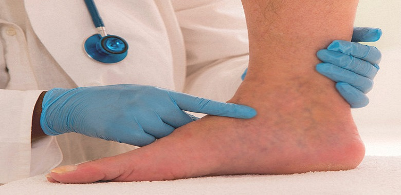
Peripheral Edema
Peripheral edema is a very common finding in daily medical practice both in outpatient and inpatient settings. It can vary from benign condition to serious medical conditions, including congestive heart failure, liver failure, and kidney failure. This activity reviews the evaluation and treatment of peripheral edema and highlights the role of the interprofessional team in evaluating and treating patients with this condition.
Objectives:
- Describe the causes of peripheral edema.
- Outline the evaluation that should be completed on a patient with peripheral edema.
- Summarize the management of peripheral edema.
- Explain the importance of coordination of care of the interprofessional team in caring for patients with peripheral edema.
Introduction
Fluid compartments in the human body are divided between the intracellular and extracellular spaces. The extracellular space constitutes about one-third of total body water, which is further divided into intravascular plasma volume (25%) and the extravascular interstitial space (75%). The fluid balance between these compartments is maintained by hydrostatic pressures and oncotic pressures described by Starling. The other two factors that play an important role in fluid balance are vessel wall permeability and the lymphatic system. The lymphatic system collects fluid and filtered proteins from the interstitial space and returns that back to the vasculature. Any disturbance in this delicate homeostasis that results in net filtration out of the vascular space or impaired return of fluid by lymphatics leads to the accumulation of fluid in the interstitial space that is called edema. Edema can affect any part of the body and ranges from local swelling to full-blown anasarca, depending on the underlying pathology. A classic example of local swelling is an insect bite. An example of anasarca can be seen in nephrotic syndrome.[1]
Edema, other than localized edema, does not become clinically apparent until the interstitial volume has increased by 2.5 to 3 liters because the tissues constituting the interstitium can easily accommodate several liters of fluid. Therefore, a patient’s weight may increase by nearly 10% before pitting edema is evident.
Etiology
Causes of peripheral edema can be divided depending on the underlying mechanism as below.[2]
Increased Capillary Hydrostatic Pressure
Regional venous hypertension (often unilateral)
- Deep vein thrombosis
- Compartment syndrome
- Chronic venous insufficiency
Systemic venous hypertension (often bilateral)
- Heart failure
- Pericarditis
- Pulmonary hypertension
- Liver failure/cirrhosis
Increased plasma volume
- Pregnancy
- Premenstrual edema
- Renal failure
- Heart failure
- Drugs
Decrease Plasma Oncotic Pressure
Protein loss
- Nephrotic syndrome
- Preeclampsia/eclampsia
Reduced protein synthesis
- Malnutrition/malabsorption
- Liver failure/cirrhosis
- Vitamin deficiencies
Increased Capillary Permeability
- Burns
- Insect bites
- Cellulitis
- Allergic reactions
Lymphatic Obstruction
- Filariasis
- Malignancy involving lymph nodes leading to obstruction
- Postsurgical after lymphadenectomy/radiation
Others
- Myxedema in hypothyroidism
- Lipedema
- Idiopathic
Epidemiology
The most common cause of peripheral edema in patients over 50 yrs of age is venous insufficiency and related to aging, but many other underlying comorbid conditions like heart failure, renal failure, liver failure, and trauma can affect any age group. Peripheral edema can also be commonly observed in pregnancy.
Pathophysiology
The two basic steps involved in edema formation are alterations in the capillary hemodynamics that favors the leakage of fluid from the vascular compartment into the interstitium and renal retention of sodium and water by the kidneys via the renin-angiotensin-aldosterone system as a compensatory mechanism. Any systemic or local venous obstruction or expansion in plasma volume leads to an increase in the hydrostatic pressure that predisposes to edema. As normal body plasma is only about 3 liters, the diffusion of large amounts of water and electrolytes into the interstitial compartment compels the renal retention of sodium and water to maintain intravascular volume and hemodynamic stability. Effective intravascular volume depletion that occurs in congestive heart failure, liver cirrhosis initiates a neurohumoral cascade to maintain effective circulating volume. This cascade works via renal vasoconstriction, reducing glomerular filtration, increases sodium reabsorption proximally mediated by angiotensin II and norepinephrine, and increases sodium and water reabsorption in the collecting tubules mediated by aldosterone and antidiuretic hormone. Additionally, endothelium-derived factors like nitric oxide and prostaglandins further limit sodium and water excretion, therefore, promoting edema.[2]
The major contributor to maintaining intravascular oncotic pressure is due to impermeant proteins, mainly albumin. Albumin is essential for maintaining plasma oncotic pressure, and a level below 2 g/dl of plasma often results in edema. Hypoproteinemia can occur in many conditions, including nephrotic syndrome, severe nutritional deficiency, and severe liver disease in which hepatic synthetic function is impaired. Some medications, like calcium channel blockers, especially dihydropyridines, are more notorious, causing peripheral edema due to more selective arteriolar vasodilatation. Other uncommon conditions are myxedema, lymphedema, and idiopathic edema. Myxedema occurs in hypothyroidism, causing more often localized edema of the eyelids, face, and hands. There is an accumulation of mucopolysaccharides and proteins in the interstitium due to an increase in capillary permeability followed by sodium and water, but the exact pathophysiology in myxedema is not fully understood. Lymphedema is caused by impaired lymphatic transport leading to the accumulation of lymphatic fluid in the interstitium mostly in extremities.[3] Idiopathic edema is poorly understood that primarily affects premenopausal women. The key features are periodic episodes of edema in hands, legs, and abdominal bloating that are not clearly related to the menstrual cycle. By definition, it is a diagnosis of exclusion. It is most common in the third and fourth decades of life. Psychologic and emotional disturbances can exacerbate this condition.
History and Physical
The history and physical is very important because of a wide range of differential diagnoses in peripheral edema. Therefore, the timing of the edema, any changes with position, whether it is unilateral or bilateral, medication history, and evaluation for systemic diseases, are very crucial. Acute swelling of the limb within 72 hours is more common in deep venous thrombosis (DVT), cellulitis, acute injury from trauma, or recent initiation of new medications like calcium channel blockers. The chronic accumulation of more generalized edema over days, weeks, and months is usually due to underlying systemic conditions like congestive heart failure (CHF), liver disease, and renal disease. Dependent edema is more common in venous insufficiency that usually improves with elevation and worsens with dependency. Edema due to a decrease in plasma oncotic pressure like malabsorption, liver failure, the nephrotic syndrome does not change much with the position. Unilateral edema suggests local insults like DVT, cellulitis, venous obstruction, or lymphatic obstruction from tumor and radiation treatment. On the other hand, bilateral edema suggests systemic diseases such as CHF, liver failure, kidney disease, or severe malabsorption syndromes.[4]
The detailed physical exam can help immensely to differentiate systemic causes such as CHF (common findings are jugular venous distension, dyspnea, bilateral crackles, history of heart disease), liver disease (jaundice, ascites, history of hepatitis, and alcohol use disorder), renal disease (proteinuria, oliguria, history of uncontrolled diabetes and hypertension), thyroid disease (fatigue, anemia, weight gain). Edema should be assessed for pitting, tenderness, and skin changes. Also, in the early stages of lymphedema, pitting occurs due to an influx of protein-rich fluid into the interstitium before fibrosis of the subcutaneous tissue; therefore, lymphedema should be considered in the differential in pitting edema. Tenderness is common in cellulitis and DVT, while edema in systemic diseases like CHF, renal, and liver disease is usually nontender. Acute DVT and cellulitis can increase the temperature of the skin over the affected area due to the activation of cellular and humoral factors. Chronic venous insufficiency is often associated with brawny reddish texture due to the deposition of hemosiderin. In later stages, patients can develop venous ulcers that may progress to deep, weeping erosions.[5] Myxedema, due to hypothyroidism, presents with generalized thick skin with yellow to orange discoloration associated with nonpitting periorbital edema.[6] In lipedema, which is a pathologic accumulation of adipose tissue in the extremities, the feet are generally spared.
Evaluation
Initial efforts in the workup should focus on excluding major systemic causes, including heart failure, liver failure, and kidney failure. Therefore, a thorough history and physical examination are very important to minimize unnecessary testing. If systemic causes are suspected, for example, in CHF, chest radiography, EKG, serum brain natriuretic peptide (BNP) should be checked. Depending on these tests, echocardiography can be obtained for further evaluation.[7] In renal disease, the basic metabolic profile, including serum creatinine and urinalysis to check proteinuria, are the basic laboratory tests. Renal ultrasound can be obtained if suspicion for renal disease is high to rule out intrinsic disease.[8] In liver disease, start with liver function tests and albumin followed by a liver ultrasound if suspicion is high or abnormal labs.[9] If hypothyroidism is suspected, then thyroid function tests followed by ultrasound are obtained if necessary. Doppler ultrasonography is the modality of choice if suspicion for DVT is high. It can also be used to confirm the diagnosis of chronic venous insufficiency. Refer to the flow chart for evaluation and work up below.
Treatment / Management
Treatment should be guided by an underlying condition that predisposes to the formation of edema.[10] Diuretics are the treatment of choice in CHF, liver, and renal disease. Loop diuretics are usually the most effective, but several doses are required daily because of their short half-life. Patients with cirrhosis often have secondary hyperaldosteronism; therefore, spironolactone is the first choice of diuretic therapy along with furosemide.[11] However, rapid diuresis should be avoided in cirrhotic patients, especially in those without much peripheral edema, to avoid hepatorenal syndrome and hemodynamic collapse in already low intravascular fluid volume. If needed, paracentesis can be performed to reduce the need for a high dose of diuretics and to avoid electrolyte imbalance. In patients with DVT, anticoagulation therapy either with low molecular weight heparin or newer anticoagulants like rivaroxaban or apixaban are used.[12] Currently, warfarin is not used much because of less risk of bleeding with newer anticoagulants.[13] Also, in addition to anticoagulation, compression stockings should be used to prevent post-thrombotic syndrome.[14] In patients with chronic venous insufficiency, mechanical therapies, including leg elevation and compression stockings, are effective. Compression therapy is contraindicated in peripheral arterial disease because it can compromise blood supply further. In lymphedema, treatment involves complex decongestive physiotherapy that is composed of manual lymphatic massage and multilayer bandages. Maintenance therapy includes compression stockings and pneumatic compression devices. In patients where medications are the offending agent, they should be discontinued if possible and switch to a different class.
Differential Diagnosis
Following are the differential diagnosis of peripheral edema:
- Congestive heart failure: Decreased cardiac out or ejection fraction leads to pulmonary and peripheral venous congestion resulting in pulmonary edema and peripheral edema, respectively. Edema is bilateral and symmetric.
- Hepatic disease: Bilateral edema caused by portal vein congestion leading to an increase in capillary permeability and a decrease in plasma oncotic pressure due to a decrease in the synthesis of albumin by the liver.
- Renal disease: Bilateral edema caused by protein loss, especially albumin such as nephrotic syndrome and an increase in plasma volume due to renal retention and activation of neurohumoral factors.
- Venous insufficiency: Usually, bilateral edema caused by the failure of venous return secondary to chronic deep venous system damage and/or incompetence.
- Deep vein thrombus: Usually acute in nature and unilateral caused by obstruction of deep veins
- Lymphedema: Chronic often following lymphatic obstruction from trauma or surgery. Another common cause in developing countries is filariasis. Location can be upper or lower extremities depending on the underlying cause.
- Myxedema: Soft tissue edema in patients with severe and advanced hypothyroidism. It is typically associated with other hypothyroid features such as bradycardia, constipation, and weight gain.
- Angioedema and urticaria: Secondary to allergic reactions such as bug bites, medications.
- Cellulitis: Edema with signs of infection like fever, elevated white blood cell count. Most common in patients who are obese and chronic medical conditions like diabetes mellitus.
- Lipedema: Chronic, begins around or after puberty. Predominantly involves thighs, legs, buttocks. Spares feet, ankles, and torso.
- Medication-induced: Onset is weeks after the start of medication. Soft, pitting edema resolves within days after stopping the offending medication.[15]
- Obstructive sleep apnea: Chronic secondary to pulmonary hypertension. Patients will usually have daytime fatigue, snoring, and obesity.[16]
Prognosis
Peripheral edema is a common manifestation of multiple disease entities ranging from advanced heart failure, liver disease to localized swelling from an allergic reaction. Therefore prognosis depends upon the underlying disease process.
Complications
Peripheral edema may be a warning sign for many systemic diseases and if not treated early leads to high morbidity and mortality. The most important diseases to rule out are heart disease, liver disease, and kidney disease. Therefore, detailed history and physical followed by relevant testing are very essential in these patients to decrease the complications.
Deterrence and Patient Education
Patient education is very crucial in peripheral edema as in other diseases. In congestive heart failure, a low salt diet is one of the most important parts of management, along with diuretic therapy. Generally speaking, patients should be educated regarding healthy lifestyles such as exercise, diet, and routine check-ups to diagnose underlying disease entities early and prevent long-term complications.
Enhancing Healthcare Team Outcomes
Peripheral edema can poses a diagnostic dilemma to family physicians due to a wide range of underlying diseases. As discussed above in detail, the causes of edema may be due to a myriad of diagnoses, including heart, liver, renal, thyroid, and other vascular etiologies. Therefore, initial efforts in work-up should focus on ruling out any major organ system failure as the underlying etiology. If the systemic disease is suspected, then it is essential to consult with an interprofessional team of specialists that includes a cardiologist, gastroenterologist, nephrologist, and so forth, depending upon underlying disease suspicion. The nurses are also vital members of the interprofessional group as they will monitor the patient’s daily progress, especially when the patient is on diuretics by monitoring daily weights, fluid input, and output. If the patient is on antibiotics or anticoagulants, then the pharmacist will ensure that the patient is on the right dosage and no underlying allergy to that specific medicine. The current recommendations have been developed after an exhaustive review of current medical literature from peer-reviewed journals and book chapters to determine the appropriateness of diagnosis and treatment.
The outcomes of edema depend on the cause. Therefore, to improve outcomes, prompt consultation with an interprofessional group of specialists is very important to prevent complications.
References
1.
Little RC, Ginsburg JM. The physiologic basis for clinical edema. Arch Intern Med. 1984 Aug;144(8):1661-4. [PubMed]2.
Trayes KP, Studdiford JS, Pickle S, Tully AS. Edema: diagnosis and management. Am Fam Physician. 2013 Jul 15;88(2):102-10. [PubMed]3.
Ely JW, Osheroff JA, Chambliss ML, Ebell MH. Approach to leg edema of unclear etiology. J Am Board Fam Med. 2006 Mar-Apr;19(2):148-60. [PubMed]4.
Shah MG, Cho S, Atwood JE, Heidenreich PA. Peripheral edema due to heart disease: diagnosis and outcome. Clin Cardiol. 2006 Jan;29(1):31-5. [PMC free article] [PubMed]5.
Hyder ON, Soukas PA. Chronic Venous Insufficiency: Novel Management Strategies for an Under-diagnosed Disease Process. R I Med J (2013). 2017 May 01;100(5):37-39. [PubMed]6.
Milkau M, Sayk F. [Thyroid Storm and Myxedema Coma]. Dtsch Med Wochenschr. 2018 Mar;143(6):397-405. [PubMed]7.
Bestetti RB, Cardinalli-Neto A, Couto LB. The history of the evolution of the knowledge about the diagnosis and the pathogenetic aspects of heart failure: From the Egyptians to James Mackenzie. Int J Cardiol. 2020 Apr 01;304:109-115. [PubMed]8.
Nephrotic Syndrome. Am Fam Physician. 2016 Mar 15;93(6):Online. [PubMed]9.
Mengi S, Phelps L, Silva TSCD, Ludman A. Clinical examination remains crucial to the correct diagnosis: a case of severe peripheral oedema referred for investigation of heart failure. BMJ Case Rep. 2019 Aug 01;12(7) [PMC free article] [PubMed]10.
Cho S, Atwood JE. Peripheral edema. Am J Med. 2002 Nov;113(7):580-6. [PubMed]11.
Tsochatzis EA, Bosch J, Burroughs AK. Liver cirrhosis. Lancet. 2014 May 17;383(9930):1749-61. [PubMed]12.
Thachil J. Deep vein thrombosis. Hematology. 2014 Jul;19(5):309-10. [PubMed]13.
Almutairi AR, Zhou L, Gellad WF, Lee JK, Slack MK, Martin JR, Lo-Ciganic WH. Effectiveness and Safety of Non-vitamin K Antagonist Oral Anticoagulants for Atrial Fibrillation and Venous Thromboembolism: A Systematic Review and Meta-analyses. Clin Ther. 2017 Jul;39(7):1456-1478.e36. [PubMed]14.
Kahn SR, Galanaud JP, Vedantham S, Ginsberg JS. Guidance for the prevention and treatment of the post-thrombotic syndrome. J Thromb Thrombolysis. 2016 Jan;41(1):144-53. [PMC free article] [PubMed]15.
Makani H, Bangalore S, Romero J, Htyte N, Berrios RS, Makwana H, Messerli FH. Peripheral edema associated with calcium channel blockers: incidence and withdrawal rate–a meta-analysis of randomized trials. J Hypertens. 2011 Jul;29(7):1270-80. [PubMed]16.
Veasey SC, Rosen IM. Obstructive Sleep Apnea in Adults. N Engl J Med. 2019 Apr 11;380(15):1442-1449. [PubMed]
Source: PubMed
What is the differential diagnosis of peripheral oedema?
Peripheral oedema is a common finding in outpatient and inpatient settings in daily medical practice. It may range from benign to life-threatening medical conditions, including congestive heart failure, liver failure, and kidney failure.
Differential diagnosis of peripheral oedema:
- Congestive heart failure
a) Reduced cardiac output or ejection fraction causes pulmonary and peripheral venous congestion, resulting in pulmonary and peripheral oedema.
b) Symmetrical and bilateral oedema.
- Hepatic disease
a) Due to a decrease in albumin production by the liver, bilateral oedema is caused by portal vein congestion, which causes an increase in capillary permeability and a
drop in plasma oncotic pressure.
- Renal disease
a) Bilateral oedema related to protein loss, particularly albumin, such as nephrotic syndrome, and an increase in plasma volume due to renal retention and activation of
neurohumoral factors.
- Venous insufficiency
a) Bilateral oedema is caused by venous return failure due to chronic damage and/or incompetence of the deep venous system.
- Deep vein thrombus
a) Acute and unilateral oedema caused by deep vein obstruction.
- Lymphoedema
a) It is chronic as a result of lymphatic obstruction due to trauma or surgery. Filariasis is another common cause in developing countries. Depending on the underlying
causes, the location can be upper or lower extremities.
- Myxoedema
a) Soft tissue oedema in patients with severe and advanced hypothyroidism, which is associated with other hypothyroid symptoms, including bradycardia, constipation,
and weight gain.
- Angioedema and urticaria
a) It is caused by allergic reactions, such as bug bites and medications.
- Cellulitis
a) Oedema associated with fever and elevated white blood cell count.
b) It is common in obese patients and patients with diabetes.
- Lipoedema
a) Chronic.
b) It starts during or after puberty.
c) It involves thighs, legs, and buttocks.
- Medication-induced
a) The onset occurs several weeks after the medicine is started.
b) Soft, pitting oedema resolves within days after the withdrawal of the offending medication.
- Obstructive sleep apnoea
a) Chronic due to pulmonary hypertension.
b) Symptoms: Daytime fatigue, snoring, and obesity
Fever of Unknown Origin
Fever of unknown origin (FUO) was first described by Dr. Petersdorf and Dr. Beesom in 1961. FUO was defined as a temperature of 101 degrees Fahrenheit (38.3 degrees Centigrade) or higher with a minimum duration of three weeks without an established diagnosis after an intensive one-week investigation in the hospital. Today, due to technological advances allowing for sophisticated outpatient evaluations, the one-week inpatient investigation is no longer required. This activity reviews the cause and presentation of fever of unknown origin and highlights the role of the interprofessional team in its management.
Objectives:
- Describe the workup of a patient with a fever of unknown origin.
- Outline the causes for fever of unknown origin.
- Summarize the treatment of patients with fever of unknown origin.
- Review the importance of improving care coordination among interprofessional team members to improve outcomes for patients affected by fever of unknown origin.
Introduction
Fever of unknown origin (FUO) was first described by Dr. Petersdorf and Dr. Beesom in 1961.[1] FUO was defined as a temperature of 101 degrees Fahrenheit (38.3 degrees Centigrade) or higher with a minimum duration of three weeks without an established diagnosis despite at least one week’s investigation in the hospital. This definition was later changed to accommodate technological advances allowing for sophisticated outpatient evaluations, increasing numbers of immunocompromised individuals including those with human immunodeficiency virus (HIV)/acquired immunodeficiency syndrome (AIDS), and more complex treatment options becoming available. The revised definition proposed by Durack and Street in 1991 divided cases into four distinct subclasses: classic FUO, nosocomial FUO, neutropenic FUO, and HIV-related FUO.[2]
A comprehensive history and physical examination can aid in diagnosis and direct diagnostic testing. Recommended investigations for work-up include complete blood count (CBC) with differential, three sets of blood cultures (from different sites, several hours apart, and before initiation of antibiotic therapy, if indicated), chest radiograph, complete metabolic panel (including hepatitis serologies if liver function tests are abnormal), urinalysis with microscopy and urine culture, erythrocyte sedimentation rate (ESR), C-reactive protein (CRP), antinuclear antibodies (ANA), rheumatoid factor (RA), cytomegalovirus IgM antibodies or virus detection in blood, heterophile antibody test, tuberculin skin test, HIV testing and computed tomography (CT) scan of the abdomen.[3]
Over 200 malignant/neoplastic, infectious, rheumatic/inflammatory, and miscellaneous disorders can cause FUO.[4] Providers often order non-clue-based imaging and specific testing early in the FUO workup, which may be misleading and is certainly not economical.[4] Despite extensive workup and diagnostic advances, up to 51% of FUO cases remain undiagnosed.[5][6] In modern medicine, FUO remains one of the most challenging diagnoses.
It is important to note that immunocompromised and HIV patients may require an entirely different approach in diagnosing and treatment of recurrent fevers. This article focuses on FUO in immunocompetent adult patients.[3]
Etiology
The causes of fever of unknown origin (FUO) are often common conditions presenting atypically. The list of causes is extensive, and it is broken down into broader categories, such as infection, noninfectious inflammatory conditions, malignancies, and miscellaneous.
Noninfectious Inflammatory Causes of FUO
- Giant cell (temporal) arteritis
- Adult Still disease (juvenile rheumatoid arthritis)
- Systemic lupus erythematosus (SLE)
- Periarteritis nodosa/microscopic polyangiitis (PAN/MPA)
- Rheumatoid arthritis (RA)
- Antiphospholipid syndrome (APS)
- Gout
- Pseudogout
- Behçet disease
- Sarcoidosis
- Felty syndrome
- Takayasu arteritis
- Kikuchi disease
- Periodic fever adenitis pharyngitis aphthous ulcer (PFAPA) syndrome
Infectious Causes of FUO
- Tuberculosis (TB)
- Q fever
- Brucellosis
- HIV infection
- Abdominopelvic abscesses
- Cat scratch disease (CSD)
- Epstein-Barr virus (EBV) infection
- Cytomegalovirus (CMV) infection
- Enteric (typhoid) fever
- Toxoplasmosis
- Extrapulmonary TB
- Organ-based infectious causes of FUO:
- Subacute bacterial endocarditis (SBE)
- Chronic sinusitis/mastoiditis
- Chronic prostatitis
- Discitis
- Vascular graft infections
- Whipple disease
- Multicentric Castleman disease (MCD)
- Cholecystitis
- Lymphogranuloma venereum (LGV)
- Tickborne infections:
- Babesiosis, Ehrlichiosis
- Anaplasmosis
- Tickborne relapsing fever (rodent-infested cabins)
- Regional infections:
- Histoplasmosis
- Coccidioidomycosis
- Leptospirosis
- Visceral leishmaniasis
- Rat-bite fever
- Louse-borne relapsing fever
Malignant and Neoplastic Causes of FUO
- Lymphoma
- Renal cell carcinoma
- Myeloproliferative disorder
- Acute myelogenous leukemia
- Multiple myeloma
- Breast/liver/pancreatic/colon cancer
- Atrial myxoma
- Metastases to brain/liver
- Malignant histiocytosis
Miscellaneous Causes of FUO
- Cirrhosis (due to portal endotoxins)
- Drug fever
- Thyroiditis
- Crohn disease
- Pulmonary emboli
- Hypothalamic syndrome
- Familial periodic fever syndromes
- Cyclic neutropenia
- Factitious fever
Common Causes of Fever in the Different Subclasses
- Classic FUO: The frequency of each category varies by both time and location, although, endocarditis, complicated urinary tract infections, abscesses, and tuberculosis (TB) are consistently reported in patients with classic FUO. In patients over the age of 65, connective tissue diseases are determined to be the cause of fever more frequently.[7] Fever in travelers is more likely to be secondary to infections such as malaria, typhoid fever, and acute HIV.[8]
- Nosocomial FUO: Healthcare-associated fevers can be due to drug fever, complications post-operatively, venous thromboembolic disease, malignancy, transfusion-related reactions, or Clostridium difficile infection.[7] Risk factors such as surgical procedures, instrumentation, intravascular devices, immobilization, and medications can help determine the diagnostic testing necessary to obtain a diagnosis.
- Neutropenic FUO: Fevers are common in this subclass and are frequently due to infection.
- HIV-related FUO: Fevers can be present during acute illness, but are also common in the setting of untreated infection signifying additional infection with opportunistic organisms.[9]
Epidemiology
Epidemiology of fever of unknown origin (FUO) varies based on etiology of fever, age group, geography, environmental exposure, and immune/HIV status. In developing countries, an infectious etiology of FUO is most prevalent whereas, in developed countries, FUO is likely due to non-infectious inflammatory disease.[6]
History and Physical
There is no clear-cut diagnostic approach to fever of unknown origin (FUO). Thorough history with a focus on the most probable etiology based on the patient’s symptoms is the key to pinpoint the origin of FUO. Information about previous illnesses, localizing symptoms, alcohol intake, home medications, occupational exposures, pets, travel, and familial disorders should not be overlooked. Constellation of patient-reported symptoms should help providers narrow down the etiology of the etiologic category of fevers as each of these has clinical hallmarks. For example, if a patient presents with B-symptoms, early satiety, and significant weight loss, the provider should pursue a malignancy workup. On the other hand, if a patient presents with rigors, an infectious etiology should be considered, while joint involvement is a hallmark of rheumatologic disorders.[4]
Important Aspects of History
- Family history
- Immunization history
- Dental history
- Occupational history
- Travel history
- Nutrition and weight history
- Drug history (over-the-counter medications, illicit substances)
- Sexual history
- Recreational habits
- Animal contacts
- Surgery, trauma, or procedures
Fever Patterns
Importantly, fevers should be verified in a clinical setting, and fever patterns should be analyzed. Fever pattern analysis can provide additional clues to specific infectious culprits.
- Tertian or quartan fever in prolonged malaria (occurring every third or fourth day)
- Undulant fever in brucellosis (fevers and sweats in the evening, resolving by morning)
- Tick-borne relapsing fever in borreliosis (week-long fevers with week-long remissions)
- Pel-Ebstein fever in Hodgkin disease (week-long high fevers with week-long remissions)
- Periodic fevers in cyclic neutropenia
- Double quotidian fever (two fever spikes a day) in adult Still disease, malaria, and typhoid
Historical Clues and Physical Examination in Infectious Causes of FUO
If an infectious etiology is likely, history of presenting illness should include prior invasive procedures/surgeries, dentition, TB exposure, pet contacts, mosquito/tick bites, rodent exposure, history of blood transfusions, and immunosuppressive drugs.
- In case of an intraabdominal abscess, perinephric abscess, psoas abscess there would be a previous history of abdominal surgery, trauma, or a history of peritonitis, endoscopy, urologic or gynecologic procedures.
- A history of exposure to unpasteurized dairy may suggest brucellosis, Q fever, Yersinia enterocolitica.
- Exposure to birds may suggest Chlamydia psittaci infection.
- Exposure to cats may suggest toxoplasmosis or cat scratch disease.
- Consider HIV, disseminated gonorrhea in travelers, and sexual encounters without barrier precautions.
- Acute Epstein-Barr virus (EBV) infection in day care centers as it spreads easily.
Important physical exam findings include a new heart murmur which could be suggestive of bacterial endocarditis, spinal tenderness indicating vertebral osteomyelitis, splenomegaly concerning for miliary TB, epstein-barr virus (EBV), and cytomegalovirus (CMV) and epididymal nodule concerning for extrapulmonary TB.[4]
Historical Clues and Physical Examination in Malignant Causes of FUO
When considering malignancy, it is important to inquire about unintentional weight loss, age-appropriate cancer screening, family history of cancer, smoking, and alcohol use. On physical exam, one could notice relative bradycardia suggestive of lymphoma/ central nervous system (CNS) malignancy, a new heart murmur pointing toward atrial myxoma, or sternal tenderness which could be concerning for a myeloproliferative disorder. Isolated hepatomegaly and FUO could be indicative of a hepatoma or liver metastases.[4]
Historical Clues and Physical Examination in non Infectious Causes of FUO
Collagen vascular and autoimmune diseases can manifest as FUO if the fever precedes other, more specific manifestations such as arthritis, pneumonitis, or renal involvement. When considering rheumatologic disorder and FUO, ask about muscle and joint pain/stiffness, oral ulcers, and family history of autoimmune conditions. Rheumatologic etiology of FUO is less likely if a patient reports symptoms of rigors or chills. Fever distribution analysis could differentiate periarteritis nodosa (morning fevers) vs. adult Still disease (double quotidian). On physical exam, it is important to look for oral ulcers (Behcet disease, systemic lupus erythematosus [SLE]), unequal pulses (Takayasu arteritis), lymphadenopathy (SLE, RA, sarcoidosis), and rashes (sarcoidosis, SLE, adult Still disease). An epididymal nodule is a clue for periarteritis nodosa, SLE, and sarcoidosis whereas hepatomegaly without splenomegaly argues against rheumatologic disorders.[4]
Historical Clues and Physical Examination in Miscellaneous Causes of FUO
Cirrhosis and Crohn disease are often overlooked as miscellaneous causes of FUO. If suspected, it is important to inquire about past medical history, history of alcohol intake, intravenous drug use, non-alcoholic hepatosteatosis (NASH), and hepatitis. On physical examination, splenomegaly is an important diagnostic clue for Crohn disease and liver cirrhosis.[4]
Evaluation
When working up the differential diagnosis for FUO, it is important to remember that the cause is more likely a subtle or atypical manifestation of a common disease rather than a rare disease. Diagnosing a cause of FUO can be a cumbersome task and requires repeated diligent and thorough history taking along with a complete physical examination.
Non-invasive Tests
Initial diagnostic testing should include:
- Complete blood count with differential
- Complete metabolic panel
- Urine analysis with microscopy and urine culture
- Three sets of blood cultures (from different sites, several hours apart, and prior to initiation of antibiotic therapy)
- Chest radiograph
- Erythrocyte sedimentation rate (ESR)
- C-reactive protein (CRP)
- Lactate dehydrogenase (LDH)
- Creatinine phosphokinase
- ANA
- Rheumatoid factor
- Cytomegalovirus IgM/PCR
- Heterophile antibody test
- Tuberculin skin test or interferon-gamma release assay
- HIV immunoassay
- CT scan of the abdomen
- CT scan of the chest
- Cardiac echocardiography can be helpful if culture-negative endocarditis or atrial myxoma is suspected.
To diagnose FUO, the non-invasive testing outlined above should have been inconclusive. At this point, a clinician should exclude surreptitious manipulation of the thermometer and analyze patients’ medication lists to evaluate for drug-induced fevers.[6]
Nuclear Medicine Tests
In the past, nuclear medicine testing was generally reserved for cases that remain undiagnosed after thorough initial evaluation. Recent European studies suggest utilizing fluorodeoxyglucose positron emission tomography (FDG-PET)/CT scan earlier in FUO workup, if available.[10][11][12] FDG-PET/CT and FDG/PET are highly sensitive and non-invasive diagnostic techniques for anatomic localization of infectious, inflammatory, or neoplastic processes and, although nonspecific, can guide further definitive tests such as biopsy or aspiration.
If FDG-PET is not available, labeled leukocyte studies could be used as an alternative; however, they might have a lower diagnostic yield. Gallium- and indium-labeled leukocyte studies are highly sensitive but not specific enough for establishing a diagnosis. However, these tests are helpful to localize the involved site for a targeted evaluation with a CT scan. Note that indium scans have a high rate of false negatives with bone infections. Positron emission tomography can aid in detecting obscure infections or malignancies.
Invasive Tests
The most common invasive tests associated with FUO are biopsies of lymph nodes, liver, bone marrow, epididymal nodule, and temporal artery. These tests are performed only if the clinical picture or initial tests reveal findings that require histopathological evaluation. Biopsies are most commonly used to diagnose malignancy, certain infections, myeloproliferative disorders, and inflammatory conditions causing FUO. For example, temporal artery biopsy should be considered in a patient older than 60 years old and with significantly elevated ESR, particularly, if there are other symptoms suggestive of giant cell arteritis. Additionally, if the physical examination reveals lymphadenopathy in a patient with FUO, lymph node biopsy is recommended and may reveal definitive etiology of fever.
Endoscopic examination of the upper and lower gastrointestinal tract should be done, including retrograde cholangiography when indicated as for Crohn disease, biliary tract disease, and gastrointestinal tumors. Crohn disease is the most common gastrointestinal cause of FUO. Diarrhea and other abdominal symptoms are sometimes absent in young adults.
Treatment / Management
There is no single standard FUO management protocol given the variety of possible etiologies. The most important thing is to investigate and rule out all possible diagnoses. Specific treatment should be started, once a diagnosis is made. Please note that empiric antibiotics are not indicated unless the patient with FUO is neutropenic. Antibiotics may delay the diagnosis of some occult infections. Empiric glucocorticoids are also not indicated unless there is strong clinical suspicion for a specific rheumatologic diagnosis. However, in patients whose condition is deteriorating empiric therapeutic trials of antibiotics, steroids, or antituberculous agents may be considered.[5] Other exceptions to start antibiotic or steroid treatment despite inconclusive are:
- Culture-negative endocarditis
- Cryptic disseminated TB (or, occasionally, other granulomatous infections)
- Temporal arteritis with suspected vision loss
Patients with FUO rarely need surgical treatment.
Specific Examples of Treatment
- In patients with hepatic granulomas, 50% respond to corticosteroid treatment while the other 50% resolve spontaneously.
- Patients with giant cell arteritis are treated with high doses of steroids, and if the patient is very ill or has a significant ocular compromise, intravenous steroids should be administered.
- In polymyalgia rheumatica, the treatment is steroid therapy.
- When drug fever is suspected, discontinue the implicated drug. The patient would be afebrile after two days of stopping the causative drug.
The naproxen test can be performed to differentiate infectious and neoplastic etiologies of FUO. The test is conducted over 3 to 4 days, during which patient temperatures are trended while a patient is given naproxen. If temperatures decrease substantially, malignant/neoplastic etiology is likely. However, if temperatures remain the same or only minimally decrease, the FUO is likely of infectious origin. The utility of the naproxen test is not well studied, and at this time, experts believe the test is not specific enough to be useful for the individual patient.
It is important to remember that up to 51% of cases remain undiagnosed. However, the prognosis for these patients is generally good, and it is highly probable that FUO will spontaneously resolve in weeks to months. In stable patients without a diagnosis, non-steroidal anti-inflammatory drugs could be used for symptomatic management.[6]
Differential Diagnosis
The differential diagnosis for FUO is broad but can be grouped into the following four categories based on etiology: infections, neoplasms, connective tissue disease, and miscellaneous.
Infection accounts for about a third of cases of FUO. The most common infections causing FUO are:
- Miliary tuberculosis (TB)
- Brucellosis
- Q fever
- Intraabdominal, pelvic, intranephric, and perinephric abscesses
- Typhoid/enteric fever
- Actinomycosis
- Amebiasis
- Atypical mycobacterial infection
- Blastomycosis
- Brain abscess
- Campylobacter infections
- Cholangitis
- Cholelithiasis
- Chagas disease
- Candidiasis
- Dengue fever
- Diabetic ulcers
- Empyema thoracis
- Empyema gallbladder
- Toxoplasmosis
- Giardiasis
- Hepatitis A-E
- Liver abscess
- Lung abscess
- Leptospirosis
- Leishmaniasis
- Libman-sacks endocarditis
- Cat scratch disease (CSD)
- Malaria
- Mycoplasma
- Mucormycosis
- SARS COVID 19
- Pelvic inflammatory disease
- HIV
- Cytomegalovirus
- Epstein-Barr virus
- Extrapulmonary (renal, central nervous system) TB
Note that in the HIV population 75% of cases of FUO are infectious but rarely due to HIV itself.[4]
Another third of FUO cases is due to rheumatologic and inflammatory disorders, such as:
- Adult Still disease
- Giant cell/temporal arteritis
- Periarteritis nodosa
- Microscopic polyangiitis
- Rheumatoid arthritis (RA)
- Systemic lupus erythematosus (SLE)
- Takayasu arteritis
- Kikuchi disease
- Sarcoidosis
- Felty syndrome
- Gaucher disease
- Polyarticular gout
- Pseudogout
- Antiphospholipid syndrome (APS)
- Behcet disease
- Marshall syndrome[4]
Neoplasms and malignancies account for up to 18% of FUO etiologies. The most common neoplasms associated with FUO are:
- Lymphoma
- Renal cell carcinoma
- Acute myeloid leukemia
- Myeloproliferative disorders
- Atrial myxoma
- Multiple myeloma
- Colon carcinoma
- Pancreatic carcinoma
- Hepatoma
- CNC metastasis
- Liver metastasis
- Systemic mastocytosis[4]
The remainder of FUO etiologies is classified as miscellaneous. These include:
- Drug-induced fevers
- Liver cirrhosis
- Subacute thyroiditis
- Crohn disease
- Deep vein thrombosis
- Pulmonary embolus
- Hematomas
- Familial Mediterranean fever
- Hypothalamic dysfunction
- Hypertriglyceridemia (type V)
- Fictitious fever[4]
Prognosis
The prognosis of fever of unknown origin (FUO) varies based on the etiology of the fever and by the nature of the underlying disease. However poorer prognosis has been reported in elderly patients and those diagnosed with a malignancy. Children without a discernible cause, eventually, do better than adults.
Complications
Complications also vary based on the diagnosis of certain diseases that may have been causing fever of unknown origin (FUO). Interestingly, patients with an undiagnosed cause of FUO have favorable outcomes and no reported complications after fever resolution.[13]
Deterrence and Patient Education
It is important for patients to note the difficulties associated with the diagnostic process when one presents with a fever of unknown origin. Patients should work together with providers by providing a thorough history to aid in directed diagnostic testing.
Enhancing Healthcare Team Outcomes
In modern medicine, FUO remains one of the most challenging diagnoses as it can be caused by over 200 neoplastic, infectious, inflammatory, and miscellaneous disorders. Diagnosing FUO requires a thorough history, repeated physical examinations, and selective diagnostic testing. Providers should avoid taking a “shot-gun” approach early in the FUO workup as it can be misleading. Directed diagnostic testing based on a patient’s history and physical is more likely to yield a diagnosis and is more cost-effective.
An interdisciplinary approach is important while pursuing work-up for FUO. It is important to communicate the importance of monitoring fevers without treatment when appropriate with nursing staff to evaluate fever curves and guide additional testing. Primary care providers and hospitalists should work together with specialists (based on etiology, but could include infectious diseases, rheumatology, or hematology/oncology) to ensure early diagnosis and treatment. Discussing the case with the pharmacy can also be beneficial in determining whether any medications the patient is taking can be causing drug-induced fevers.
References
1.
PETERSDORF RG, BEESON PB. Fever of unexplained origin: report on 100 cases. Medicine (Baltimore). 1961 Feb;40:1-30. [PubMed]2.
Durack DT, Street AC. Fever of unknown origin–reexamined and redefined. Curr Clin Top Infect Dis. 1991;11:35-51. [PubMed]3.
Arnow PM, Flaherty JP. Fever of unknown origin. Lancet. 1997 Aug 23;350(9077):575-80. [PubMed]4.
Cunha BA, Lortholary O, Cunha CB. Fever of unknown origin: a clinical approach. Am J Med. 2015 Oct;128(10):1138.e1-1138.e15. [PubMed]5.
Bleeker-Rovers CP, Vos FJ, de Kleijn EMHA, Mudde AH, Dofferhoff TSM, Richter C, Smilde TJ, Krabbe PFM, Oyen WJG, van der Meer JWM. A prospective multicenter study on fever of unknown origin: the yield of a structured diagnostic protocol. Medicine (Baltimore). 2007 Jan;86(1):26-38. [PubMed]6.
Mulders-Manders C, Simon A, Bleeker-Rovers C. Fever of unknown origin. Clin Med (Lond). 2015 Jun;15(3):280-4. [PMC free article] [PubMed]7.
Hayakawa K, Ramasamy B, Chandrasekar PH. Fever of unknown origin: an evidence-based review. Am J Med Sci. 2012 Oct;344(4):307-16. [PubMed]8.
Speil C, Mushtaq A, Adamski A, Khardori N. Fever of unknown origin in the returning traveler. Infect Dis Clin North Am. 2007 Dec;21(4):1091-113, x. [PubMed]9.
Sepkowitz KA. Effect of prophylaxis on the clinical manifestations of AIDS-related opportunistic infections. Clin Infect Dis. 1998 Apr;26(4):806-10. [PubMed]10.
Bleeker-Rovers CP, Vos FJ, Corstens FH, Oyen WJ. Imaging of infectious diseases using [18F] fluorodeoxyglucose PET. Q J Nucl Med Mol Imaging. 2008 Mar;52(1):17-29. [PubMed]11.
Schönau V, Vogel K, Englbrecht M, Wacker J, Schmidt D, Manger B, Kuwert T, Schett G. The value of 18F-FDG-PET/CT in identifying the cause of fever of unknown origin (FUO) and inflammation of unknown origin (IUO): data from a prospective study. Ann Rheum Dis. 2018 Jan;77(1):70-77. [PubMed]12.
Kouijzer IJE, Mulders-Manders CM, Bleeker-Rovers CP, Oyen WJG. Fever of Unknown Origin: the Value of FDG-PET/CT. Semin Nucl Med. 2018 Mar;48(2):100-107. [PubMed]13.
Mansueto P, Di Lorenzo G, Rizzo M, Di Rosa S, Vitale G, Rini G, Mansueto S, Affronti M. Fever of unknown origin in a Mediterranean survey from a division of internal medicine: report of 91 cases during a 12-year-period (1991-2002). Intern Emerg Med. 2008 Sep;3(3):219-25. [PubMed]
Source: PubMed
