The road to Alzheimer’s disease could start with brain changes in the womb


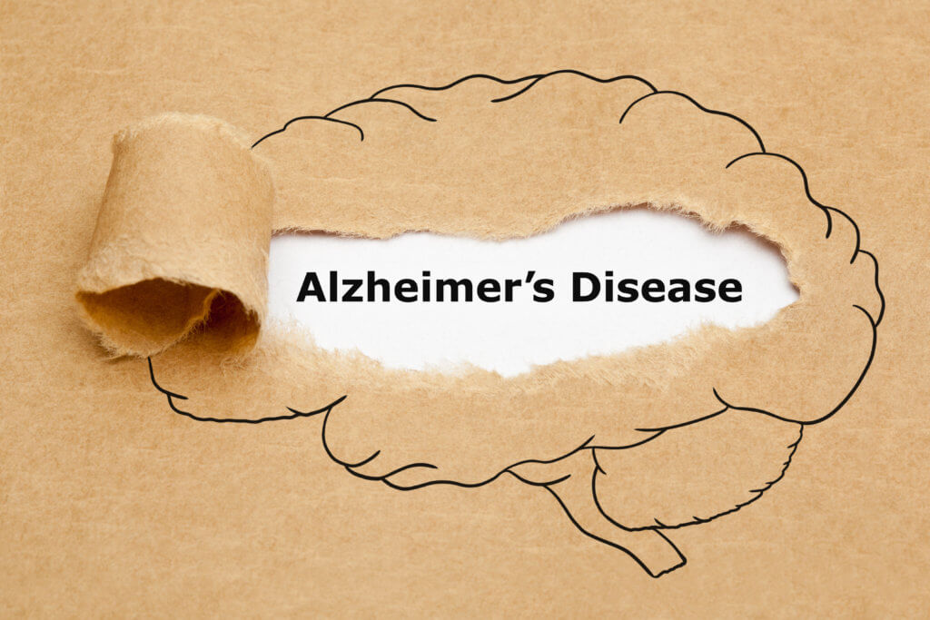
New research suggests that the origins of Alzheimer’s disease may go all the way back to when a patient is still in the womb, where abnormalities in brain development may lay the groundwork for this memory-depriving illness. These findings could potentially lead to a screening program that identifies at-risk individuals at birth.

Although there is currently no cure for Alzheimer’s, increasing attention is being paid to protective lifestyle changes, which include maintaining physical fitness and consuming a diet rich in fish, fruit, and vegetables. Neurodegenerative diseases are generally diagnosed between the ages of 40 and 60. However, it is believed that clinical symptoms emerge several decades after the onset of decline in specific brain cell connections.

A French team of scientists explain that this decline might stem from molecular-scale anomalies present from childhood, or potentially even earlier.

“We were interested in the amyloid precursor protein, or APP, which is highly expressed throughout the development of the nervous system,” says lead author Bassem Hassan of the Paris Brain Institute.

“It is an exciting research target as its fragmentation produces the famous amyloid peptides, whose toxic aggregation is associated with neuronal death observed in Alzheimer’s disease. We, therefore, suspect that APP may play a central role in the early stages of the disease,” Hassan continues in a media release.

Alzheimer's Disease

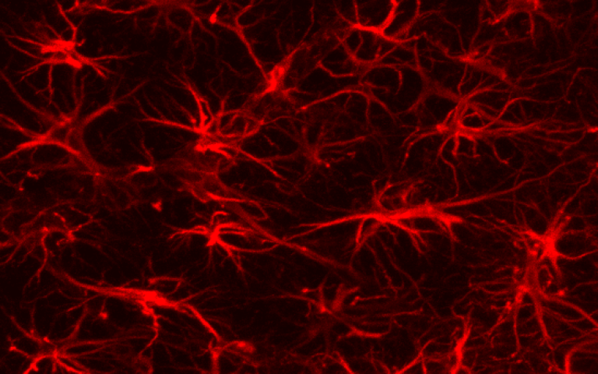
In many species, APP is involved in various biological processes, such as repairing cerebral lesions, orchestrating cellular response after oxygen deprivation or controlling brain plasticity.

APP is highly expressed during the differentiation and migration of cortical neurons, which are responsible for functions such as speech and swallowing – functions often compromised by dementia. The complex process of formation from stem cells begins in the fetus from five weeks gestation and is almost complete by 28 weeks.

“In humans, neurogenesis lasts particularly long compared with other species,” explains Khadijeh Shabani, a post-doctoral researcher at Paris Brain Institute. “Neural stem cells remain in a progenitor state for an extended period. Only later do they differentiate into glial cells, astrocytes, or oligodendrocytes that will form the architecture of the brain and spinal cord.”

Until now, it remained unclear how this balance between stem cell proliferation and differentiation into various cell types was regulated. Moreover, it was unknown whether the extended duration of human neurogenesis could potentially contribute to neurodegenerative diseases.

Historically, Alzheimer’s drugs have proved unsuccessful due to their prescription at a stage when the disease has already taken hold. However, this study, published in the journal Science Advances, paves the way for the development of medications that target APP in middle age – or even earlier.

The researchers utilized cell sequencing data from fetuses at 10 weeks and 18 weeks gestation to track APP expression during human brain development. They found that the protein was initially expressed in six cell types and later in 16 types.

By using gene editing to produce neural stem cells lacking APP expression and comparing these modified cells with those from fetuses, the researchers gathered important data.

“This comparison provided us with valuable data,” Shabani explains. “We observed that in the absence of APP, neural stem cells produced many more neurons, more rapidly, and were less inclined to proliferate in the progenitor cell state.”

brain astrocytes
Image of brain cell astrocytes

Specifically, APP regulates neurogenesis timing by influencing two finely-tuned genetic mechanisms – one chemical pathway controlling stem cell proliferation and another triggering the production of new neurons.

“In mouse models, neurogenesis is already very fast – too fast for APP deprivation to accelerate it further. We can imagine that the regulatory role of this protein is negligible in mice, while it is essential in the neurodevelopment of our species: to acquire its final form, our brain needs to generate huge quantities of neurons over a very long period, and according to a definite plan. APP-related abnormalities could cause premature neurogenesis and significant cellular stress, the consequences of which would be observable later,” suggests Hassan.

“Moreover, the brain regions in which early signs of Alzheimer’s disease appear also take the longest to mature during childhood and adolescence.”

There could be a direct link between the timing of human neurogenesis and the mechanisms of neurodegeneration. Given that the number of dementia cases worldwide is expected to triple to over 150 million by 2050 due to ageing populations, finding a therapy that targets the root cause is paramount in medical research.

APP appears to play a critical role in this respect, although further studies are necessary to confirm its centrality. Such research could significantly advance our understanding of Alzheimer’s disease and the brain’s development, potentially leading to early identification methods and new treatments.

Medicine, pills on top of brain MRI scans
(© Katsiaryna – stock.adobe.com)

The Paris Brain Institute team’s findings have added a crucial piece to the Alzheimer’s puzzle. This work suggests a paradigm shift in our understanding, positioning the disease’s origins much earlier in life than previously assumed. If their hypothesis holds true, we may be able to detect those at risk at a much earlier stage, offering the potential for targeted preventative measures and therapies.

In a world where Alzheimer’s cases are set to skyrocket, the implications of this research are vast and could significantly alter our approach to tackling this debilitating disease. The promise of a therapy that intervenes at the cause, rather than just managing symptoms, represents a “holy grail” in medical research. With Alzheimer’s’ seeds potentially sown in the womb, this research offers a new understanding that could bring us closer to achieving this goal.

“These disturbances lead to the formation of a brain that functions normally at birth but is particularly vulnerable to certain biological events – such as inflammation, excitotoxicity or somatic mutations – and certain environmental factors such as a poor diet, lack of sleep, infections, etc.,” the study authors conclude.

“Over time, these different stresses could lead to neurodegeneration – a phenomenon specific to the human species and made particularly visible by the increase in life expectancy.”

Early Sound Exposure in the Womb Shapes the Auditory System


Summary: Muffled sounds experienced in the womb prime the brain’s ability to interpret some sounds and may be key for auditory development.

Source: MIT

Inside the womb, fetuses can begin to hear some sounds around 20 weeks of gestation. However, the input they are exposed to is limited to low-frequency sounds because of the muffling effect of the amniotic fluid and surrounding tissues.

A new MIT-led study suggests that this degraded sensory input is beneficial, and perhaps necessary, for auditory development.

Using simple computer models of the human auditory processing, the researchers showed that initially limiting input to low-frequency sounds as the models learned to perform certain tasks actually improved their performance.

Along with an earlier study from the same team, which showed that early exposure to blurry faces improves computer models’ subsequent generalization ability to recognize faces, the findings suggest that receiving low-quality sensory input may be key to some aspects of brain development.

“Instead of thinking of the poor quality of the input as a limitation that biology is imposing on us, this work takes the standpoint that perhaps nature is being clever and giving us the right kind of impetus to develop the mechanisms that later prove to be very beneficial when we are asked to deal with challenging recognition tasks,” says Pawan Sinha, a professor of vision and computational neuroscience in MIT’s Department of Brain and Cognitive Sciences, who led the research team.

In the new study, the researchers showed that exposing a computational model of the human auditory system to a full range of frequencies from the beginning led to worse generalization performance on tasks that require absorbing information over longer periods of time — for example, identifying emotions from a voice clip.

From the applied perspective, the findings suggest that babies born prematurely may benefit from being exposed to lower-frequency sounds rather than the full spectrum of frequencies that they now hear in neonatal intensive care units, the researchers say.

Marin Vogelsang and Lukas Vogelsang, currently both students at EPFL Lausanne, are the lead authors of the study, which appears in the journal Developmental Science. Sidney Diamond, a retired neurologist and now an MIT research affiliate, is also an author of the paper.

Low-quality input

Several years ago, Sinha and his colleagues became interested in studying how low-quality sensory input affects the brain’s subsequent development. This question arose in part after the researchers had the opportunity to meet and study a young boy who had been born with cataracts that were not removed until he was four years old.

This boy, who was born in China, was later adopted by an American family and referred to Sinha’s lab at the age of 10. Studies revealed that his vision was nearly normal, with one notable exception: He performed very poorly in recognizing faces. Other studies of children born blind have also revealed deficits in face recognition after their sight was restored.

The researchers hypothesized that this impairment might be a result of missing out on some of the low-quality visual input that babies and young children normally receive. When babies are born, their visual acuity is very poor — around 20/800, 1/40 the strength of normal 20/20 vision. This is in part because of the lower packing density of photoreceptors in the newborn retina. As the baby grows, the receptors become more densely packed and visual acuity improves.

“The theory we proposed was that this initial period of blurry or degraded vision was very important. Because everything is so blurry, the brain needs to integrate over larger areas of the visual field,” Sinha says.

To explore this theory, the researchers used a type of computational model of vision known as a convolutional neural network. They trained the model to recognize faces, giving it either blurry input followed later by clear input, or clear input from the beginning. They found that the models that received fuzzy input early on showed superior generalization performance on facial recognition tasks.

Additionally, the neural networks’ receptive fields — the size of the visual area that they cover — were larger than the receptive fields in models trained on the clear input from the beginning.

After that study was published in 2018, the researchers wanted to explore whether this phenomenon could also be seen in other types of sensory systems. For audition, the timeline of development is slightly different, as full-term babies are born with nearly normal hearing across the sound spectrum. However, during the prenatal period, while the auditory system is still developing, babies are exposed to degraded sound quality in the womb.

To examine the effects of that degraded input, the researchers trained a computational model of human audition to perform a task that requires integrating information over long time periods — identifying emotion from a voice clip. As the models learned the task, the researchers fed them one of four different types of auditory input: low frequency only, full frequency only, low frequency followed by full frequency, and full frequency followed by low frequency.

Low frequency followed by full frequency most closely mimics what developing infants are exposed to, and the researchers found that the computer models exposed to that scenario exhibited the most generalized performance profile on the emotion recognition task. Those models also generated larger temporal receptive fields, meaning that they were able to analyze sounds occurring over a longer time period.

This shows a baby and sound waves
A new MIT-led study suggests that degraded sensory input is beneficial, and necessary, for auditory development.

https://0d962d78169d15560a8b763aea670abc.safeframe.googlesyndication.com/safeframe/1-0-38/html/container.html

This suggests, just like the vision study, that degraded input early in development actually promotes better sensory integration abilities later in life.

“It supports the idea that starting with very limited information, and then getting better and better over time might actually be a feature of the system rather than being a bug,” Lukas Vogelsang says.

Effects of premature birth

Previous research done by other labs has found that babies born prematurely do show impairments in processing low-frequency sounds. They perform worse than full-term babies on tests of emotion classification, later in life. The MIT team’s computational findings suggest that these impairments may be the result of missing out on some of the low-quality sensory input they would normally receive in the womb.

“If you provide full-frequency input right from the get-go, then you are taking away the impetus on the part of the brain to try to discover long range or extended temporal structure. It can get by with just local temporal structure,” Sinha says. “Presumably that is what immediate immersion in full-frequency soundscapes does to the brain of a prematurely born child.”

The researchers suggest that for babies born prematurely, it could be beneficial to expose them to primarily low-frequency sounds after birth, to mimic the womb-like conditions they’re missing out on.

The research team is now exploring other areas in which this kind of degraded input may be beneficial to brain development. These include aspects of vision, such as color perception, as well as qualitatively different domains such as linguistic development.

“We have been surprised by how consistent the narrative and the hypothesis of the experimental results are, to this idea of initial degradations being adaptive for developmental purposes,” Sinha says.

“I feel that this work illustrates the gratifying surprises science offers us. We did not expect that the ideas which germinated from our work with congenitally blind children would have much bearing on our thinking about audition. But, in fact, there appears to be a beautiful conceptual commonality between the two domains. And, maybe that common thread goes even beyond these two sensory modalities. There are clearly a host of exciting research questions ahead of us.”

Funding: The research was funded by the National Institutes of Health.


Abstract

Prenatal auditory experience and its sequelae

Towards the end of the second trimester of gestation, a human fetus is able to register environmental sounds. This in-utero auditory experience is characterized by comprising strongly low-pass filtered versions of sounds from the external world.

Here, we present computational tests of the hypothesis that this early exposure to severely degraded auditory inputs serves an adaptive purpose – it may induce the neural development of extended temporal integration.

Such integration can facilitate the detection of information carried by low-frequency variations in the auditory signal, including emotional or other prosodic content.

To test this prediction, we characterized the impact of several training regimens, biomimetic and otherwise, on a computational model system trained and tested on the task of emotion recognition.

We find that training with an auditory trajectory recapitulating that of a neurotypical infant in the pre-to-post-natal period results in temporally-extended receptive field structures and yields the best subsequent accuracy and generalization performance on the task of emotion recognition.

This strongly suggests that the progression from low-pass-filtered to full-frequency inputs is likely to be an adaptive feature of our development, conferring significant benefits to later auditory processing abilities relying on temporally-extended analyses.

Additionally, this finding can help explain some of the auditory impairments associated with preterm births, suggests guidelines for the design of auditory environments in neonatal care units, and points to enhanced training procedures for computational models.

  • A human fetus’ auditory experience comprises strongly low-pass-filtered versions of sounds in the environment. We examine the potential consequences of these degradations of incident sounds.
  • Results of our computational simulations strongly suggest that, rather than being epiphenomenal limitations, these degradations are likely to be an adaptive feature of our development.
  • These findings have implications for auditory impairments associated with preterm births, the design of auditory environments in neonatal care units, and enhanced computational training procedures.

Unborn Baby Partially Removed from Womb for Heart Surgery, Placed Back, Born 10 Weeks Later


In a rare, delicate, and ultimately lifesaving surgery, an unborn baby was partially removed from his mother’s womb at 26 weeks so that a tumor growing on his heart could be removed. He was then placed back in his mom’s womb for a further 10 weeks before being born at nearly full term.

Doctors at Cleveland Clinic, Ohio, said in a press release the baby is only the second person in the world to undergo this unique surgery, and survive. Today, he is thriving.

Baby Rylan Harrison Drinnon was diagnosed in the spring of 2021 with intrapericardial teratoma with fetal hydrops in utero, an extremely rare condition leading to heart failure if left untreated, according to the statement.

Epoch Times Photo
Dr. Hani Najm, who led the heart surgery team, inserts an IV line in the fetus’s right arm to deliver fluids and medications as needed.

“As far as we know, Cleveland Clinic is the second academic medical center in the world to have performed this fetal surgery successfully with continued pregnancy and delivery,” said Dr. Darrell Cass, director of Cleveland Clinic’s Fetal Surgery and Fetal Care Center.

“In this case, time was of the essence. Shortly after the patient arrived at Cleveland Clinic, imaging tests showed that the tumor kept growing and the fetus’s heart function was deteriorating.”

The malignant mass was compressing the left side of unborn baby Rylan’s heart, cutting off circulation and leading to an accumulation of fluid around his heart and other organs.

Parents Sam and Dave Drinnon of Pittsburgh were referred to Cleveland for their expertise, said Cass. A multidisciplinary team from Cleveland Clinic and Children’s Clinic performed surgery in May 2021 to remove Rylan’s tumor.

After making a “Caesarean section-like incision” to expose the mother’s uterus, the team, led by Dr. Hani Najm, Cleveland Clinic’s chair of pediatric and congenital heart surgery, used ultrasound to locate the placenta and fetus. They opened the uterus and lifted out Rylan’s arms to expose his chest. Najm removed the tumor from the baby’s beating heart before placing him back in the uterus, in a surgery lasting 3 1/2 hours.

“As soon as the tumor was removed, the compression of the left atrium disappeared, and there was a nice blood flow that was almost back to normal,” Najm reported.

Epoch Times Photo
Baby Rylan Harrison Drinnon.

Both mom and baby recovered well, and Rylan was able to remain in the womb until near full term, according to the statement.

Maternal-fetal medicine specialist Dr. Amanda Kalan, who attended the surgery, oversaw Sam’s aftercare and the delivery of her healthy baby boy by C-section on July 13, 10 weeks after the surgery.

Cass expressed pride in his team for their massive success.

Epoch Times Photo

“This tumor was growing rapidly in the exact wrong spot,” Cass explained. “We needed to act quickly and decisively to rescue the fetus … as far as we know, Cleveland Clinic is the second academic medical center in the world to have performed this fetal surgery successfully, with continued pregnancy and delivery.”

Only one previous incidence, said Cass, has ever been documented in the world’s medical literature.

Najm claimed that such innovative fetal surgery “provides hope to other families who may receive a similar devastating diagnosis.”

Looking to the future, Rylan will likely need surgery to reposition his sternum, which did not heal properly in the womb. Doctors will monitor his heart health as he grows to ensure the tumor does not reappear.

The Drinnons are beyond grateful for the lifesaving intervention.

“Now they have this beautiful boy, Rylan, and they think he’s going to be special,” Cass, told Cleveland.com. “He’s going to grow up to be a completely normal kid that just had a really unique odyssey to get to where he is now.”

In Addition to Testosterone, Another Hormone Is Vital for Early Male Development


A hormone called androsterone, produced in the placenta and other organs, plays a role in fetal development in the womb

Baby
While testosterone plays a significant role in fetal development, it is not the only hormone that influences masculinization. 

Often the first question parents are asked after the birth of their child is “congratulations, girl or boy?” For parents of one in 2,000 to 4,000 births, however, there is not an easy answer. This is when the baby has “ambiguous” genitalia, where it is not clear which sex they belong to. In baby boys, this was long thought to be caused by problems linked to testosterone—as were more common disorders such as undescended testicles and malformed penises, which respectively occur in 9 percent and 1 percent of births.

But now it is clear that the reality is slightly different. According to new research in which I am a co-author, another hormone known as androsterone—which originates in the placenta and fetal adrenal gland—is also vital to the process that turns fetuses in boys. These insights have the potential to make a big difference to how we treat sexual disorders in male babies in future—and are also relevant to the whole debate about male and female identity.

Even small children are aware that men and women usually look different. It is common knowledge that boys become men because the testes of the man produce the “male” hormone testosterone and, in turn, testosterone makes men masculine. We know this thanks to the French endocrinologist Alfred Jost’s groundbreaking studies in the early 1950s.

There are several times in boys’ lives in which bursts of testosterone play a key role in their development as males. The most well known is of course puberty, in which the testes start making much more testosterone. This makes boys hairier, grows their genitals and makes their voices break.

The other times are the “mini-puberty” that takes place at around three months after birth, which leads certain changes in the testes and brain; and when a boy is still a fetus in the womb, around three months into his mother’s pregnancy. While all these bursts of testosterone are probably very important in making a normal male, it is the one in the womb that affects whether the child will be a boy at all. What is now clear is that testosterone and the testes have been hogging the podium when in fact we need to share the honors around.

Testosterone and super-testosterone

Testosterone is part a family of male sex hormones called androgens. To get a normal male, testosterone needs to be turned into another androgen called dihyrotestosterone or DHT, a “super-testosterone” that is five times more potent than its cousin. This conversion is done in the tissue of what will become the penis, along with the other parts of the body that develop male characteristics. The consequences of the process are clear: boys who cannot turn testosterone into DHT are born looking female and only become more obviously male at puberty.

These include the Guevedoces in the Dominican Republic, who, due to a genetic mutation, lack the enzymes to make the DHT conversion. Studying these extraordinary children in the early 1970s led the American researcher Julianne Imperato-McGinley to develop the drug finasteride to treat prostate cancer.

For years, this story was considered complete—masculinization was due to testosterone and the conversion of testosterone to DHT. Then an Australian zoologist named Marilyn Renfree, in an elegant series of studies in the 2000s, published the first evidence that things may not be that simple. She was actually studying wallabies, since the young in the pouch were easily accessible for experimental purposes and they mimic much of the period of pregnancy in humans and other mammals with placentas. Renfree found that the genitals of the young male wallabies made DHT even without testosterone from their testes. The only reliable conclusion was that they were converting other androgens to DHT.

It became clear that there are two ways to make a “male signal” in a wallaby fetus, both of which are necessary to normal sexual development. The first is by testosterone from the testes. The second is through different androgens that can also be made by other organs in the human, including the fetus’s adrenal glands, liver and the placenta. These other processes came to be known as the “backdoor” pathway.

But was the same thing true in humans? It was later shown that it was, by studying male human newborns who were not properly masculinized; they had undescended testes and ambiguous genitals, despite having testes that made testosterone. It turned out they were unable to make the backdoor androgens because they had mutations in the genes of enzymes that were key to the process of the conversion into DHT.

As further evidence that both types of male signal are essential to normal development of human male fetuses, it was also discovered that fetuses whose placentas are not working properly are around twice as likely to be born with undescended testes or with malformed penises—especially if they are also born abnormally small (for their gestational age).

What we have shown

In our research, which also involved the University of Glasgow and French and Swedish collaborators, we have been able to explain why. We measured the levels of different male sex hormones in the blood of male and female fetuses, and were surprised to find that only two androgens were higher in males than females: testosterone and androsterone. The relevance to the placenta is that it is up to 6,000 times heavier than the fetus and it makes large amounts of a hormone called progesterone, which it can convert into androsterone—as can the fetal liver and adrenal glands. The human fetuses’ testes have no ability to make this conversion.

Fetus Development
Fetal development.

We then also showed that the testosterone and androsterone were converted into DHT in male target tissues like the penis. And not only are both androgens required to masculinize the fetus, there can be abnormalities where levels are lower than normal: for example, a good index of the degree of masculinization is the distance between the anus and genitals, and this is shorter than usual in newborns with malformed penises.

People affected by disorders of sexual development, including malformed penises, can have a very difficult time and face delicate surgery, hormone therapy and other treatments. Every new piece of information into how masculinization happens raises the prospect of improving when and how these disorders are detected and treated in future. Early enough diagnosis of reduced placental function related to androgen production in early pregnancy might enable treatment before penis formation is complete, avoiding the need for corrective surgery later in life.

A final take-home message from our study is that while testosterone and androsterone are indeed higher on average in male than female fetuses, the difference is quite small. There is also considerable overlap between the lowest levels in boys and the highest levels in girls. Those in society who are adamant that the only choice for people is a binary choice of man or woman are not basing their views on biological reality. Treasured beliefs about the supremacy of testosterone and the testes in making a man are also obviously flawed.

Big Pharma now wants to vaccinate babies while they’re still in the womb


It’s bad enough when pregnant mothers get vaccinated, but now Big Pharma wants to take vaccination to a whole new level. Instead of just vaccinating mothers-to-be, the industry wants to be able to inject unborn babies with their latest concoctions, too.

Image: Big Pharma now wants to vaccinate babies while they’re still in the womb

The 21st Century Cures Act is poised to pass any day now. While this legislation claims to bring medicine into the 21st century, it honestly seems intended to do the exact opposite. Instead of looking forward to new horizons, this bill would merely double down on conventional methods that have been in use for decades. The 21st Century Cures bill managed to pass the House last year, and after being broken up into smaller sections by the Senate, is set to see approval any day now.

One of the more concerning aspects of this bill lies in the Senate bill S. 2742 (the Promoting Biomedical Research and Public Health for Patients Act), which appears to be laying down the groundwork for amending the current vaccination schedule to include expectant mothers and their still-developing, unborn children. This particular provision contains the following language:

Notwithstanding any other provision of law, for purposes of this subtitle, both a woman who received a covered vaccine while pregnant and any child who was in utero at the time such woman received the vaccine shall be considered persons to whom the covered vaccine was administered and persons who received the covered vaccine.

Now, this could be interpreted in any number of ways. It may, for example, simply be a form of liability coverage. But, it could also be paving the way for unborn children to be considered eligible for vaccination. If this is the case, it won’t be long before we see manufacturers developing new lines of vaccines designed with babies in utero in mind.

The Centers for Disease Control already suggests that pregnant women get a whole host of vaccinations – whooping cough, flu and hepatitis B vaccines are all pushed on mothers-to-be as the epitome of protection for their child – but many question the necessity and use of such injections.

In addition to these concerns, the new bill may also open doors for the current vaccination schedule to be shifted or expanded, and create a slew of opportunities for the industry to increase their profit margins. As the Alliance for Natural Health (ANH) suggests, “Maybe the first DTaP (diphtheria, tetanus, and pertussis) shot will be given to the mother and the fetus, rather than to the child at one month. Perhaps the schedule will be expanded.” This is a great example of what the 21st Century Cures act could allow to take place.

There are many potential risks to doing this; for example, the aluminum adjuvants found in many vaccines have never been tested for safety in children, let alone unborn babies. And, as the ANH notes, there doesn’t seem to be any research on the safety of multiple vaccinations being administered during pregnancy, nor any kind of comparison between the potential risks and benefits.

This is far from the only concerning aspect of the bill; many other parts of it have also been raising a few eyebrows. For example, many critics say that the bill goes way too far when it comes to relaxing approval standards for drugs and devices. For example, one provision in the Cures Act would allow a company to obtain approval for a second use of an FDA-approved drug, without having to conduct a randomized clinical trial. Instead, companies could use something known as “real world evidence.” Real world evidence is a much lower standard of evidence – and it lacks any assessment of risk versus benefits. It’s more subject to bias, more easily manipulated, and in general, can be very misleading.

Heritage Action, a conservative group, commented that the Act had expanded from 300 pages last year, to “an almost 1,000-page omnibus health care spending bill.”

Heritage Action went on to state, “In Washington terms, back-room negotiators have turned the Cures bill into a Christmas Tree, loaded with handouts for special interests, all at the expense of the taxpayer.”

Across the aisle, Senator Elizabeth Warren has vehemently opposed the bill and lambasted the legislation as being nothing more than a license for corporate “fraud,” “bribery” and “extortion.” Just seven Democratic senators voted against the legislation, while 70 Republicans voted to shoot it down.

In her floor speech, Senator Warren stated, “Pushing treatments without scientific evidence that they work is fraud—fraud that can hurt people.” Hopefully, she and the rest of the bill’s opponents from both political parties can band together and stop the bill, and the establishment, before it’s too late.