Diabetes and liver cancer.


A Stanford Medicine study identifies an easily measured biophysical property that can identify Type 2 diabetics at increased risk for liver cancer who don’t meet current screening guidelines.
test

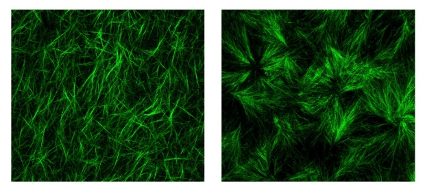
Collagen fibers in a three-dimensional hydrogel (left panel). When advanced glycation end products — a byproduct of poorly controlled diabetes — are added (right), the fibers are shorter and more cross-linked, creating conditions that favor cancer progression.
Weiguo Fan

For centuries, doctors have used their hands as essential diagnostic tools — exploring joints and palpating abdomens to assess a patient’s health. Often a cancer will reveal itself as a lump or unusual stiffness in a normally bouncy tissue or organ.

More recently, the relationship between stiffness and cancer has been documented through biophysical studies and clinical trials, particularly in liver and breast cancer. For example, stiffness is a primary hallmark of liver cirrhosis, which can progress to liver cancer.

Now researchers at Stanford University have shown that another biophysical characteristic known as viscoelasticity — think of how stretching a ball of Silly Putty or a clump of bread dough is met at first with resistance, and then with release — is even more tightly correlated with liver cancer than stiffness, particularly in people with Type 2 diabetes.

The distinction matters because people with Type 2 diabetes are two to three times as likely as people without diabetes to develop liver cancer, which often occurs in the absence of cirrhosis. Liver cancer rates are increasing in part because the prevalence of diabetes is growing worldwide, particularly in marginalized communities where healthy food choices and opportunities for regular exercise are scarce.

“This is the first time that the dogma of matrix stiffness as a primary predictor of liver cancer is being challenged,” said professor of gastroenterology and hepatology Natalie Torok, MD. “Current guidelines recommend routine liver cancer screening only for people with cirrhosis. As a result, many people with Type 2 diabetes are not screened at all. These new findings have major implications not just for liver cancer, but also other cancers for which diabetes is a risk factor, including breast cancers.”

Torok is the senior author of the study, which was published online Jan. 31 in Nature. Postdoctoral scholar Weiguo Fan, PhD, is the lead author.

Torok and her colleagues collaborated with researchers in the laboratory of associate professor of mechanical engineering Ovijit Chaudhuri, PhD, to investigate the role of viscoelasticity in liver cancer in patient samples, animal models and cells grown in the laboratory in a Jell-O-like tissue scaffolding called a hydrogel.

“This study is the first on the role of viscoelasticity in cancer with data that spans from humans and mouse models to in vitro 3D culture studies and computational simulations,” Chaudhuri said. “It definitively establishes the role of viscoelasticity in liver cancer progression.”

The study was supported by Stanford Medicine’s SPARK Program in Translational Research and its Innovative Medicines Accelerator, complementary programs meant to streamline the path of promising academic laboratory discoveries to clinical applications that benefit patients.

Stiffness test

Liver stiffness is measured non-invasively with imaging techniques called transient elastography or MR elastography involving a vibrating pad placed on the abdomen. The vibrations are transmitted from the imaging probe to the organ; the wave of vibration moving through a stiff medium differs from one moving through something more malleable. People with a liver stiffness that exceeds a certain threshold are diagnosed with liver cirrhosis; current guidelines recommend that they be screened every six months for liver cancer with an abdominal ultrasound and blood tests.

Measurements such as stiffness arise from what’s called the extracellular matrix — the space between and around an organ’s cells that is chock-a-block with proteins, sugars and minerals.

“Our organs are not just clumps of cells,” Chaudhuri said. “The cells exist in a scaffolding called the extracellular matrix that gives them physical support but also affects their maturation, specialization and functioning.”

Like a grade-school teacher, the matrix provides a physical framework that supports and organizes the cells, gentling and channeling them to harmoniously create a functional tissue. When the matrix is disrupted, any bad-apple cancerous or pre-cancerous cells more readily lose their way, spreading to places they shouldn’t; dividing uncontrollably; or morphing in to other, more dangerous versions of themselves.

People with diabetes have elevated levels of what are called advanced glycation end products, or AGEs. AGEs arise when blood sugar is poorly controlled and elevated levels of sugar molecules known as glucose begin to glom onto nearby proteins including collagen — a key structural component of the extracellular matrix. (AGEs are also present in protein- or fat-rich foods or in foods prepared at high heat such as frying or broiling.)

The researchers found that liver samples from people with Type 2 diabetes had higher levels of AGEs and were more viscoelastic — but no more stiff — than liver samples from people without Type 2 diabetes. A closer look in laboratory mice showed animals fed a diet high in AGEs had shorter and less interconnected collagen fibers in the liver’s extracellular matrix than those found in animals fed standard chow. 

Next, the researchers studied how cells behaved when grown in the laboratory in a three-dimensional gel to mimic liver matrix structure. Tinkering with the cells outside the body allowed them to assess the effect of various changes in their growth and behavior.

“In our engineered hydrogels, we can tune one biophysical property such as viscoelasticity or stiffness at a time to understand how each property impacts the cells,” Chaudhuri said. “We saw that a change in viscoelasticity alone is enough to drive a more invasive behavior in the cells.”

In particular, the researchers noted that a more viscoelastic matrix promotes changes in liver cell shape and allows the formation of invasive protrusions on their membranes that help them escape natural barriers meant to keep cells in their rightful places.

Paradigm shift

Finally, Torok and her colleagues went one step further, dissecting a series of cellular signals that promote liver cancer progression in viscoelastic conditions that includes a cancer-associated protein called YAP.

“This is the first time that changes in collagen structure have been proven to promote viscoelasticity and liver cancer progression independent of stiffness,” Torok said. “It’s a complete change in paradigm that could explain the greater risk of liver cancer in people with Type 2 diabetes and may help select people should undergo regular liver cancer screening.”

Fortunately, like stiffness, viscoelasticity can be assessed non-invasively with MR elastography by changing a few parameters in vibration frequency and measurement. Torok is planning to launch a clinical trial to further study viscoelasticity, Type 2 diabetes and liver cancer progression.

“One of the major questions in medicine today is why people with diabetes and fatty liver disease are so prone to liver cancer, and how we can address this,” Torok said. “Our research suggests that many more people, particularly those with diabetes, should be screened for cancer. If we did that, perhaps we could act earlier and save lives.”

Researchers from Purdue University; Tsinghua University in Beijing; the University of Pittsburgh; the University of California, Davis; Albert Einstein College of Medicine; Keio University in Yokohama; and the University of Pennsylvania contributed to the work.

Machine Learning-Based Models Predict HCC Treatment Response


Applying machine learning-based models to presently underutilized imaging features could help construct more reliable criteria for organ allocation and liver transplant eligibility in patients with hepatocellular carcinoma (HCC), according to a study published in the American Journal of Roentgenology.

“The findings suggest that machine learning-based models can predict recurrence before therapy allocation in patients with early-stage hepatocellular carcinoma initially eligible for liver transplant,” wrote corresponding author Julius Chapiro, MD, from the department of radiology and biomedical imaging at Yale University School of Medicine in New Haven, CT.

Dr Chapiro and colleagues’ proof-of-concept study included 120 patients (88 men, 32 women; median age, 60 years) diagnosed with early-stage HCC between June 2005 and March 2018, who were initially eligible for liver transplant and underwent treatment by transplant, resection, or thermal ablation. Patients underwent pretreatment MRI and posttreatment imaging surveillance, and imaging features were extracted from postcontrast phases of pretreatment MRI examinations using a pretrained convolutional neural network (VGG-16). Pretreatment clinical characteristics (including laboratory data) and extracted imaging features were integrated to develop three ML models—clinical, imaging, combined—for recurrence prediction within 1–6 years posttreatment.

Ultimately, all three models predicted posttreatment recurrence for early-stage HCC from pretreatment clinical (AUC 0.60–0.78, across all six time frames), MRI (AUC 0.71–0.85), and both data combined (AUC 0.62–0.86). Using imaging data as the sole model input yielded higher predictive performance than clinical data alone; however, combining both data types did not significantly improve performance over use of imaging data alone.

Heavy alcohol use increases liver impairment, risk for HCC in hepatitis-induced cirrhosis


Among patients with hepatitis-induced cirrhosis, heavy alcohol consumption increased liver function impairment and hepatocellular carcinoma prevalence, according to research.

“The interaction between alcohol and hepatitis virus B or C has been widely studied recently with non-unanimous conclusions. Heavy alcohol consumption had been shown to accelerate the development of liver fibrosis and to increase the prevalence of HCC and mortality in HCV infected patients. Concerning the interaction between alcohol and HBV, less conclusive studies have been done,” Kodjo-Kunale Abassa, from the department of gastroenterology at The Third Affiliated Hospital of Sun Yat-Sen University in China, and colleagues wrote. “Nevertheless, more needs to be explored about the prevalence of other liver cirrhosis complications such as esophageal and gastric variceal bleeding (EGVB) among these groups of patients.”

Patients with combined alcohol-induced liver disease and HBV had an increased risk for: Hepatocellular carcinoma; OR = 2.01; Esophageal and gastric variceal bleeding; OR = 1.74

To investigate the influence of alcohol on clinical outcomes in patients with HBV and HCV-induced liver cirrhosis, researchers retrospectively reviewed medical records of 22,287 patients (mean age, 52 years; 84.1% men) diagnosed with liver cirrhosis from January 2010 to December 2019. They divided patients into groups by etiology (alcohol-induced liver disease [ALD]: 1,652; HBV: 18,079; HCV: 682; combined ALD and HBV: 1,594; and combined ALD and HCV: 280) and compared liver function impairment severity and liver cirrhosis complications using laboratory data.

Compared with the HBV group, a greater proportion of patients in the combined ALD and HBV group had a Child Pugh grade C (18.8% vs. 28%; < .001) or Model for End-Stage Liver Disease (MELD) score greater than 18 (18.5% vs. 24.1%; P < .001) with a 2.01-fold and 1.74-fold higher risk for HCC (OR = 2.01; 95% CI, 1.58-2.55) and EGVB (OR = 1.74; 95% CI, 1.3-2.33). Similarly, compared with the HCV group, a greater proportion of patients in the combined ALD and HCV group had a MELD score greater than 18 (7.6% vs. 13.3%; P < .05).

Researchers noted a decreased risk for HCC and EGVB following alcohol abstinence; patients who abstained from alcohol use and received antiviral treatment had the lowest risk for HCC (OR = 0.1; 95% CI, 0.05-0.2) and EGVB (OR = 0.17; 95% CI, 0.06-0.45).

“Alcohol increased significantly the severity of liver function impairment and the prevalence of liver cirrhosis complications, particularly HCC and EGVB, in hepatitis virus-induced liver cirrhosis patients (HBV and HCV),” Abassa and colleagues concluded. “Remarkably, long-term abstinence from alcohol coupled with efficient antiviral treatment effectively decreased the prevalence of HCC and EGVB in these populations, thus the importance of stressing not only on the importance of antiviral treatment in patients with coexisting ALD and viral cirrhosis, but also on long-term alcohol abstinence.”

Dual hepatitis B and C-associated hepatocellular carcinoma: clinical characteristics, outcome, and prognostic role of albumin–bilirubin grade


Abstract

Background

Albumin–bilirubin (ALBI) grade is used to evaluate the outcome of patients with hepatocellular carcinoma (HCC) which is often associated with chronic hepatitis B virus (HBV) or hepatitis C virus (HCV) infection. This study aimed to investigate the clinical characteristics, outcome, and prognostic role of ALBI grade in dual HBV/HCV-related HCC.

Methods

A total 3341 HCC patients with viral etiology were prospectively enrolled and retrospectively analyzed. Multivariate Cox proportional hazards model was used to identify independent prognostic predictors.

Results

Of all patients, 2083 (62%), 1068 (32%), and 190 (6%) patients had HBV, HCV, and dual HBV/HCV infection, respectively. The mean age of HBV, HCV, and dual virus group was 60, 68, and 64 years (p < 0.001), respectively. There was no significant survival difference between HBV, HCV, and dual HBV/HCV-related HCC group (p = 0.712). Multivariate Cox analysis in dual HBV/HCV-related HCC showed that multiple tumors [hazard ratio (HR): 1.537, p = 0.044], tumor size >3 cm (HR 2.014, p = 0.044), total tumor volume (TTV) >50 cm3 (HR 3.050, p < 0.001), vascular invasion (HR 3.258, p < 0.001), performance status 2–4 (HR 2.232, p < 0.001), ALBI grade 2–3 (HR 2.177, p < 0.001), and BCLC stage B–D (HR 2.479, p < 0.001) were independent predictors of poor survival.

Conclusions

Dual viral infection does not accelerate the development of HCC in HBV carriers. Patient survival is similar between dual HBV/HCV-related HCC and single HBV- or HCV-related HCC group. The ALBI grade is a robust prognostic model in dual virus-related HCC to discriminate patient long-term survival.

Glasgow Prognostic Score as a useful prognostic factor after hepatectomy for hepatocellular carcinoma.


Abstract

Background

Several previous studies have revealed that the Glasgow Prognostic Score (GPS) is a clinically useful scoring system to predict the prognosis of patients with various kinds of advanced cancers. However, there have been few reports on the relationship between the GPS and prognosis after hepatectomy for hepatocellular carcinoma (HCC). Therefore, we performed an analysis of the relationship between the GPS and prognosis after hepatectomy for HCC.

Methods

Between January 2005 and December 2009, 352 HCC patients underwent hepatectomy at Kumamoto University Hospital. Nineteen clinicopathologic factors were analyzed, using univariate and multivariate analyses.

Results

Univariate analysis showed that significant risk factors for poor survival included serum albumin level (<3.5 g/dL), tumor size (>35 mm), presence of ascites, portal vein invasion, operation time (>400 min), blood loss (>360 mL), requirement for blood transfusion, and GPS. Multivariate analysis revealed that tumor size [hazard ratio (HR) 3.355; p = 0.003], operation time (HR 2.634; p = 0.006), portal vein invasion (HR 2.419; p = 0.009), and GPS (HR 3.796; p < 0.001) were independent factors for poor prognosis.

Conclusion

The GPS was demonstrated to be a statistically significant prognostic factor after hepatectomy for HCC.

Source: International Journal of Clinical Oncology

Researchers Identify Liver Cancer Progenitor Cells Before Tumors Become Visible.


Stained liver biopsy micrograph showing hepatocellular carcinoma cells with Mallory bodies (reds and blacks).
For the first time, researchers at the University of California, San Diego School of Medicine have isolated and characterized the progenitor cells that eventually give rise to malignant hepatocellular carcinoma (HCC) tumors – the most common form of liver cancer. The researchers found ways to identify and isolate the HCC progenitor cells (HcPC) long before actual tumors were apparent.

<p>Stained liver biopsy micrograph showing hepatocellular carcinoma cells with Mallory bodies (reds and blacks).</p>

Writing in the October 10, 2013 issue of the journal Cell, principal investigator Michael Karin, PhD, Distinguished Professor of Pharmacology and Pathology, and colleagues report that HcPC take form within dysplastic or abnormal lesions often found in damaged or cirrhotic livers. The liver damage can be due to viral infections like hepatitis or from chronic alcohol abuse.

“It was never established whether dysplastic lesions are just a regenerative (healing) response of the liver triggered by tissue damage or are actually pre-malignant lesions that harbor tumor progenitor cells,” said study co-author Debanjan Dhar, PhD, a postdoctoral researcher in Karin’s lab. “Here we show that HcPC are likely derived from dysplastic lesions, can progress to malignant tumors and further demonstrate that the malignant progression of HcPC to full-blown liver cancer depends upon the microenvironment that surrounds them.”

The researchers were able to characterize HcPC based on several biomarkers that distinguish them from normal cells. They also identified cellular signaling pathways activated in HcPC that are critical “to their malignant potential,” said Dhar.

The findings may have profound implications for treating HCC which, while relatively rare in the United States compared to other types of cancer, is difficult to diagnose and treat, with poor prognoses for patients. HCC is usually fatal within three to six months of diagnosis, according to National Institutes of Health data. An estimated 30,000 new cases of liver cancer are diagnosed annually in the U.S., predominantly among men. More than 21,600 Americans die from liver cancer each year, a rate that has been rising slowly for several decades. In other parts of the world, HCC is a major cause of cancer-related deaths.

Most cancers are best detected and treated at the earliest possible stage. HCC is problematic because it develops slowly and frequently displays no symptoms. By the time it is detected, said Dhar, it is usually at an advanced stage with no effective therapy.

“Our findings can be translated into both early detection and therapeutic intervention,” he said. “Better understanding of HcPC cellular networks will provide us with new and effective therapeutic targets.”

For example, the researchers were able to detect “potential” malignant lesions in needle biopsies of a subset of patients infected with the hepatitis C virus, but who hadn’t yet developed HCC. Hepatitis C is a major risk factor for HCC development.

Dhar said identifying premalignant lesions in high-risk patients based on HcPC markers would allow for earlier detection and therapeutic interventions. “Furthermore, in future, therapies can be developed to specifically eliminate the HcPC even before a tumor has developed.”

Co-authors include Guobin He, Joan Font-Burgada, Yuhong Jiang and Shabnam Shalapour, Laboratory of Gene Regulation and Signal Transduction, Departments of Pharmacology and Pathology, UCSD; Hayato Nakagawa, Laboratory of Gene Regulation and Signal Transduction, Departments of Pharmacology and Pathology, UCSD and Department of Gastroenterology, University of Tokyo; Hisanobu Ogata, Laboratory of Gene Regulation and Signal Transduction, Departments of Pharmacology and Pathology, UCSD and Department of Medicine and Clinical Science, Kyushu University, Japan; Ekihiro Seki, Department of Medicine, UCSD; Shawn E. Yost, Bioinformatics Graduate Program and Rady Children’s Hospital-San Diego and Department of Pediatrics, UCSD; Kristen Jepsen, Rady Children’s Hospital-San Diego and Department of Pediatrics, UCSD; Kelly A. Frazer, Rady Children’s Hospital-San Diego and Department of Pediatrics, UCSD, UC San Diego Moores Cancer Center, Clinical and Translational Research Institute, UCSD and Institute for Genomic Medicine, UCSD; Olivier Harismendy, Rady Children’s Hospital-San Diego and Department of Pediatrics, UCSD, UC San Diego Moores Cancer Center, Clinical and Translational Research Institute, UCSD; Maria Hatziapostolou and Dimitrios Iliopoulos, Center for Systems Biomedicine, Geffen School of Medicine, UCLA; Atsushi Suetsugu and Robert M. Hoffman, Department of Surgery, UCSD and Anticancer, Inc., San Diego; and Ryosuke Tateishi  and Kazuhiko Koike, Department of Gastroenterology, University of Tokyo.

Funding support for this research came, in part, from the Superfund Basic Research Program, the National Institutes of Health (CA118165 and CA155120), Wellcome Trust, American Diabetes Association, the Center for Translational Science, the National Center for Research Resources IMAT program, postdoctoral research fellowships from the Damon Runyon Cancer Research Foundation, the American Liver Foundation, Daiichi Sankyo Foundation of Life Science, the California Institute for Regenerative Medicine Stem Cell Training Grant II, Kanzawa Medical Research Foundation, the German Research Foundation and a Young Investigator Award from the National Childhood Cancer Foundation “CureSearch.”

<p>Stained liver biopsy micrograph showing hepatocellular carcinoma cells with Mallory bodies (reds and blacks).</p>
Stained liver biopsy micrograph showing hepatocellular carcinoma cells with Mallory bodies (reds and blacks).
For the first time, researchers at the University of California, San Diego School of Medicine have isolated and characterized the progenitor cells that eventually give rise to malignant hepatocellular carcinoma (HCC) tumors – the most common form of liver cancer. The researchers found ways to identify and isolate the HCC progenitor cells (HcPC) long before actual tumors were apparent.

Writing in the October 10, 2013 issue of the journal Cell, principal investigator Michael Karin, PhD, Distinguished Professor of Pharmacology and Pathology, and colleagues report that HcPC take form within dysplastic or abnormal lesions often found in damaged or cirrhotic livers. The liver damage can be due to viral infections like hepatitis or from chronic alcohol abuse.

“It was never established whether dysplastic lesions are just a regenerative (healing) response of the liver triggered by tissue damage or are actually pre-malignant lesions that harbor tumor progenitor cells,” said study co-author Debanjan Dhar, PhD, a postdoctoral researcher in Karin’s lab. “Here we show that HcPC are likely derived from dysplastic lesions, can progress to malignant tumors and further demonstrate that the malignant progression of HcPC to full-blown liver cancer depends upon the microenvironment that surrounds them.”

The researchers were able to characterize HcPC based on several biomarkers that distinguish them from normal cells. They also identified cellular signaling pathways activated in HcPC that are critical “to their malignant potential,” said Dhar.

The findings may have profound implications for treating HCC which, while relatively rare in the United States compared to other types of cancer, is difficult to diagnose and treat, with poor prognoses for patients. HCC is usually fatal within three to six months of diagnosis, according to National Institutes of Health data. An estimated 30,000 new cases of liver cancer are diagnosed annually in the U.S., predominantly among men. More than 21,600 Americans die from liver cancer each year, a rate that has been rising slowly for several decades. In other parts of the world, HCC is a major cause of cancer-related deaths.

Most cancers are best detected and treated at the earliest possible stage. HCC is problematic because it develops slowly and frequently displays no symptoms. By the time it is detected, said Dhar, it is usually at an advanced stage with no effective therapy.

“Our findings can be translated into both early detection and therapeutic intervention,” he said. “Better understanding of HcPC cellular networks will provide us with new and effective therapeutic targets.”

For example, the researchers were able to detect “potential” malignant lesions in needle biopsies of a subset of patients infected with the hepatitis C virus, but who hadn’t yet developed HCC. Hepatitis C is a major risk factor for HCC development.

Dhar said identifying premalignant lesions in high-risk patients based on HcPC markers would allow for earlier detection and therapeutic interventions. “Furthermore, in future, therapies can be developed to specifically eliminate the HcPC even before a tumor has developed.”

Co-authors include Guobin He, Joan Font-Burgada, Yuhong Jiang and Shabnam Shalapour, Laboratory of Gene Regulation and Signal Transduction, Departments of Pharmacology and Pathology, UCSD; Hayato Nakagawa, Laboratory of Gene Regulation and Signal Transduction, Departments of Pharmacology and Pathology, UCSD and Department of Gastroenterology, University of Tokyo; Hisanobu Ogata, Laboratory of Gene Regulation and Signal Transduction, Departments of Pharmacology and Pathology, UCSD and Department of Medicine and Clinical Science, Kyushu University, Japan; Ekihiro Seki, Department of Medicine, UCSD; Shawn E. Yost, Bioinformatics Graduate Program and Rady Children’s Hospital-San Diego and Department of Pediatrics, UCSD; Kristen Jepsen, Rady Children’s Hospital-San Diego and Department of Pediatrics, UCSD; Kelly A. Frazer, Rady Children’s Hospital-San Diego and Department of Pediatrics, UCSD, UC San Diego Moores Cancer Center, Clinical and Translational Research Institute, UCSD and Institute for Genomic Medicine, UCSD; Olivier Harismendy, Rady Children’s Hospital-San Diego and Department of Pediatrics, UCSD, UC San Diego Moores Cancer Center, Clinical and Translational Research Institute, UCSD; Maria Hatziapostolou and Dimitrios Iliopoulos, Center for Systems Biomedicine, Geffen School of Medicine, UCLA; Atsushi Suetsugu and Robert M. Hoffman, Department of Surgery, UCSD and Anticancer, Inc., San Diego; and Ryosuke Tateishi  and Kazuhiko Koike, Department of Gastroenterology, University of Tokyo.

Funding support for this research came, in part, from the Superfund Basic Research Program, the National Institutes of Health (CA118165 and CA155120), Wellcome Trust, American Diabetes Association, the Center for Translational Science, the National Center for Research Resources IMAT program, postdoctoral research fellowships from the Damon Runyon Cancer Research Foundation, the American Liver Foundation, Daiichi Sankyo Foundation of Life Science, the California Institute for Regenerative Medicine Stem Cell Training Grant II, Kanzawa Medical Research Foundation, the German Research Foundation and a Young Investigator Award from the National Childhood Cancer Foundation “CureSearch.”

Keratin 19: a key role player in the invasion of human hepatocellular carcinomas.


Abstract

Objective Keratin (K)19, a biliary/hepatic progenitor cell (HPC) marker, is expressed in a subset of hepatocellular carcinomas (HCC) with poor prognosis. The underlying mechanisms driving this phenotype of K19-positive HCC remain elusive.

Design Clinicopathological value of K19 was compared with EpCAM, and α-fetoprotein, in a Caucasian cohort of 242 consecutive patients (167 surgical specimens, 75 needle biopsies) with different underlying aetiologies. Using microarrays and microRNA profiling the molecular phenotype of K19-positive HCCs was identified. Clinical primary HCC samples were submitted to in vitro invasion assays and to side population analysis. HCC cell lines were transfected with synthetic siRNAs against KRT19 and submitted to invasion and cytotoxicity assays.

Results In the cohort of surgical specimens, K19 expression showed the strongest correlation with increased tumour size (p<0.01), decreased tumour differentiation (p<0.001), metastasis (p<0.05) and microvascular invasion (p<0.001). The prognostic value of K19 was also confirmed in a set of 75 needle biopsies. Profiling showed that K19-positive HCCs highly express invasion-related/metastasis-related markers (eg, VASP, TACSTD2, LAMB1, LAMC2, PDGFRA), biliary/HPC markers (eg, CD133, GSTP1, NOTCH2, JAG1) and members of the miRNA family 200 (eg, miR-141, miR-200c). In vitro, primary human K19-positive tumour cells showed increased invasiveness, and reside in the chemoresistant side population. Functionally, K19/KRT19 knockdown results in reduced invasion, loss of invadopodia formation and decreased resistance to doxorubicin, 5-fluorouracil and sorafenib.

Conclusions Giving the distinct invasive properties, the different molecular profile and the poor prognostic outcome, K19-positive HCCs should be considered as a seperate entity of HCCs.

Source: BMJ

Local Therapies for Unresectable Primary Hepatocellular Carcinoma.


Objectives. To characterize the comparative effectiveness and harms of various local hepatic therapies for patients with unresectable primary hepatocellular carcinoma (HCC) who are not candidates for surgical resection or liver transplantation. Local hepatic therapies include those related to ablation, embolization, and radiotherapy. Data sources. We searched MEDLINE® and Embase® from January 2000 to July 2012. We also searched for gray literature in databases with regulatory information, clinical trial registries, abstracts and conference papers, as well as information from manufacturers. Review methods. We sought studies reporting two final health outcomes—overall survival and quality of life—and various adverse events related to the different interventions. Data were dually abstracted by a team of four reviewers. A third reviewer resolved conflicts when necessary. We assessed the quality of individual studies and graded the strength of the body of evidence according to prespecified methods. Results. We identified 1,707 articles through the literature search, excluded 1,665 at various stages of screening, and included 42 articles. To these we added 6 hand-searched articles for a total of 48 articles included in this review. Our searches of gray literature sources did not yield any additional published studies. The included literature was comprised of 6 randomized controlled trials (RCTs), 4 nonrandomized comparative studies, 35 case series, and 3 case reports. One RCT was rated as good, three were rated as fair, and two were rated as poor quality. We included 13 local hepatic therapies in this review; however, there was sufficient comparative evidence (three RCTs) to assess only one direct comparison: radiofrequency ablation (RFA) versus percutaneous ethanol injection (PEI)/percutaneous acetic acid injection (PAI). Three-year survival when treated with RFA was superior to that for PEI/PAI for unresectable HCC, with a moderate grade of evidence. Time to progression (TTP) and local recurrence were better for RFA than PEI/PAI, but length of stay (LOS) was longer after RFA than PEI/PAI. Strength of evidence for all other comparisons was rated insufficient. There was a low level of evidence to support longer overall survival following RFA than PEI/PAI for the subgroup of patients with larger lesion size. Conclusions. Of the 13 interventions included in this report, only 1 comparison had sufficient evidence to receive a rating above insufficient. There was moderate strength of evidence demonstrating better overall survival at 3 years, a low level of evidence supporting improved overall survival for patients with larger lesion sizes, and low strength of evidence for improved TTP and local control for RFA than PEI/PAI for the treatment of unresectable HCC. A low level of evidence also supports a longer LOS following RFA than PEI/PAI. For all other outcomes and comparisons, there is insufficient evidence to permit conclusions on the comparative effectiveness of local hepatic therapies for unresectable HCC. Additional RCTs are necessary for all comparisons. Focusing on comparisons with RFA may allow for the greatest integration of new data with the current body of evidence.

Oncology – Radiation

An apparently exhaustive (and exhausting) review. However, the authors ultimately include only 42 of 1707 papers identified in their literature search (plus 6 identified by hand search). I am aware of at least 3 other publications (of prospective case series), published within the time span reviewed, which I would consider meet the criteria for inclusion. Why they were excluded is not apparent since the full list of publications screened is not given. Others may be aware of additional publications in their own area of special interest which have not been included in the review. Nevertheless, it is not likely that any number of additional papers, other than unidentified randomised trials, would change the conclusions of the authors; there is a lack of well conducted studies, and of randomized trials in particular.

Source: Agency for Healthcare Research and Quality (US); 2013 May


Exercise Could Hold Key to Successful Cancer and Mental Health Treatment.


prevent-cancer

Mounting evidence continues to show that exercise may be a key component in successful cancer prevention and treatment. Studies have also found that it can help keep cancer from recurring, so it’s really a triple-win.

Yet not surprisingly few oncologists ever tell their patients to engage in exercise beyond their simple daily, normal activities, and many cancer patients are reluctant to exercise, or even discuss it with their oncologist. Hopefully, you will not be one of them.

Most recently, research announced at the 2013 International Liver Congress1found that mice who exercised on a motorized treadmill for an hour each day, five days a week for 32 weeks, experienced fewer incidents of liver cancer than sedentary mice.

Exercise may also be absolutely crucial in the treatment of depression, according to recent research.2 I’ve often stated this, and the science continues to support this advice.

Meanwhile, mounting evidence condemns the “evidence-based” drug paradigm, as reviews keep finding that large amounts of published drug research is either seriously flawed or outright fraudulent — motivated of course by the financial interests of the funding party.

Might Exercise Be a Key to Cancer Cure?

Hepatocellular carcinoma (HCC) is a cancer that originates in your liver cells, and is one of the most common types of cancers. According to the featured article inMedical News Today,3 HCC accounts for just over five percent of all cancers worldwide, and causes an estimated 695,000 deaths annually.

According to the reported research,4 the first of its kind for this type of tumor, regular exercise may be the key to significantly reducing your chances for developing liver cancer.

The study involved two groups of mice: One group was fed a high fat diet, and then divided into two sub-groups — one that exercised and one that did not. The second group was fed a controlled diet, and also divided into sub-groups of exercise and non-exercise. According to the featured article:

“After 32 weeks of regular exercise, 71 percent of mice on the controlled diet developed tumors larger than 10mm versus 100 percent in the sedentary group. The mean number and volume of HCC tumors per liver was also reduced in the exercise group compared to the sedentary group.”

In the high-fat diet group, exercise decreased the development of non-alcoholic fatty liver disease. Professor Jean-Francois Dufour told Medical News Today:

“We know that modern, unhealthy lifestyles predispose people to non-alcoholic fatty liver disease which may lead to liver cancer; however it’s been previously unknown whether regular exercise reduces the risk of developing HCC. This research is significant because it opens the door for further studies to prove that regular exercise can reduce the chance of people developing HCC.

The results could eventually lead to some very tangible benefits for people staring down the barrel of liver cancer and I look forward to seeing human studies in this important area in the future. The prognosis for liver cancer patients is often bleak as only a proportion of patients are suitable for potentially curative treatments so any kind of positive news in this arena is warmly welcomed.”

Exercise Needs to be Part of the New Standard of Care for Cancer

But the benefits of exercise are not limited to prevention alone. It can also help you recuperate faster and help prevent recurrence of cancer. A report issued by the British organization Macmillan Cancer Support5 just last year argues that exercise really should be part of standard cancer care. It recommends that all patients getting cancer treatment should be told to engage in moderate-intensity exercise for two and a half hours every week, stating that the advice to rest and take it easy after treatment is an outdated view.

According to Ciaran Devane, chief executive of Macmillan Cancer Support:7

Cancer patients would be shocked if they knew just how much of a benefit physical activity could have on their recovery and long term health, in some cases reducing their chances of having to go through the grueling ordeal of treatment all over again…”

Indeed, the reduction in risk for recurrence is quite impressive. For example, previous research has shown that breast and colon cancer patients who exercise regularly have half the recurrence rate than non-exercisers.8 Macmillan Cancer Support also notes that exercise can help you to mitigate some of the common side effects of conventional cancer treatment, including:

Reduce fatigue and improve your energy levels Manage stress, anxiety, low mood or depression Improve bone health
Improve heart health (some chemotherapy drugs and radiotherapy can cause heart problems later in life) Build muscle strength, relieve pain and improve range of movement Maintain a healthy weight
Sleep better Improve your appetite Prevent constipation

Exercise Can Also Benefit Your Mental Health — Even When Forced

Many recent studies have shown that exercise provides a level of protection against stress-related disorders and depression. But could it still work if it was prescribed and forced upon you, by doctor’s orders, for example; or if part of a mandatory program, such as high school students or military, who are required to participate whether they like it or not?

To find out, researchers at the University of Colorado Boulder devised an animal study to determine whether rats that were forced to exercise would experience the same stress- and anxiety-reduction as those who were free to choose if and when to exercise.

The rats exercised either voluntarily or forcibly for six weeks, after which they were exposed to a stressor. The following day, their anxiety levels were tested by measuring how long they froze when placed in an environment they’d been conditioned to fear. The longer the rats remained frozen, like “a deer in headlights,” the greater the residual anxiety from the previous day’s stressor. According to the lead author:9

“Regardless of whether the rats chose to run or were forced to run they were protected against stress and anxiety. The sedentary rats froze for longer periods of time than any of the active rats. The implications are that humans who perceive exercise as being forced — perhaps including those who feel like they have to exercise for health reasons — are maybe still going to get the benefits in terms of reducing anxiety and depression.”

Could 89 Percent of ‘Landmark’ Cancer Research Be Untruthful?

Findings such as the ones above, which demonstrate the significant benefits of lifestyle changes like exercise on your physical and mental health, become all the more important in light of mounting evidence showing that conventional drug treatment research has been sorely compromised by industry funding. As discussed in a recent GreenMedInfo article,10 the alleged “groundbreaking” results of nearly nine out of 10 cancer studies cannot be reproduced by any means!

“This means that to an extent, we have based our healthcare and clinical guidelines on fake studies that reported untruthful results in order to accommodate the interests of industrial corporations,” Eleni Roumeliotou writes.

“Cancer is a major killer in US. The American Cancer Society reports that in 2012, more than half a million Americans died from cancer, while more than 1.6 million new cases were diagnosed. Given the seriousness of these statistics and the necessity of evidence-based medicine, it would make sense to trust that honest, objective research is tirelessly trying to find the best cancer therapies out there.”

Alas, this trust in the scientific rigor of medical research appears to have been misplaced. First of all, nearly three-quarters of all retracted drug studies are due to falsification of data,11 meaning it’s not even a matter of misinterpretation of data; rather the data used to draw conclusions are pure fiction. Large numbers of patients can be affected when false findings are published, as the average lag time between publication of the study and the issuing of a retraction is 39 months. And that’s if it’s ever caught at all.

Last year, former drug company researcher Glenn Begley also showed that the vast majority of the “landmark” studies on cancer are unreliable — and a high proportion of those unreliable studies come from respectable university labs. Begley looked at 53 papers in the world’s top journals, and found that he and a team of scientists could NOT replicate 47 of the 53 published studies — all of which were considered important and valuable for the future of cancer treatments!

Part of the problem, they said, is that scientists often ignore negative findings in their results that might raise a warning. Instead, they opt for cherry-picking conclusions in an effort to put their research in a favorable light. The allegations appeared in the March 28 issue of the prestigious journal Nature.12

“It was shocking,” Begley said.13 “These are the studies the pharmaceutical industry relies on to identify new targets for drug development. But if you’re going to place a $1 million or $2 million or $5 million bet on an observation, you need to be sure it’s true. As we tried to reproduce these papers we became convinced you can’t take anything at face value.”

As if that’s not disturbing enough, Roumeliotou points out that Begley was not permitted to disclose which 53 cancer studies he evaluated and found to be without scientific merit. She writes:14

“…when they contacted the original authors and asked for details of the experiments, they had to sign an agreement that they would not disclose their findings or sources. This shows that the scientists, who published the tainted research, were all along, fully aware of the discrepancies of their articles and criminally conscious of the fact that they were misleading the medical and public opinion.”

Your Lifestyle has Tremendous Influence Over Your Health and Cancer Risk…

In light of the evidence supporting the notion that lifestyle changes, such as exercise, have a profound impact on human health and diseases of both mind and body, it would be foolish in the extreme to ignore such advice. Especially when you consider that the conventional drug paradigm is riddled with unreliable and outright fraudulent research — courtesy of the financial influence of the drug industry itself, which funds the vast majority of drug research.

Studies on exercise and other lifestyle changes however are less likely to be fraudulent simply because there’s no money to be made by coming to the conclusion that exercise may be helpful — unless it was funded by a gym franchise, perhaps…

Whether you’re trying to address your mental or physical health, I would strongly recommend you read up on my Peak Fitness program, which includes high-intensity exercises that can reduce your exercise time while actually increasing your benefits.

Now, if you have cancer or any other chronic disease, you will of course need to tailor your exercise routine to your individual circumstances, taking into account your fitness level and current health. Often, you will be able to take part in a regular exercise program — one that involves a variety of exercises like strength training, core-building, stretching, aerobic and anaerobic — with very little changes necessary. However, at times you may find you need to exercise at a lower intensity, or for shorter durations.

Always listen to your body and if you feel you need a break, take time to rest. But even exercising for just a few minutes a day is better than not exercising at all, and you’ll likely find that your stamina increases and you’re able to complete more challenging workouts with each passing day. In the event you are suffering from a very weakened immune system, you may want to exercise at home instead of visiting a public gym. But remember that exercise will ultimately help to boost your immune system, so it’s very important to continue with your program, even if you suffer from chronic illness or cancer.

Source: mercola.com

Cross-stage and combination treatment for Barcelona Clinic of Liver Cancer stage B (intermediate stage) hepatocellular carcinoma.


The American Association for the Study of Liver Diseases (AASLD) has approved practice guideline for patients with hepatocellular carcinoma (HCC) staging, originally developed by the Barcelona Clinic of Liver Cancer.1 The guidelines recommend that liver transplantation, surgical resection, and local ablation therapy, including percutaneous ethanol injection (PEI) and radiofrequency ablation (RFA), can all be considered as curative treatments for patients in the very early and early stages (stages 0 and A). Patients classified in the intermediate stage (stage B) should be treated by transcatheter arterial chemoemolization (transarterial chemoembolization [TACE]). Sorafenib, a multikinase inhibitor with both anti-angiogenic and antiproliferative properties, has been shown to prolong the median overall survival and the median time to progression (TTP) compared to placebo in two randomized, controlled trials (RCT).2,3 Thus, in the 2010 revision of the AASLD guidelines, it was proposed as the current standard of care (SOC) for patients in advanced-stage (stage C) HCC.

In actual fact, among the 22 recommendations in the AASLD guidelines for the management of HCC, only five (21%) can be categorized as supported by level I evidence, according to evidence-based medicine clinical practice guidelines. These five recommendations supported by level I evidence are: (i) HCC surveillance is recommended in high-risk patients; (ii) comparison between results of PEI and RFA; (iii) benefit of TACE; (iv) effects of sorafenib; and (v) no benefit of tamoxifen, anti-androgens, octreotide, or hepatic artery ligation/embolization. The proportion of level I evidence in this HCC guideline is lower than for AASLD chronic hepatitis B (CHB) practice guideline (28/90, or 31%).1 One reason for this is the acknowledged greater difficulty to conduct RCT for HCC than for CHB. Therefore, only adopting results from RCT is not feasible or practical for HCC management. Instead, information from well-conducted longitudinal outcomes research is important, although this notionally only provides level II evidence.

In this issue of the Journal of Gastroenterology and Hepatology, Kim et al. reported observations on 264 patients with stage B HCC who received TACE. Approximately one-third (n = 97) were free of progressive disease (PD[–]) during the first 6 months after TACE, and only one-eighth (n = 33) were PD(–) after a mean 18-month follow up.4 Although TACE is the only recommended treatment in AASLD guidelines for stage B patients, its efficacy remains unsatisfactory. We need more optimal treatment for patients with stage B HCC. In particular, we need to know whether cross-stage or combination use of existing treatment modalities can improve clinical outcomes.

The first issue is whether curative treatment could be used in the management for stage B patients. Conservative Milan criteria and expanded University of California, San Francisco (UCSF) criteria are the two most widely accepted indications for liver transplantation for patients with HCC.5 Some cases of stage B HCC might meet UCSF criteria and undergo liver transplantation. Although surgical resection is the treatment of choice for stage 0/A cases, it is impossible to conduct a RCT comparing this traditional treatment and placebo. The same argument applies to stage B patients. Several clinical outcome studies, including our own,6 have provided evidence of benefit from surgical resection for stage B patients. In particular, such studies provide undeniable evidence that stage B HCC patients selected for surgical resection obtained better survival than those who were treated by TACE. Several recent reviews in surgical literature also showed a better overall survival in cases with surgical resection than in those treated by non-surgical therapies, even though these patients were beyond stage 0/A7. However, surgical resection should not be the ordinary choice to treat stage B patients, as its application is limited to several factors. These include patients’ choice, liver functional reserve, skillful surgeons, and experienced hospitals for postoperative care.

Treating multiple nodular HCC patients with nodule numbers slightly more than three by RFA can sometimes be feasible.8 A meta-analysis of 10 RCT showed TACE combined with percutaneous ablation therapy, especially PEI, improved overall survival for large HCC.9 Mid-term outcomes of an RCT also showed that RFA combined with TACE was more effective than RFA alone in extending the ablated area; it required fewer treatment sessions and decreased local tumor progression rate for patients with intermediate-sized HCC.10 However, current settings for RFA or for combination treatment of TACE and RFA in the treatment of stage B patients are the same as for surgical resection. Therefore, the indications for the above treatment modalities in stage B patients should be documented in the future guidelines.

However, oral medication can be used more conveniently and widely than either surgical or percutaneous procedures. The only approved molecular target therapeutic agent, sorafenib, is currently recommended as the SOC for patients with stage C HCC. Several RCT have been or are being conducted to prove the benefits of combining sorafenib and SOC for patients with earlier stages.

A phase I trial has clarified possible adverse effects experienced by patients treated with a combination of TACE and sorafenib. The incidence of these is generally comparable with those with sorafenib alone; an exception is grade III thrombocytopenia, which might be more frequently noted in the former group.11 Phase II trials also showed that the combination of sorafenib and drug-eluting bead–TACE in patients with unresectable HCC is safe and well tolerated, with a majority of toxicities related to sorafenib. Preliminary data concerning efficacy are also promising.12 In an interim analysis of a phase III RCT in patients before transplantation, a potential superiority in TTP was disclosed in patients with combined treatment of TACE and sorafenib over TACE alone;13 the final results are anticipated soon. Another phase III RCT conducted in Japan and Korea concluded that sorafenib did not significantly prolong TTP in patients who responded to TACE. The result might have been due to delays in starting sorafenib after TACE and/or a low daily dose of sorafenib.14 Furthermore, two ongoing large-scaled RCT in stage B patients, that is, the Eastern Cooperative Oncology Group 1208 and Sorafenib or Placebo in Combination with Transarterial Chemoembolization for Intermediate-Stage Hepatocellular Carcinoma (SPACE), are currently exploring the benefits of combination therapy.

If the results of the afore-mentioned RCT favor combination treatment, should all patients be treated with a combination of TACE and sorafenib instead of TACE alone? The answer is absolutely “no”. Although TACE is now categorized as a non-curative treatment, some patients can be very well controlled or even cured by it. Thus, we should identify those patients with “TACE refractory” or “TACE failure” and then switch to sorafenib monotherapy, or add this agent to ongoing TACE.

Kim et al. proposed the term “stage progression” (SP),4 which they defined as the development of either vascular invasion or extrahepatic metastasis, or progression from stage B to stage C HCC during the course of TACE treatment. Their conclusion is that SP might be the end-point of TACE, so that cases with SP can be defined as “TACE refractory”. However, on the basis of the AASLD guidelines, stage C should not represent TACE refractory, and it is actually defined as out of the indications of TACE. “SP-free survival” should be the end-point of TACE in current practice. Thus, declaring that SP is representative as TACE refractory must be too late. They also concluded that the development of progression or the need for three sessions of TACE within the first 6 months could be predictive of TACE refractoriness. This finding is closer to the situation of “TACE refractory”.

The above issue has been documented in the 2010 version the Japan Society of Hepatology (JSH) guidelines,8 who clearly defined TACE failure or refractory as: (a) an intrahepatic lesion; (a-i) more than two consecutive incomplete necroses (depositions of lipiodol < 50%) are seen by response evaluation computed tomography (CT) within the treated tumors 4 weeks after adequately-performed TACE; (a-ii) more than two consecutive appearances of a new lesion (recurrence) are seen in the liver by response evaluation CT at 4 weeks after adequately-performed TACE; (b) appearance of vascular invasion; (c) appearance of extrahepatic spread; and (d) tumor marker: continuous elevation of tumor markers, even immediately after TACE. The JSH also gave the following recommendation: as hepatic arterial infusion chemotherapy is not effective for TACE failure patients, molecular-targeted therapy is the first choice of treatment.

Cases with tumor progression, followed by intensive TACE, should switch to sorafenib. Kudo and Ueshima reported 15 cases with complete response by sorafenib,15 and two of 90 cases in their center achieved complete response. In a literature review, more than 10 case reports like this can be found from PubMed. We have also experienced a case with complete response. It is believed that if patients are not suitable for TACE, the treatment modality should be switched to sorafenib. However, the indication of combination therapy of TACE and sorafenib is still controversy now. There was not evidence enough to use the combination therapy in all stage B patients initially as a purpose in preventing TAE refractory/failure. Repeated TACE could give significant survival benefits to metastatic HCC patients with conserved liver function and intrahepatic HCC T3 stage.16 Combination therapy should be considered in patients who can get any benefit from TACE for tumor control. However, TACE still has some serious adverse effects, such as deteriorating liver function and intolerance of post-TACE syndrome. For TACE-refractory cases without any contraindications of combination therapy of TACE and sorafenib, an RCT to compare sorafenib with and without TACE should be conducted to elucidate the difference between switching and adding on. Both overall survival and quality of life should be assessed in these studies.

In summary, the combination treatment of TACE and sorafenib is currently a hot issue. In the future, it could become an option for SOC for stage B HCC cases and might improve patient survival. However, more information from RCT and outcome research, such as the interesting data reported by Kim et al. in this issue,4 is required.

Jump to…

References

  • 1

Bruix J, Sherman M. Management of hepatocellular carcinoma: an update. Hepatology 2011; 53: 10202.

Direct Link:

Llovet JM, Ricci S, Mazzaferro V et al. Sorafenib in advanced hepatocellular carcinoma. N. Engl. J. Med. 2008; 359: 37890.

Cheng AL, Kang YK, Chen Z et al. Efficacy and safety of sorafenib in patients in the Asia–Pacific region with advanced hepatocellular carcinoma: a phase III randomised, double-blind, placebo-controlled trial. Lancet Oncol. 2009; 10: 2534.

Kim HY, Park JW, Joo J et al. Severity and timing of progression predict refractoriness to transarterial chemoembolization in hepatocellular carcinoma. J. Gastroenterol. Hepatol. 2012; 27: 10516.

Direct Link:

Mazzaferro V, Chun YS, Poon RT et al. Liver transplantation for hepatocellular carcinoma. Ann. Surg. Oncol. 2008; 15: 10017.

Wang JH, Changchien CS, Hu TH et al. The efficacy of treatment schedules according to Barcelona Clinic Liver Cancer staging for hepatocellular carcinoma—Survival analysis of 3892 patients. Eur. J. Cancer 2008; 44: 10006.

Chow PK. Resection for hepatocellular carcinoma: is it justifiable to restrict this to the American Association for the Study of the Liver/Barcelona Clinic for Liver Cancer criteria? J. Gastroenterol. Hepatol. 2012; 27: 4527.

Direct Link:

Kudo M, Izumi N, Kokudo N et al. Management of hepatocellular carcinoma in Japan: Consensus-Based Clinical Practice Guidelines proposed by the Japan Society of Hepatology (JSH) 2010 updated version. Dig. Dis. 2011; 29: 33964.

Wang W, Shi J, Xie WF. Transarterial chemoembolization in combination with percutaneous ablation therapy in unresectable hepatocellular carcinoma: a meta-analysis. Liver Int. 2010; 30: 7419.

Direct Link:

Morimoto M, Numata K, Kondou M, Nozaki A, Morita S, Tanaka K. Midterm outcomes in patients with intermediate-sized hepatocellular carcinoma: a randomized controlled trial for determining the efficacy of radiofrequency ablation combined with transcatheter arterial chemoembolization. Cancer 2010; 116: 545260.

Direct Link:

Dufour JF, Hoppe H, Heim MH et al. Continuous administration of sorafenib in combination with transarterial chemoembolization in patients with hepatocellular carcinoma: results of a phase I study. Oncologist 2010; 15: 1198204.

Pawlik TM, Reyes DK, Cosgrove D, Kamel IR, Bhagat N, Geschwind JF. Phase II trial of sorafenib combined with concurrent transarterial chemoembolization with drug-eluting beads for hepatocellular carcinoma. J. Clin. Oncol. 2011; 29: 39607.

Hoffmann K, Glimm H, Radeleff B et al. Prospective, randomized, double-blind, multi-center, Phase III clinical study on transarterial chemoembolization (TACE) combined with sorafenib versus TACE plus placebo in patients with hepatocellular cancer before liver transplantation—HeiLivCa [ISRCTN24081794]. BMC Cancer 2008; 8: 349.

Kudo M, Imanaka K, Chida N et al. Phase III study of sorafenib after transarterial chemoembolisation in Japanese and Korean patients with unresectable hepatocellular carcinoma. Eur. J. Cancer 2011; 47: 211727.

Kudo M, Ueshima K. Positioning of a molecular-targeted agent, sorafenib, in the treatment algorithm for hepatocellular carcinoma and implication of many complete remission cases in Japan. Oncology 2010; 78 (Suppl. 1): 15466.

Yoo DJ, Kim KM, Jin YJ et al. Clinical outcome of 251 patients with extrahepatic metastasis at initial diagnosis of hepatocellular carcinoma: does transarterial chemoembolization improve survival in these patients? J. Gastroenterol. Hepatol. 2011; 26: 14554.

Direct Link: