The Harms of Electromagnetic Fields & How to Protect Yourself


Electromagnetic fields (EMFs), from extremely low-frequency non-ionizing radiation all the way to high-frequency x-rays and gamma rays, have been consistently demonstrated to cause harm to living organisms, including humans, animals, insects, plants, and microbes. Although the profit-driven telecommunications industry continues to deny this long-proven truth, thousands of studies over several decades have demonstrated the negative effects of EMFs on biology.

EMFs are produced by a wide variety of technological infrastructure and devices, as well as natural sources. Many devices that emit EMFs are wireless, and transmit frequencies that carry information from one device to another through the air. Many others require being plugged into power sources.

As a result of governments all throughout the developed world refusing to regulate exposure to non-ionizing radiation, electromagnetic pollution in the environment is increasing at an exponential rate. It is a type of pollution that we cannot get away from, since it is now everywhere in our modern cities.

The industry continues to deny the existence of this harm by stating that the non-ionizing radiation emitted from their devices and infrastructure is too low to cause a heating effect in the body. This is based on the assumption that a heating effect is the only possible mechanism of harm, which is a bold and ignorant assumption that has been consistently proven false by scientists around the world.

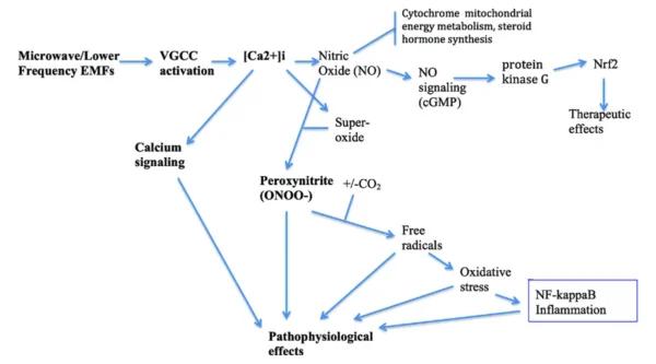
It has been found that the primary mechanism of radiation damage is NOT due to any heating effect. The negative effects are mainly caused by the way that EMFs interact with the electromagnetically sensitive membranes that surround all of our cells, which wreaks havoc throughout every system in our body.

Natural electromagnetic fields emitted from the Earth and its atmosphere have constantly changing frequencies that all life forms on Earth have evolved to live within. The natural background radiation on Earth is recognizable by our cells as a property of our environment, which we are well adapted to.

Non-native (manmade, artificial) electronic infrastructure and devices emit vastly different patterns than natural sources. Each of these artificial EMF sources runs on repetitive, pulsed frequencies—if these were audible to us, we would hear something like an alarm clock or car alarm. Nothing in nature operates this way, so because of this, our bodies are constantly in ‘alarm mode’ due to the unnatural signature. If we are being exposed to the same frequency over and over again, day after day, our cells will become overstimulated.

Like a virus, our body attempts to “attack” the EMFs that it perceives as a foreign invader. Unfortunately, there is nothing physically present to attack and defeat, so the battle goes on forever, or until the harmful stimulus is removed. In this ongoing battle, our immune system can exhaust itself, and have less energy to respond to actual invaders, such as viruses and bacteria, or to repair and regenerate the body.

This constant work results in a weakened immune system and reduced overall vitality, which can result in a variety of unpleasant and potentially serious symptoms, including:

• fatigue and lack of focus • inflammation • depression and anxiety • viral and bacterial infections • heart arrhythmias • digestive problems • insomnia • reduced fertility • allergies and autoimmune issues • weakness, nausea, tremors

Numerous studies have linked long-term EMF exposure to an increased risk of many health conditions, including Alzheimer’s, Parkinson’s, autism, heart disease, miscarriage, brain tumors, leukemia, immune system disorders, learning disabilities, memory loss, sleep disorders, headaches and migraines, lowered sperm counts, increased blood pressure, hormonal imbalance, and DNA damage.

The long-term ramifications of EMF exposure are also significant. The immunosuppressive, hormone-disruptive, and DNA-damaging effects of long-term EMF exposure could have harmful epigenetic consequences on the vitality of our children and grandchildren.

The most comprehensive EMF protection solution is to generate a field that’s based on the infinitely varied and highly structured fields found in nature, but at a higher amplitude, so the body’s cells can perceive and entrain with this field over the harmful EMFs.

This is how Blushield works.

Blushield EMF Protection utilizes the power sources in your house, car, or a battery to create strong harmonious scalar fields based on ever-varying natural frequencies, which is what we’re biologically adapted to. Because these fields are strong enough to compete against the high-powered EMFs around us, and the frequencies are natural and recognizable to our bodies, we will automatically begin to “entrain” with the Blushield frequencies, and stop reacting to the harmful EMFs.

It’s like changing radio stations—switching from heavy metal to a classical symphony, calming your entire nervous system.

Can Herbs Combat Inflammation?


Can Herbs Combat Inflammation?
Boswellia Capsules

Question: I take ibuprofen for inflammation, but I’d rather use a natural product. Is there an anti-inflammatory herb that is just as effective?

Ibuprofen is effective against inflammation, but like other non-steroidal anti-inflammatory drugs (NSAIDs) it has adverse effects and can lead to long-term complications. There are several well-studied anti-inflammatory herbs that I recommend you consider trying instead of taking an NSAID. The most important are:

• Turmeric (Curcuma longa): Turmeric accounts for the yellow color of curry and American mustard and has a distinctive sharp flavor. I recommend using turmeric to address all inflammatory disorders, including arthritis, tendinitis, and autoimmune conditions. Take 400 to 600 milligrams of turmeric extracts (available in tablets or capsules) three times per day or as directed on the product label. Whole turmeric is more effective than isolated curcumin, its major constituent. Look for products standardized for 95 percent curcuminoids.

Don’t use turmeric if you have gallstones or bile duct dysfunction. Pregnant women shouldn’t use it without their doctors’ approval. In rare cases, extended use can cause stomach upset or heartburn. As with ginger, you won’t get the full effect for two months. Make sure the product includes black pepper extract (piperine, Bioperine) to help ensure optimal absorption from the gut.

• Ginger: Powdered dry ginger is an effective anti-inflammatory agent. Take one to two capsules (500 to 1,000 milligrams) twice a day with food. A 2020 review of scientific research found that ginger could lower blood levels of C-reactive protein, high sensitivity C-reactive protein, and tumor necrosis factor-alpha, all of which are markers for inflammation. Be patient—the full benefit takes two months to develop.

• Boswellin: This is an extract of the herb Boswellia, used in Ayurvedic medicine and available in capsule form. It may be useful for generalized inflammatory conditions such as fibromyalgia. A 2020 review of the research also confirmed that Boswellia can be effective against the pain and inflammation of osteoarthritis. The dosage is two capsules twice a day unless the product label directs differently.

You can take all of these herbs in combination.

In addition to herbs, you can also take dietary steps to reduce inflammation. The specific fats in your diet affect the way the body makes prostaglandins, a group of hormones that regulate inflammation. Some prostaglandins intensify the inflammatory response while others reduce it. To help your body reduce inflammation eliminate polyunsaturated vegetable oils, margarine, vegetable shortening, all partially hydrogenated oils, and all foods that contain trans-fatty acids (read food labels to check for the presence of these oils). Instead, use extra-virgin olive oil and avocado oil as your main fats and increase your intake of omega-3 fatty acids, found in oily, cold-water fish, freshly ground flaxseeds or expeller pressed flaxseed oil, and walnuts.

Pulsed Electromagnetic Field Therapy May Relieve Osteoarthritis


Pulsed electromagnetic field (PEMF) therapy shows promise in reducing pain and improving quality of life for osteoarthritis patients.

As the U.S. population grows older, an increase in agonizing joint conditions lurks on the horizon. Chief among them is osteoarthritis, a debilitating disease caused by the natural breakdown of cartilage.

But new research suggests that a solution may lie in the power of magnetic fields—a drug-free therapy that could offer relief without the risks of common painkillers.

Pulsed Electromagnetic Fields Shown to Significantly Reduce Pain

A recent systematic review published in the Journal of Clinical Medicine sheds light on the potential of pulsed electromagnetic field (PEMF) therapy, introduced into clinical practice in the 1970s with a device approved by the Food and Drug Administration to help speed up the healing of broken fractures to alleviate symptoms of osteoarthritis.

For the new review, researchers analyzed 17 randomized controlled trials involving 1,197 patients with osteoarthritis who underwent PEMF therapy. They assessed the data to evaluate the effectiveness of PEMF across different anatomical areas, focusing on levels of pain reduction and improvements in patients’ quality of life.

The findings suggest that PEMF therapy significantly improved pain reduction, as assessed through the Visual Analog Scale (VAS) and Western Ontario and McMaster Universities Osteoarthritis Index (WOMAC) scores. VAS scores decreased by 60 percent, and WOMAC scores improved by 42 percent.

The review also noted participants’ reduced medication usage and enhanced physical function.

Despite these positive outcomes, the review highlighted the variability in treatment duration and the types of PEMF devices used, with the study authors calling for further investigation and standardization in these areas.

How Magnetic Therapy Works

PEMF therapy has gained popularity for its ability to alleviate pain and support healing across various conditions, Dr. Peter C. Lascarides, assistant professor of physical medicine and rehabilitation at the Zucker School of Medicine at Hofstra/Northwell Health, told The Epoch Times.

Easing Osteoarthritis Discomfort: Try These Tips for Symptom Relief

“It’s especially useful for people with chronic pain conditions like fibromyalgia, known for causing widespread pain and fatigue and osteoarthritis, which involves significant joint pain and inflammation,” he said.

Beyond speeding up the healing of broken bones, there is evidence that PEMF therapy helps to improve physical function and decrease stiffness, according to Dr. Lascarides.

PEMF is currently also used to stimulate the body in ways that aid in pain relief and tissue regeneration. This electromagnetic-based therapy is already commonly used for bone fractures that fail to heal within three to six months.

Patients are prescribed a small, battery-powered pulse generator connected to a coil placed next to the injury for eight to 10 hours per day. Alternatively, an electrical stimulator may be implanted near the break. Either way, the PEMF device generates a magnetic field that induces currents to flow in the nearby tissues.

“By using electromagnetic fields, PEMF therapy stimulates the body’s own healing mechanisms,” Dr. Lascarides said, “which encourages ’self-repair’ that can lead to marked improvements in comfort and mobility.”

PEMF Considered Safe With Minimal Side Effects

Overall, PEMF therapy is considered a safe treatment with minimal significant side effects.

Most people experience no issues during therapy, although a few may feel slight, brief discomfort at the point of application, Dr. Lascarides said. However, he noted that people with electronic implants, such as pacemakers, should avoid PEMF therapy to prevent potential interference with their devices.

Because of limited research on the effect of PEMF therapy on pregnant women, they should forego this treatment as a safety precaution, he said.

The existing research in this area raises some concerns. A small 2019 study published in Environmental Health found that prenatal exposure to an extremely low magnetic field had measurable adverse effects on female infants, although not on males. Effects included smaller head, upper arm, and abdomen circumferences, suggesting reduced fetal growth.

Pain Relief Without the Risks Linked to NSAIDs

Current research on PEMF therapy is “encouraging,” suggesting that it may be an effective, noninvasive method for managing pain and aiding healing without relying on drugs or surgery.

This is significant, as nonsteroidal anti-inflammatory drugs (NSAIDs) come with a broad range of side effects, including increased risk of gastrointestinal bleeding, heart attack, and stroke.

Chronic NSAID use has also been associated with an elevated risk of peptic ulcers and acute kidney failure.

Dr. Lascarides said PEMF therapy is promising as a possible treatment option to alleviate patients’ discomfort and improve their quality of life without the usual risks and side effects of traditional pharmaceutical-based pain treatments. However, there is still much to learn about it, he said.

“Ongoing and additional studies are crucial to further understand how PEMF therapy can be best applied, who stands to benefit most from it, and how it can be integrated into more comprehensive care strategies to optimize its advantages for those seeking relief,” Dr. Lascarides said.

Immunogenic cell death in cancer: targeting necroptosis to induce antitumour immunity


Abstract

Most metastatic cancers remain incurable due to the emergence of apoptosis-resistant clones, fuelled by intratumour heterogeneity and tumour evolution. To improve treatment, therapies should not only kill cancer cells but also activate the immune system against the tumour to eliminate any residual cancer cells that survive treatment. While current cancer therapies rely heavily on apoptosis — a largely immunologically silent form of cell death — there is growing interest in harnessing immunogenic forms of cell death such as necroptosis. Unlike apoptosis, necroptosis generates second messengers that act on immune cells in the tumour microenvironment, alerting them of danger. This lytic form of cell death optimizes the provision of antigens and adjuvanticity for immune cells, potentially boosting anticancer treatment approaches by combining cellular suicide and immune response approaches. In this Review, we discuss the mechanisms of necroptosis and how it activates antigen-presenting cells, drives cross-priming of CD8+ T cells and induces antitumour immune responses. We also examine the opportunities and potential drawbacks of such strategies for exposing cancer cells to immunological attacks.

Sustained-release bimatoprost implant shows safety, efficacy in early study


A sustained-release bimatoprost drug delivery platform shows promise in the treatment of ocular hypertension and open-angle glaucoma, according to a poster at the American Society of Cataract and Refractive Surgery meeting.

Gregory J. Katz, MDABO, and co-authors presented 12-month interim safety and efficacy data from a first-in-human study in which 23 eyes were implanted with the SpyGlass Pharma IOL-based drug delivery platform with bimatoprost, at doses of 75 µg, 150 µg or 300 µg, at the time of cataract surgery.

A statistically significant IOP reduction was achieved in all cohorts at all timepoints. Mean IOP decreased from 25.1 mm Hg at baseline to 13.9 mm Hg at 12 months. No additional IOP-lowering medication was required. All eyes achieved best corrected visual acuity of 20/40 or better, with minimal adverse events that were unrelated to the drug delivery device.

This method of administering medication has the potential to become a valuable option for managing ocular hypertension and open-angle glaucoma in eyes requiring cataract extraction, according to the study authors.

SpyGlass is enrolling patients in a phase 1/2 study with a larger patient pool and is working with the FDA to advance the program to commercial approval.

“The compelling clinical data … suggest we are on track to bringing our innovative platform to patients,” Patrick Mooney, CEO of SpyGlass Pharma, said in a press release.

Presence of neurofilament light chain linked to sleep, cognition in Parkinson’s disease


Key takeaways:

  • 31 patients, with and without mild cognitive impairment, underwent overnight polysomnogram with blood draw for biomarkers.
  • Neurofilament light chain levels were elevated in those with MCI by morning.

DENVER — Cognitive impairment in Parkinson’s is a mitigating factor for presence of blood-based biomarkers during sleep as indicator of disease progression, according to a poster at the American Academy of Neurology annual meeting.

“The glymphatic system is very active during sleep and we’re looking to see if sleep could be some sort of intervention for people with Parkinson’s disease,” Caileigh Dintino, BS, a research assistant in the Parkinson’s and Movement Disorders Center at Virginia Commonwealth University, told Healio.

Picture of an illuminated brain, a hand, pills and neurons in Parkinson's disease
New research suggests that presence of biomarker neurofilament light chain impacts both sleep and cognition in those with Parkinson’s disease.Image: Adobe Stock

Prior research suggests sleep disturbances in PD can contribute to disease progression and increase risk of dementia, but it is unknown whether disrupted sleep in PD alters neuroinflammatory and neurodegenerative biomarkers, which typically indicate disease progression and cognitive decline.

Dintino and colleagues aimed to investigate whether sleep in PD is associated with changes in blood-based biomarker levels such as neurofilament light chain, over the course of a single night.

Their study included 31 individuals with PD (mean age 67.4±6.0 years; 58% male), including 23 with normal cognition (52.1% female, mean age 67.4 ±6.0 years) and eight with mild cognitive impairment (87.5% male, mean age 68.4 ±6.6 years). All patients underwent an overnight polysomnogram with blood draws twice during the session, once at 8 p.m. and once at 6:30 a.m.

All participants also submitted to a full cognitive testing battery. Mild cognitive impairment (MCI) was defined by Z score on any two tests among five categories: executive function, memory, language, attention, visuospatial).

Plasma samples were analyzed using Meso Scale Discovery immunoassays for neuroinflammatory biomarkers interleukin-6 (IL-6), monocyte chemoattractant protein 1 (MCP-1) and tumor necrosis factor alpha (TNF-a), as well as the U-PLEX assay for alpha-synuclein.

Neurofilament light chain (NfL), which is released from axons upon injury or neuronal death, was measured using Quanterix Simoa assays, while T-tests were employed to compare overnight changes in biomarker levels. Subsequent correlations were tested with Pearson coefficients and a significance threshold of P < 0.05.

Dintino and colleagues reported that morning NfL levels were 16.5% higher in those with PD and MCI (PM: 13.70 ±4.15, AM: 16.38 ±7.79; P = 0.043), but not significantly different for those without cognitive issues (PM: 17.24 ±7.88, AM: 18.82 ±7.93; = 0.091).

Morning levels of TNF-a were also elevated in the PD-MCI (PM: 1.83 ±0.88, AM: 1.86 ±0.87; P = 0.02) population but not for those without MCI (PM: 1.65 ±0.54, AM: 1.71 ±0.5; P = 0.38).

“Sleep is a big complaint [among those with Parkinson’s disease],” Dintino said. “Harnessing sleep, as a way to not only to improve quality of life, but to eventually slow disease progression … could be life-changing for these people.”

Anlotinib inhibits growth of human esophageal cancer TE-1 cells by negative regulating PI3K/Akt signaling pathway


Abstract

Anlotinib is effective in treatment of many kinds of malignant cancer, but its antineoplastic effects on esophageal cancer remains unclear. This study aims to investigate its impact on esophageal cancer and the underlying mechanisms. Anlotiniband 5-fluorouracil + cisplatin (5-FU + DDP) was administered separately to human esophageal cancer TE- 1 cells tumor xenograft mouse models every 3 days. Tumor size and body weight were measured before each treatment and at the end of the experiment. In vitro studies were conducted using TE- 1 cells to examine the effects of Anlotinib. Cell viability, migration, proliferation, apoptosis, cell cycle, their regulatory proteins and the transcriptomic changes were analyzed. Anlotinib reduced tumor size, tumor weight, and the ratio of tumor weight to body weight in vivo. It decreased the viability of TE- 1 cells, with a 50% growth-inhibitory concentration of 9.454 μM for 24 h, induced apoptosis, and arrested TE- 1 cell cycle in the S phase. It inhibited migration and proliferation while negatively regulating the PI3K/Akt signaling pathway. Enhanced expressions of P21, Bax, and lowered expressions of cyclin A1, cyclin B1, CDK1, PI3K, Akt, p-Akt, and Bcl-2 were observed after Anlotinib treatment. Anlotinib exhibits antineoplastic activity against human esophageal cancer TE- 1 cells by negatively regulating the PI3K/Akt signaling pathway, consequently altering the expressions of proteins related to proliferation, apoptosis, and the cell cycle.

1 Introduction

Esophageal cancer (EC) is characterized by high morbidity and mortality [12]. It consists of two major histologic subtypes: adenocarcinoma and squamous cell carcinoma(SCC), with SCC being the predominant subtype. EC exhibits significant regional variations globally. Certain regions in Asia, East Africa, and South America, such as China, Iran, Kenya, and Brazil, have higher incidence rates of esophageal cancer. This is associated with local factors such as diet, lifestyle, cooking methods, food choices, and environmental factors. Despite significant advancements in surgery, preoperative chemotherapy, and radiotherapy, the survival rate remains low, with a 5-year survival rate ranging from 15 to 20%, and a median survival time of 1.5 years [3]. This is despite the considerable progress in surgery, preoperative chemotherapy, and radiotherapy. The therapy regimen primarily depends on the physical condition and tumor stage, typically classified by the TMN stage of EC patients. Chemotherapy is widely applied to patients who are not suitable for surgery.

Anlotinib is a recently approved chemotherapeutic agent that has gained approval for the treatment of advanced non-small-cell lung cancer as a third-line therapy option in China [45]. It functions as a multikinase inhibitor, targeting vascular endothelial growth factor receptors (EGFR) 1–3, fibroblast growth factor receptors 1–4, the platelet-derived growth factor receptor, and the stem cell factor receptor to inhibit neoangiogenesis and tumor progression [6,7,8]. Anlotinib also inhibits the activity of basic fibroblast growth factor receptor (bFGFR). bFGFR is another crucial receptor associated with tumor growth and angiogenesis. By affecting the bFGFR signaling pathway, Anlotinib can regulate cell growth, differentiation, and migration, thereby influencing the development of tumors. Anlotinib has been found to induce apoptosis in tumor cells, which refers to programmed cell death. This is an essential part of the normal cell lifecycle but is often disrupted in tumors. By prompting tumor cells to undergo programmed cell death, Anlotinib helps inhibit the growth of tumors.

Furthermore, studies have demonstrated the efficacy of anlotinib against intrahepatic cholangiocarcinoma [9], soft tissue sarcoma [10], thyroid cancer [11], and colorectal cancer [12]. It exhibited notable therapeutic effects against esophageal squamous cell carcinoma (ESCC) on patient-derived xenograft models when combined with chemoradiotherapy, while the exact mechanism remains unclear. Therefore, our study was designed to investigate the efficacy and underlying mechanism of anlotinibon ESCC using tumor xenograft animal models and TE- 1 cells.

Discussion

In China, approximately 240,000 new cases of EC are diagnosed each year, with EC ranking as the fourth leading cause of cancer-related mortality [13,14,15]. Many patients receive diagnoses at advanced stages, leading to 5-year survival rate of merely 5%. The discovery of new medicines for treating EC is an imperative need. Current studies on anlotinib have mostly focused on its anticancer activity against advanced non-small-cell lung cancer [7]. Jingzhen Shi combined anlotinib with chemoradiotherapy to treat EC with good achievements [16]. These results have inspired us to investigate the underlying mechanisms behind anlotinib’s effects on EC.

In our study, anlotinib demonstrated excellent antineoplastic activity against EC in vivo, with less body weight loss in comparison to the 5-FU + DDP treatment. This effect was corroborated by the outcomes observed in the TE-1 cell line, where anlotinib exhibited an IC50 value of 9.454 μM. Furthermore, anlotinib induced TE- 1 cell apoptosis and notably arrested the cell cycle in the S phase, thereby inhibiting migration and proliferation in a dose-dependent manner. These findings indicate that Anlotinib has potential therapeutic capabilities against esophageal cancer, exhibiting significant anti-tumor effects both in vivo and in vitro. Furthermore, Anlotinib effectively inhibits the migration and proliferation of esophageal cancer cells by inducing apoptosis and arresting the cell cycle, providing important evidence for its further research and clinical application as an anti-cancer treatment.

The PI3K/Akt signaling pathway is a crucial cellular signaling cascade involved in regulating various biological processes such as cell survival, proliferation, differentiation, and metabolism [1718]. Its name derives from two key proteins involved in the pathway: phosphoinositide 3-kinase (PI3K) and protein kinase B (Akt), also known as protein kinase B activated kinase (PKB). PI3K is an enzyme that catalyzes the conversion of phosphatidylinositol 4,5-bisphosphate (PIP2) to phosphatidylinositol 3,4,5-trisphosphate (PIP3) on the cell membrane [1920]. Upon activation, PIP3 can bind and activate Akt, thereby initiating downstream signaling cascades. Akt, a critical regulatory protein in the PI3K/Akt pathway, promotes cell survival and proliferation while inhibiting apoptosis. Akt exerts its effects by phosphorylating various cellular factors, cell cycle proteins, and transcription factors, thereby modulating cellular physiology. Aberrant activation of the PI3K/Akt signaling pathway is closely associated with the onset and progression of various diseases, including cancer [2122]. PI3K/Akt signaling pathway plays a crucial role in the proliferation, survival, and invasion of tumor cells [2324] including TE- 1 cells [25,26,27]. The activation of PI3K promotes Akt to be phosphorylated intop-Akt, p-Akt acts on its downstream proteins to enhance TE-1 cell survival, proliferation and invasion. The PI3K/Akt signaling pathway can also participate in cell survival and inflammatory responses by activating NF-κB (nuclear factor-kappa B). NF-κB is a transcription factor that regulates the expression of various genes, including those associated with cell survival, proliferation, and immune responses. The excessive activation of the PI3K/Akt signaling pathway is closely associated with the occurrence and development of various diseases, including cancer, diabetes, and neurological disorders. Therefore, this signaling pathway has become a crucial target for drug development, and drugs targeting its abnormal activation are being investigated for the treatment of diseases such as cancer. Our findings indicated that anlotinib treatment notably downregulated PI3K, Akt, and p-Akt, aligning with the results of other studies [910].

Survivin plays a significant role in regulating cell division, apoptosis (programmed cell death), and cell survival. It is a member of the inhibitor of apoptosis (IAP) family of proteins, which are characterized by their ability to inhibit apoptosis and promote cell survival [2829]. Survivin is overexpressed in various types of human tumor cells, including lung cancer, breast cancers, EC, as well as TE-1 cells, as indicated by our research. The suppression of survivin promotes tumor cells apoptosis and enhances radiosensitivity of esophageal cancer cells [30,31,32]. Besides, Bax serves as an apoptosis promoter, while Bcl-2, an important homolog of Bax, plays the inverse effects of Bax [3334]. Our results demonstrated that anlotinib treatment significantly promoted TE- 1 cells apoptosis through suppression of survivin, Bcl-2 and enhancement of Bax expressions. By understanding how Anlotinib regulates key genes such as survivin, Bcl-2, and Bax to promote apoptosis in TE-1 cells, we can gain deeper insights into its mechanism of action in anticancer processes, which can guide further clinical research and the development of more effective treatment strategies.

Cyclin A1 is the product of CCNA1 gene expression. Except Akt as one of the two central proteins affectd by anlotinib treatment, Cyclin A1 was the other central protein which interacting with other proteins to regulate cell cycle. Cyclin A1 which working together with cyclinB1 to promote S to G2/M phase transition. The combination of cyclin A1 or cyclinB1 to CDK1 triggers cell cycle into mitosis [35,36,37]. P21 is currently recognized as a potent universal CDK inhibitor which forms complexes with CDKs and cyclins to arrest cell cycle [3839]. Anlotinib negatively regulated cyclin A1, cyclin B1 and CDK1 expressions while positively up regulated P21 expression in TE- 1 cells. These observations elucidate the mechanism underlying its effects on cell cycle arrest.

This article has some shortcomings. Firstly, although the study elucidated the negative regulation of the PI3K/Akt signaling pathway by Anlotinib, the underlying mechanisms of this regulation have not been fully explored. Further mechanistic studies are needed to analyze the molecular pathways involved and confirm the observed effects. Secondly, TE-1 cells may not fully represent the heterogeneity of esophageal cancer. Including other esophageal cancer cell lines or samples originating from patients could provide a more comprehensive understanding of the effects of Anlotinib in different molecular subtypes of the disease.

5 Conclusions

Anlotinib can induce apoptosis and cell cycle arrest, inhibit migration and proliferation of TE- 1 cells by negatively regulating PI3K/Akt signaling pathway, and consequently regulating expressions of apoptosis- related and cell-cycle-related proteins. The detailed underlying mechanism may be further elucidated in future research.

Brain Blood Flow Syncs with Visual Stimuli


Summary: Researchers pioneered a new method to monitor blood vessel dynamics in the mouse brain, revealing that visual stimuli can synchronize vasomotion, potentially improving brain function. By exposing mice to a specific pattern of horizontally moving stripes, the team observed that vasomotion matched the stimulus pattern’s speed and spread throughout the brain.

This synchronization suggests a mechanism by which the brain enhances its circulation of nutrients and clears waste, possibly boosting cognitive abilities. The findings could have implications for treating and preventing neurological conditions.

Key Facts:

  1. Innovative Monitoring Technique: The study introduces a novel method to observe blood vessel dynamics through the intact skull or deep within the brain using optical fibers.
  2. Vasomotion Synchronization: Visual stimuli can entrain the brain’s vasomotion, leading to synchronized blood vessel activity that enhances nutrient delivery and waste removal across the entire brain.
  3. Potential Health Benefits: This synchronized vasomotion may improve learning, aid in stroke recovery, and potentially delay neurodegenerative diseases like dementia by improving the brain’s metabolic efficiency.

Source: Tohoku University

Compared with computers, the brain can perform computations with a very low net energy supply. Yet our understanding surrounding how the biological brain manages energy is still incomplete.

What is known, however, is that the dilation and constriction cycles of blood vessels, or vasomotion, spontaneously occur in the brain, a process that likely contributes to enhancing the circulation of energetic nutrients and clearing wasteful materials.

Now, researchers from Tohoku University have developed a method that easily observes and monitors blood vessel dynamics in the mouse brain. This can be done either through the intact skull of a mouse, or deep into the brain using an implanted optical fiber.

Since it has been reported that sensory stimuli can cause dilation of blood vessels or hyperemia, researchers sought to induce vasomotion via presenting mice with visual stimuli.

What they discovered was when a mouse was shown a horizontally moving stripe pattern that changed direction every 2 to 3 seconds, it caused a reaction in the mouse’s blood vessels that matched the pattern’s speed.

Mice were presented with 15-minute visual training sessions interleaved with 1-hour resting periods for 4 times per day. With such spaced training, the amplitude of the synchronized vasomotion gradually increased.

Interestingly, the visually induced vasomotion was not confined to the area of the cerebral cortex responsible for visual information processing. In other words, synchronized vasomotion spread throughout the whole brain.

“Synchronized vascular motion can be entrained with slowly oscillating visual stimuli,” says Professor Ko Matsui of the Super-network Brain Physiology lab at Tohoku University, who led the research.

“Such enhancement of circulation mechanisms may benefit the information processing capacity of the brain.”

While it’s long been known that changes in neuron connections support learning and memory, the plasticity of vasomotion hasn’t been described before.

Matsui and his colleagues found that a specific visual pattern makes the eyes move more, and this eye movement improvement depends on changes in the brain’s cerebellum. The researchers also observed that blood vessel activity in the cerebellum synchronized with this optokinetic motor learning.

Lead study investigator, Daichi Sasaki, believes that synchronized vasomotion, which efficiently delivers oxygen and glucose, could improve learning abilities

He states, “Our next step is to explore the advantages of vasomotion synchronization. It might help clear waste like amyloid beta, potentially delaying or preventing dementia.

“Stroke recovery could also benefit from better energy supply and waste removal. Additionally, synchronized vasomotion might even enhance intelligence beyond our natural capabilities.”

Maternal Cytokine Levels Linked to Child’s Mental Health


Summary: Researchers discovered a critical role for the cytokine XCL1 in fetal brain development and the emotional behavior of offspring, challenging previous assumptions about its low impact due to minimal circulating levels during pregnancy. The study shows that a temporary spike in maternal XCL1 is essential for proper placental development and regulating fear behavior in male offspring.

Disruptions in this cytokine level were linked to increased anxiety and stress reactions due to neuronal abnormalities in the ventral hippocampus. These findings provide new insights into how maternal immune responses during pregnancy might influence psychiatric conditions in children.

Key Facts:

  1. Critical XCL1 Spike: A transient increase in XCL1 during pregnancy is crucial for healthy placental development and managing offspring emotional behavior, particularly fear responses.
  2. Neuronal Impact: Blocking or neutralizing this cytokine spike leads to neuronal abnormalities in the ventral hippocampus and increased anxiety behaviors in male offspring.
  3. Long-Term Effects: Although the immune and neuronal abnormalities observed in offspring normalize by adulthood, the early-life inflammatory state linked to XCL1 deficiency may set the stage for adult anxious behavior.

Source: Weill Cornell University

Researchers at Weill Cornell Medicine have discovered in a preclinical model that cytokines, proteins that control immune response, circulating in maternal blood during pregnancy may mitigate an offspring’s risk for psychiatric conditions.

The findings are surprising because circulating maternal cytokines are at such low levels that they were not implicated in fetal brain development and offspring behavior before.

This shows a pregnant woman.
Dr. Toth will explore other chemokines that may regulate placenta development and impact offspring emotional behavior

The study published online in Brain, Behavior, and Immunity on Feb. 29, reported that cytokine XCL1 produced by maternal immune cells can function as a pregnancy hormone and is required for the proper development of placenta and male offspring fear behavior.

These results support epidemiological studies which have long suggested a link between human maternal infection and inflammation during pregnancy and offspring developing psychiatric disorders later life.

“Using mouse models, we found that circulating XCL1 normally remained at the same low pre-pregnancy level throughout gestation except for a short rise and fall in the middle period,” said corresponding author Dr. Miklos Toth, professor of pharmacology at Weill Cornell Medicine.

“This temporary rise is essential for the proper development of the placenta and offspring emotional behavior.” First author Dr. Rosa Chen was a graduate student in the Toth lab during the study, which was a collaboration with Dr. Heidi Stuhlmann, acting chair of Biochemistry and also of Cell and Developmental Biology and the Harvey Klein Professor of Biomedical Sciences, Cell and Developmental Biology at Weill Cornell Medicine

When this spike in XCL1 in maternal blood was blocked genetically or neutralized by anti-XCL1 antibodies, the researchers found increased production of factors associated with tissue damage in the fetal placenta which led to increased innate anxiety and stress reactions in male mouse offspring.

The researchers also found a neuronal abnormality in the developing brains of these offspring, specifically in the ventral hippocampus, a region that has been linked to anxiety and anxious behavior.

The immune and neuronal abnormalities observed when the cytokine spike was blocked were normalized by adulthood, suggesting that the adult anxious behavior of the offspring could be related to the early life proinflammatory state caused by the absence of elevated XCL1.

Dr. Toth will explore other chemokines that may regulate placenta development and impact offspring emotional behavior. The team plans to collaborate with researchers who have access to blood samples from pregnant women to see if the profile of XCL1, a protein also found in humans, corresponds to the observations in mouse models.

Is obesity raising your risk of dementia?


Obesity damages the tiny vessels supplying blood to the brain, and is a major cause of high blood pressure, diabetes and chronic inflammation, all of which have been repeatedly linked to dementia risk

Is obesity raising your risk of dementia?

Although there was a clear association between higher levels of obesity and lower cognitive skills in midlife, this probably wasn’t because one was causing the other. Image used for representational purpose/Pixabay

Many dementia charities advise people to maintain a healthy weight to reduce their risk of dementia. But some studies have suggested that obesity might actually protect against dementia. What does the science say?

The evidence linking obesity to dementia does at first appear to be convincing. For example, we know that being obese in middle age is associated with an increased risk of developing dementia in later life.

We also know that obesity damages the tiny vessels supplying blood to the brain, and is a major cause of high blood pressure, diabetes and chronic inflammation, all of which have been repeatedly linked to dementia risk.

However, the picture is not that clear. For example, if obesity really does cause dementia, why have dementia rates been falling in the west in recent decades at the same time as the number of obese people has been increasing? And why have several studies reported evidence of something termed an “obesity paradox” , where being obese appears to be associated with a reduced risk of dementia?

Putting aside the longstanding problem of defining what we mean when we refer to obesity and dementia (both of which are relatively broad terms for conditions that can be defined in different ways), much of the difficulty in establishing whether one causes the other arises from limitations in the type of data available to scientists trying to answer this question.

In an ideal world, we would look to test the question by designing a randomised trial . In this trial, thousands of people would be randomly assigned to an intervention that would result in half of them being obese for an extended period, while the other half are not.

Maintaining a healthy weight will reduce your risk for a wide range of other major health problems, and it may even reduce your risk of dementia.

If the obese people were then found to be more likely to have dementia in later life, we could be fairly confident that this must be the cause. These types of trials are rarely possible to conduct, however, as not only are they extremely time-consuming and expensive, they are unethical, too. (Imagine the outcry if you randomly assigned a group of people to purposefully be obese for an extended period of time.)

Most studies therefore rely on data from observational studies . This involves following a large group of people for a long time so that the long-term associations between obesity and dementia can be studied.

Although observational studies are a valuable resource for scientists, these studies can be plagued with biases that can often make it difficult to interpret the results.

One such bias relevant to dementia research is “reverse causation”, particularly if the people being studied are old and the follow-up time is short. In this situation, it is possible that people already in the early stages of developing dementia when the study begins may lose weight over time as a result of the disease, rather than the other way around. This is what is suspected to underlie the obesity paradox .

Another common issue is “confounding bias”. This is where the apparent link between obesity and dementia is caused by a different measure that is related to both. One such example is childhood intelligence, a factor that is rarely measured in observational studies, but, when available, has been shown to potentially explain associations that could otherwise be blamed on obesity in later life .

For example, recent work from my lab using data from three separate groups, each followed for 50 years from birth, has shown that lower childhood intelligence probably explains why middle-aged people with obesity are often found to already have slightly worse cognitive skills than those with normal weight.

although there was a clear association between higher levels of obesity and lower cognitive skills in midlife, this probably wasn’t because one was causing the other.

We found that although there was a clear association between higher levels of obesity and lower cognitive skills in midlife, this probably wasn’t because one was causing the other.

Instead, it was likely because a third factor (childhood intelligence) was associated with both. That is, individuals with low intelligence in childhood not only had a higher risk of becoming obese as they grew up, but were also more likely to continue to be of lower intelligence (and therefore consistently have slightly worse cognitive skills).

Without knowing the intelligence levels of children in childhood, we may have interpreted this association between midlife obesity and cognitive skills as being one of cause and effect. In reality though, both are probably just a result of this third factor from earlier in the lifespan.

Nature’s randomised trial

So how can we try to tackle these bias issues? One recent clever technique is to conduct something called a Mendelian randomisation study – sometimes referred to as “nature’s randomised trial”.

In this type of study, scientists separate a large population into two groups based solely on whether or not they have a gene (or genes) that cause obesity. As these genes are randomly inherited from both parents, this results in a “natural randomisation” of the population into two groups who are collectively balanced for every factor except for their obesity status.

Although not without its own potential biases , any differences in dementia risk are therefore interpreted to be directly caused by this obesity.

At least 10 studies have used this technique to test if obesity might cause Alzheimer’s disease – the most common form of dementia. Only one has suggested a link between the two.

So, returning to the question: does obesity really increase your risk of dementia? As scientists like to say, an absence of evidence is not the same as evidence of absence. Or put another way, just because we don’t yet have enough data yet to say that it does, it doesn’t mean that it doesn’t.

Science is an incremental process. In time, a clearer answer to this question will emerge as more data is collected and better techniques are developed.

Until then, my advice would be to follow the suggestion of dementia charities and try to maintain a healthy weight, anyway. At the very least, it will reduce your risk for a wide range of other major health problems, and it may even reduce your risk of dementia.