Anlotinib inhibits growth of human esophageal cancer TE-1 cells by negative regulating PI3K/Akt signaling pathway


Abstract

Anlotinib is effective in treatment of many kinds of malignant cancer, but its antineoplastic effects on esophageal cancer remains unclear. This study aims to investigate its impact on esophageal cancer and the underlying mechanisms. Anlotiniband 5-fluorouracil + cisplatin (5-FU + DDP) was administered separately to human esophageal cancer TE- 1 cells tumor xenograft mouse models every 3 days. Tumor size and body weight were measured before each treatment and at the end of the experiment. In vitro studies were conducted using TE- 1 cells to examine the effects of Anlotinib. Cell viability, migration, proliferation, apoptosis, cell cycle, their regulatory proteins and the transcriptomic changes were analyzed. Anlotinib reduced tumor size, tumor weight, and the ratio of tumor weight to body weight in vivo. It decreased the viability of TE- 1 cells, with a 50% growth-inhibitory concentration of 9.454 μM for 24 h, induced apoptosis, and arrested TE- 1 cell cycle in the S phase. It inhibited migration and proliferation while negatively regulating the PI3K/Akt signaling pathway. Enhanced expressions of P21, Bax, and lowered expressions of cyclin A1, cyclin B1, CDK1, PI3K, Akt, p-Akt, and Bcl-2 were observed after Anlotinib treatment. Anlotinib exhibits antineoplastic activity against human esophageal cancer TE- 1 cells by negatively regulating the PI3K/Akt signaling pathway, consequently altering the expressions of proteins related to proliferation, apoptosis, and the cell cycle.

1 Introduction

Esophageal cancer (EC) is characterized by high morbidity and mortality [12]. It consists of two major histologic subtypes: adenocarcinoma and squamous cell carcinoma(SCC), with SCC being the predominant subtype. EC exhibits significant regional variations globally. Certain regions in Asia, East Africa, and South America, such as China, Iran, Kenya, and Brazil, have higher incidence rates of esophageal cancer. This is associated with local factors such as diet, lifestyle, cooking methods, food choices, and environmental factors. Despite significant advancements in surgery, preoperative chemotherapy, and radiotherapy, the survival rate remains low, with a 5-year survival rate ranging from 15 to 20%, and a median survival time of 1.5 years [3]. This is despite the considerable progress in surgery, preoperative chemotherapy, and radiotherapy. The therapy regimen primarily depends on the physical condition and tumor stage, typically classified by the TMN stage of EC patients. Chemotherapy is widely applied to patients who are not suitable for surgery.

Anlotinib is a recently approved chemotherapeutic agent that has gained approval for the treatment of advanced non-small-cell lung cancer as a third-line therapy option in China [45]. It functions as a multikinase inhibitor, targeting vascular endothelial growth factor receptors (EGFR) 1–3, fibroblast growth factor receptors 1–4, the platelet-derived growth factor receptor, and the stem cell factor receptor to inhibit neoangiogenesis and tumor progression [6,7,8]. Anlotinib also inhibits the activity of basic fibroblast growth factor receptor (bFGFR). bFGFR is another crucial receptor associated with tumor growth and angiogenesis. By affecting the bFGFR signaling pathway, Anlotinib can regulate cell growth, differentiation, and migration, thereby influencing the development of tumors. Anlotinib has been found to induce apoptosis in tumor cells, which refers to programmed cell death. This is an essential part of the normal cell lifecycle but is often disrupted in tumors. By prompting tumor cells to undergo programmed cell death, Anlotinib helps inhibit the growth of tumors.

Furthermore, studies have demonstrated the efficacy of anlotinib against intrahepatic cholangiocarcinoma [9], soft tissue sarcoma [10], thyroid cancer [11], and colorectal cancer [12]. It exhibited notable therapeutic effects against esophageal squamous cell carcinoma (ESCC) on patient-derived xenograft models when combined with chemoradiotherapy, while the exact mechanism remains unclear. Therefore, our study was designed to investigate the efficacy and underlying mechanism of anlotinibon ESCC using tumor xenograft animal models and TE- 1 cells.

Discussion

In China, approximately 240,000 new cases of EC are diagnosed each year, with EC ranking as the fourth leading cause of cancer-related mortality [13,14,15]. Many patients receive diagnoses at advanced stages, leading to 5-year survival rate of merely 5%. The discovery of new medicines for treating EC is an imperative need. Current studies on anlotinib have mostly focused on its anticancer activity against advanced non-small-cell lung cancer [7]. Jingzhen Shi combined anlotinib with chemoradiotherapy to treat EC with good achievements [16]. These results have inspired us to investigate the underlying mechanisms behind anlotinib’s effects on EC.

In our study, anlotinib demonstrated excellent antineoplastic activity against EC in vivo, with less body weight loss in comparison to the 5-FU + DDP treatment. This effect was corroborated by the outcomes observed in the TE-1 cell line, where anlotinib exhibited an IC50 value of 9.454 μM. Furthermore, anlotinib induced TE- 1 cell apoptosis and notably arrested the cell cycle in the S phase, thereby inhibiting migration and proliferation in a dose-dependent manner. These findings indicate that Anlotinib has potential therapeutic capabilities against esophageal cancer, exhibiting significant anti-tumor effects both in vivo and in vitro. Furthermore, Anlotinib effectively inhibits the migration and proliferation of esophageal cancer cells by inducing apoptosis and arresting the cell cycle, providing important evidence for its further research and clinical application as an anti-cancer treatment.

The PI3K/Akt signaling pathway is a crucial cellular signaling cascade involved in regulating various biological processes such as cell survival, proliferation, differentiation, and metabolism [1718]. Its name derives from two key proteins involved in the pathway: phosphoinositide 3-kinase (PI3K) and protein kinase B (Akt), also known as protein kinase B activated kinase (PKB). PI3K is an enzyme that catalyzes the conversion of phosphatidylinositol 4,5-bisphosphate (PIP2) to phosphatidylinositol 3,4,5-trisphosphate (PIP3) on the cell membrane [1920]. Upon activation, PIP3 can bind and activate Akt, thereby initiating downstream signaling cascades. Akt, a critical regulatory protein in the PI3K/Akt pathway, promotes cell survival and proliferation while inhibiting apoptosis. Akt exerts its effects by phosphorylating various cellular factors, cell cycle proteins, and transcription factors, thereby modulating cellular physiology. Aberrant activation of the PI3K/Akt signaling pathway is closely associated with the onset and progression of various diseases, including cancer [2122]. PI3K/Akt signaling pathway plays a crucial role in the proliferation, survival, and invasion of tumor cells [2324] including TE- 1 cells [25,26,27]. The activation of PI3K promotes Akt to be phosphorylated intop-Akt, p-Akt acts on its downstream proteins to enhance TE-1 cell survival, proliferation and invasion. The PI3K/Akt signaling pathway can also participate in cell survival and inflammatory responses by activating NF-κB (nuclear factor-kappa B). NF-κB is a transcription factor that regulates the expression of various genes, including those associated with cell survival, proliferation, and immune responses. The excessive activation of the PI3K/Akt signaling pathway is closely associated with the occurrence and development of various diseases, including cancer, diabetes, and neurological disorders. Therefore, this signaling pathway has become a crucial target for drug development, and drugs targeting its abnormal activation are being investigated for the treatment of diseases such as cancer. Our findings indicated that anlotinib treatment notably downregulated PI3K, Akt, and p-Akt, aligning with the results of other studies [910].

Survivin plays a significant role in regulating cell division, apoptosis (programmed cell death), and cell survival. It is a member of the inhibitor of apoptosis (IAP) family of proteins, which are characterized by their ability to inhibit apoptosis and promote cell survival [2829]. Survivin is overexpressed in various types of human tumor cells, including lung cancer, breast cancers, EC, as well as TE-1 cells, as indicated by our research. The suppression of survivin promotes tumor cells apoptosis and enhances radiosensitivity of esophageal cancer cells [30,31,32]. Besides, Bax serves as an apoptosis promoter, while Bcl-2, an important homolog of Bax, plays the inverse effects of Bax [3334]. Our results demonstrated that anlotinib treatment significantly promoted TE- 1 cells apoptosis through suppression of survivin, Bcl-2 and enhancement of Bax expressions. By understanding how Anlotinib regulates key genes such as survivin, Bcl-2, and Bax to promote apoptosis in TE-1 cells, we can gain deeper insights into its mechanism of action in anticancer processes, which can guide further clinical research and the development of more effective treatment strategies.

Cyclin A1 is the product of CCNA1 gene expression. Except Akt as one of the two central proteins affectd by anlotinib treatment, Cyclin A1 was the other central protein which interacting with other proteins to regulate cell cycle. Cyclin A1 which working together with cyclinB1 to promote S to G2/M phase transition. The combination of cyclin A1 or cyclinB1 to CDK1 triggers cell cycle into mitosis [35,36,37]. P21 is currently recognized as a potent universal CDK inhibitor which forms complexes with CDKs and cyclins to arrest cell cycle [3839]. Anlotinib negatively regulated cyclin A1, cyclin B1 and CDK1 expressions while positively up regulated P21 expression in TE- 1 cells. These observations elucidate the mechanism underlying its effects on cell cycle arrest.

This article has some shortcomings. Firstly, although the study elucidated the negative regulation of the PI3K/Akt signaling pathway by Anlotinib, the underlying mechanisms of this regulation have not been fully explored. Further mechanistic studies are needed to analyze the molecular pathways involved and confirm the observed effects. Secondly, TE-1 cells may not fully represent the heterogeneity of esophageal cancer. Including other esophageal cancer cell lines or samples originating from patients could provide a more comprehensive understanding of the effects of Anlotinib in different molecular subtypes of the disease.

5 Conclusions

Anlotinib can induce apoptosis and cell cycle arrest, inhibit migration and proliferation of TE- 1 cells by negatively regulating PI3K/Akt signaling pathway, and consequently regulating expressions of apoptosis- related and cell-cycle-related proteins. The detailed underlying mechanism may be further elucidated in future research.
Esophageal Cancer: One of Least Studied Common Cancers, 5 Ways to Prevent


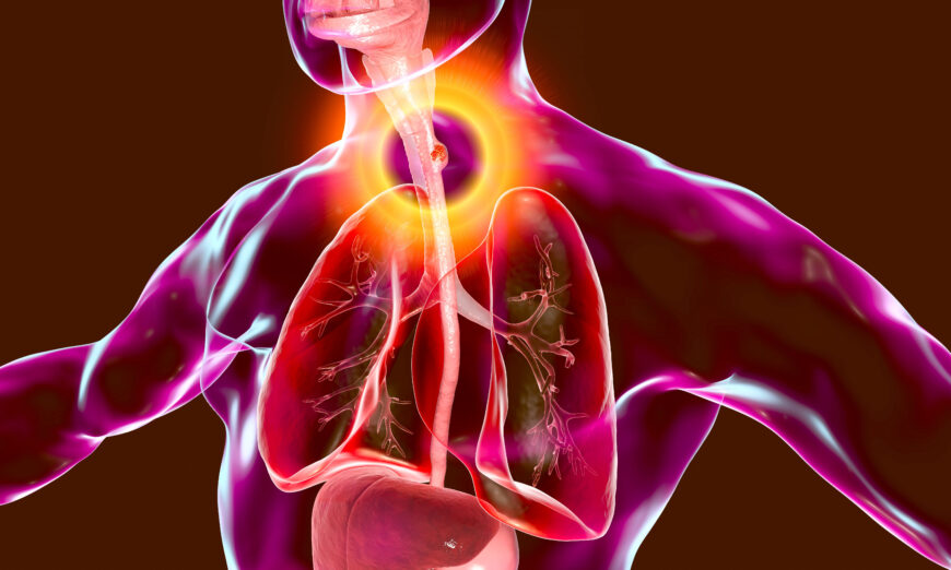
This 3D illustration shows a malignant tumor in the human esophagus. (Shutterstock)

This 3D illustration shows a malignant tumor in the human esophagus.

While esophageal cancer is the eighth most common cancer worldwide, and the sixth leading cause of cancer-related deaths, it is one of the least studied cancers.

It sounds counterintuitive. However, there are many reasons why there is a lack of study, including this cancer’s aggressive nature, poor survival rate, and the great differences in how it presents across countries and even between sexes.

Esophageal cancer is not as common in the United States. However, a 2022 analysis presented at a conference found that from 2012 to 2019, the rate of esophageal cancer for people ages 45 to 64 almost doubled, based on 5 million people’s data in Florida.

Esophageal Cancer Is One of the Least Studied Common Cancers, 3 Reasons

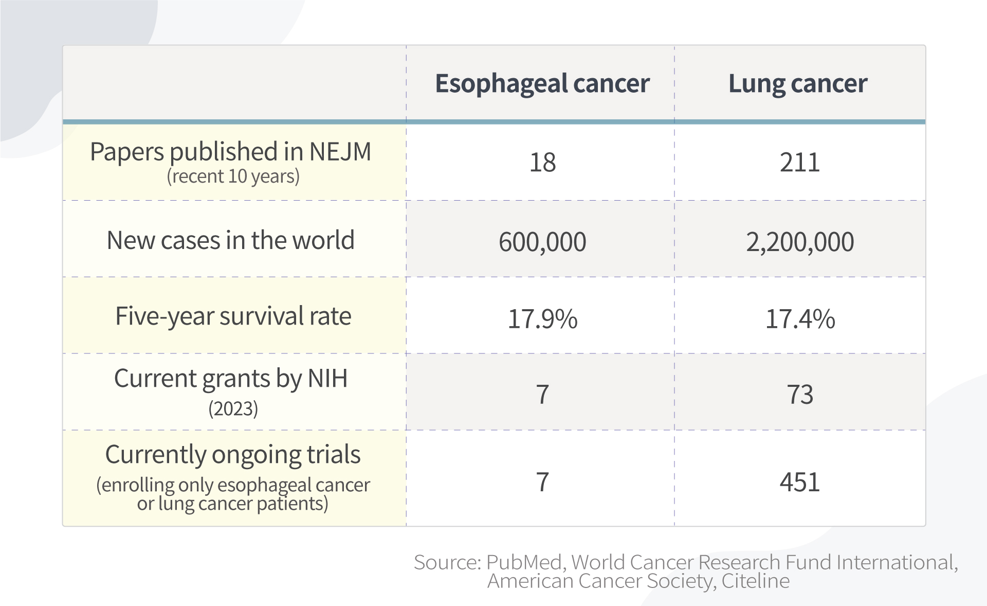
By searching publications in the New England Journal of Medicine, the world’s leading medical journal, studies in esophageal cancer are far fewer than those in lung cancer.

Epoch Times Photo
Studies in esophageal cancer are far fewer than those in lung cancer. Information accessed February 2023.

There are multiple reasons for this:

1. Incidence rates are relatively low in the United States

The majority of esophageal cancer cases are outside of the United States. Esophageal cancer makes up only about 1 percent of cancer cases in the United States.

The National Institutes of Health (NIH) is the world’s largest public funder of biomedical research. However, because esophageal cancer is so uncommon in the United States, it is difficult to be granted funding from institutions like NIH to research it.

Also, because there aren’t many esophageal cancer patients in the United States, there is less motivation for pharmaceutical companies to develop new drugs targeting esophageal cancer.

2. Presents differently between sexes and among races, but root causes unknown

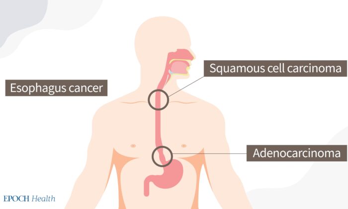
Most esophageal cancers can be classified as one of two types: adenocarcinoma or squamous cell carcinoma.

Squamous cell carcinoma is the predominant histological type worldwide. This form mainly occurs in the “Asian Esophageal Cancer Belt,” which includes China.

However, in countries like the United States, Australia, the UK, and Western Europe, adenocarcinoma incidence is higher.

Two types of esophageal cancers. (The Epoch Times)

Similar to other cancer types, esophageal cancer exhibits sex disparities in occurrence. The incidence of esophageal adenocarcinoma is about eight times higher (pdf) in men than in women. Sex is known to be an independent prognostic marker for squamous cell carcinoma but not for adenocarcinoma, with survival rates being higher in women.

Esophageal cancer affects races differently, as well. While nonwhite patients were more likely to develop squamous cell cancer or a tumor in the middle esophagus, white patients were more likely to develop adenocarcinoma or a tumor in the lower esophagus. The root causes for these differences are still unknown.

Esophageal cancer is very complicated and differs across sex, race, region, and socioeconomic status. Thus, as the number of patients in the United States is limited, and with so many factors to consider, it is hard to get statistically significant results.

3. High death rate

On average, according to a paper published in the World Journal of Gastroenterology, about 40 to 45 percent of patients die within a year after diagnosis, so it is hard to recruit patients for studies if they are too sick. This contributes to a limited number of eligible patients.

Can Esophageal Cancer Be Identified Early?

The deadliness of cancer depends on when it is found. If esophageal cancer is found while it is still localized, the patient has a 46 percent chance of survival for five more years. If it is found while it is regional, that chance drops to 26 percent, and if found while distant, only 5 percent of patients will survive five more years.

  • Localized means that the cancer is growing only in the esophagus.
  • Regional means that the cancer has spread to nearby lymph nodes or tissues.
  • Distant means that the cancer has spread to organs or lymph nodes away from the main tumor.

Unfortunately, in the earliest stages when it’s easiest to treat, esophageal cancer has very few symptoms. Therefore, it is called a silent killer.

Can screening be done? In the United States, screening for esophageal cancer is not recommended, partially because the incidence is low; therefore, for society overall, it is not cost-effective at lowering risk.

Will There Be a Sharp Increase in Esophageal Cancer in the US?

The researchers who presented their findings at the 2022 Digestive Disease Week conference argue that more middle-aged persons should be checked for esophageal cancer in light of their study, as esophageal cancer may be increasing among middle-aged people.

That being said, the study’s findings should be analyzed with caution, as the study only included adults residing in Florida, meaning it’s not wholly representative of the population of the United States. For instance, the increase might be due to Florida’s growing population (it has grown by 14.6 percent in the last decade and growth accelerated during the pandemic). Florida’s late-night eating habits may also influence the increase; Miami is a late-night eating town compared to the rest of the country. More on this later.

What Are Common Risk Factors of Esophageal Cancer?

As mentioned, nearly half of the patients die within a year once they are diagnosed—which is why prevention is very important.

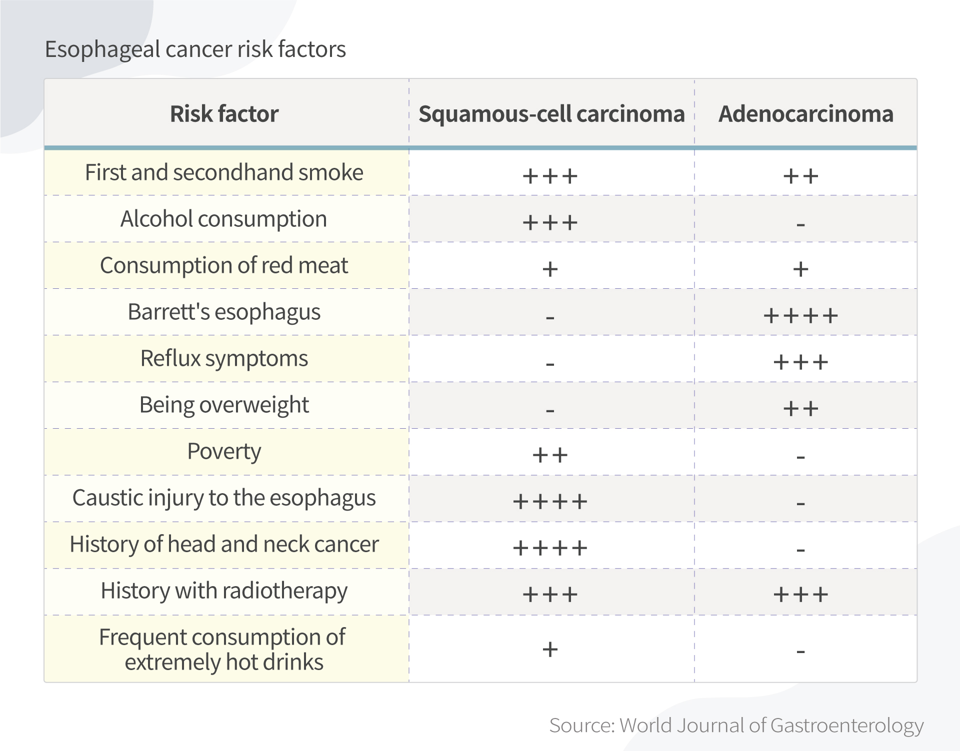
Smoking, alcohol, obesity, drinking hot tea, red meat consumption, poor oral health, low intake of fresh fruit and vegetables, and low socioeconomic status have all been associated with a higher risk of esophageal cancer, according to the World Journal of Gastroenterology paper.

Epoch Times Photo
Risk factors of adenocarcinoma and squamous cell carcinoma. (The Epoch Times)

5 Ways to Prevent Esophageal Cancer

There are a few things that you can do now to lower your chance of developing esophageal cancer.

1. Avoid eating late

When you eat late at night and then lie down, the contents of your stomach press harder against the lower esophageal sphincter. This can trigger gastroesophageal reflux disease (GERD), or acid reflux. Some ways to avoid this include:

  • Waiting two to three hours after eating to go to bed.
  • Not snacking late at night.
  • Eating larger meals earlier in the day. Try to make your last meal small.

Many people love to have late-night dinners and drinks, especially while on vacation. Although having to restrain yourself from delicious food at night might be hard, it is rewarding to control your weight and prevent other diseases beyond esophageal cancer.

2. Avoid hot foods and liquids

Different people have different eating habits. I hear discussions about why Chinese people like drinking hot water. It can be relaxing and soothing. However, drinking very hot beverages may be associated with esophageal cancer risk.

Thermal irritation has long been known to be a risk factor. Back in the 1930s, after reviewing clinical records of 771 cases of esophageal cancer, New York physician W.L. Watson wrote that “thermal irritation is probably the most constant factor predisposing to the cancer of the esophagus.” This prevalence could explain why a large proportion of all cases of esophageal cancer affects populations in which drinking tea, coffee, or maté, or eating hot foods is common.

Decades later, this conclusion is supported by multiple studies. More recently, the World Health Organization, a 2018 Chinese study, and a 2019 Iranian study all hinted at the connection.

It should be noted that some studies used a temperature greater than which most people would normally drink liquids. Still, drink liquids at a reasonable temperature.

3. Avoid smoke and alcohol

Drinking alcohol and smoking are major risk factors for esophageal cancer, especially in Western populations, as established by many retrospective studies.

The observed association between smoking and adenocarcinoma risk is weaker than that for squamous cell carcinoma, while the effect of alcohol on adenocarcinoma is uncertain.

Notably, any level of alcohol consumption increases esophageal cancer risk. The more a person drinks, the higher their risk. Compared to no alcohol consumption, heavy drinking leads to a five-fold increased risk.

4. Beware of Barrett’s esophagus

One of my studies on esophageal cancer started with a patient with a family history of esophageal cancer. He originally had Barrett’s esophagus and later developed esophageal cancer.

Barrett’s esophagus—which usually results from acid reflux—is recognized as a risk factor for esophageal cancer. Acid reflux damages the esophagus, resulting in the lining thickening and turning red. Between 5 and 8 percent of persons with acid reflux develop cancer.

The sole indicator still effective for determining whether people are at an elevated risk of developing cancer is the presence of precancerous cells (dysplasia) in Barrett’s esophagus.

But don’t worry. The diagnosis of Barrett’s esophagus should not be a reason for alarm. If it coincides with high-grade dysplasia, your doctor might recommend a minimally invasive esophagectomy.

Furthermore, recent research (pdf) has shown that under the right conditions, Barrett’s esophagus can be made to regress after endoscopic ablative therapy.

5. Boost your immune system

Before I started my research in gastrointestinal cancer, I worked at a Chinese medicine hospital and heard a story from my supervisor about his friend, who happened to be my previous neighbor. He had developed esophageal cancer at a young age and was hopeless. He then took ginseng and had a fever for days. He lost consciousness and after he woke up, he was cancer free.

Ginseng is known to strengthen the immune system. But while my anecdote sounds like a fairy tale, I did find some studies that showed there is mounting evidence that ginseng has anticancer effects.

I am not encouraging readers to take ginseng to prevent esophageal cancer without consulting Chinese medicine doctors. Plus, different ginseng has different effects.

Takeaway

According to the study using electronic health records in Florida, esophageal cancer is on the rise among middle-aged Americans. Since this is a deadly cancer that is hard to diagnose early, be aware of any early signs, such as difficulty swallowing (dysphagia), weight loss without known reasons, chest pain, pressure or burning in your esophagus, and talk to your doctors as early as you can.

The most important thing you can do today is to have a healthy lifestyle and a positive outlook, get rid of bad habits, and manage stress to ensure you have a good immune system.

Chemoradiotherapy Followed by Active Surveillance Versus Standard Esophagectomy for Esophageal Cancer: A Systematic Review and Individual Patient Data Meta-analysis.


OBJECTIVE: To compare overall survival of patients with a cCR undergoing active surveillance versus standard esophagectomy.

SUMMARY OF BACKGROUND DATA: One-third of patients with esophageal cancer have a pathologically complete response in the resection specimen after neoadjuvant chemoradiotherapy. Active surveillance may be of benefit in patients with cCR, determined with diagnostics during response evaluations after chemoradiotherapy.

METHODS: A systematic review and meta-analysis was performed comparing overall survival between patients with cCR after chemoradiotherapy undergoing active surveillance versus standard esophagectomy. Authors were contacted to supply individual patient data. Overall and progression-free survival were compared using random effects meta-analysis of randomized or propensity score matched data. Locoregional recurrence rate was assessed. The study-protocol was registered (PROSPERO: CRD42020167070).

RESULTS: Seven studies were identified comprising 788 patients, of which after randomization or propensity score matching yielded 196 active surveillance and 257 standard esophagectomy patients. All authors provided individual patient data. The risk of all-cause mortality for active surveillance was 1.08 [95% confidence interval (CI): 0.62-1.87, P = 0.75] after intention-to-treat analysis and 0.93 (95% CI: 0.56-1.54, P = 0.75) after per-protocol analysis. The risk of progression or all-cause mortality for active surveillance was 1.14 (95% CI: 0.83-1.58, P = 0.36). Five-year locoregional recurrence rate during active surveillance was 40% (95% CI: 26%-59%). 95% of active surveillance patients undergoing postponed esophagectomy for locoregional recurrence had radical resection.

CONCLUSIONS: Overall survival was comparable in patients with cCR after chemoradiotherapy undergoing active surveillance or standard esophagectomy. Diagnostic follow-up is mandatory in active surveillance and postponed esophagectomy should be offered to operable patients in case of locoregional recurrence.

Drinking hot tea linked with an increased risk of esophageal cancer


https://speciality.medicaldialogues.in/drinking-hot-tea-linked-with-an-increased-risk-of-esophageal-cancer/

Esophageal Cancer: Five Things You Need to Know.


Although it is not a common disease, esophageal cancer affects about 18,000 new patients each yearin the United States. Typically, the disease is found more often in men than in women, with men having about a ten-fold higher risk of developing esophageal cancer.

“Esophageal and gastric cancers are some of the most stubborn and aggressive cancers that we treat in the United States today,” explains Peter Enzinger, MD, director of the Center for Esophageal and Gastric Cancer at Dana-Farber. “Therapies must be quite aggressive to treat these cancers, but we must know how to effectively treat any side effects as well.”

Here are five key facts you should know about esophageal cancer:

1.      What are the risk factors associated with esophageal cancer?

While smoking and drinking can increase the likelihood of developing esophageal cancer, the disease can affect anyone.

Some common risk factors are:

2.      What is Barrett’s esophagus and how does it relate to esophageal cancer?

Peter Enzinger, MD, sees patients in the Center for Esophageal and Gastric Cancer at Dana-Farber.

Barrett’s esophagus is a pre-cancerous or, in some cases, early form of esophageal cancer. It is often due to chronic inflammation from acid reflux and appears as abnormal cells lining the esophagus. When detected early, there is a better chance of preventing the further development of cancer.

If diagnosed, it is important to treat Barrett’s esophagus to prevent the development of esophageal cancer. People diagnosed with an early form of Barrett’s esophagus will take medication and make lifestyle changes to reverse symptoms. If it is a high-grade form of Barrett’s esophagus, doctors may use a procedure called radiofrequency ablation, which removes abnormal tissue and allows normal tissue to grow back.

3.      What are the signs and symptoms of esophageal cancer?

The early stages of esophageal cancer typically have no symptoms. As the disease advances, symptoms start to become more noticeable. The most common symptoms include painful/difficult swallowing, weight loss, and regurgitation of food.

4.      How is esophageal cancer diagnosed?

A physician will conduct several tests to determine whether someone has esophageal cancer. The tests typically include a combination of chest X-rays, barium swallow, esophagoscopy, endoscopy,blood chemistry studies, complete blood count (CBC), or endoscopic ultrasound (EUS).

5.      What are the treatment options for esophageal cancer?

Esophageal cancer is difficult to treat because it is usually diagnosed in later stages of the disease. For many patients, doctors will perform surgery to remove the cancer. If the disease is diagnosed in a later stage, doctors may also recommend chemotherapy and/or radiation therapy prior to surgery.

Patients may also consider clinical trials for treatment. Dana-Farber currently offers several clinical trials for esophageal cancer and a national list is maintained at clinicaltrials.gov.

Mixed Results With MET Inhibition in Gastroesophageal Ca


Two thirds of patients with advanced, MET-amplified gastroesophageal cancer had objective responses to treatment with an investigational MET inhibitor, a subgroup analysis of a phase I trial showed.

Eight of 13 patients had partial to near-complete responses to the small-molecule inhibitor AMG-337. One patient’s primary tumor volume decreased by more than 90%. Analysis of response data for all 90 patients enrolled in the trial showed no benefit of treatment with the anti-MET agent.

“We observed dramatic responses to AMG-337 in a subset of patients with MET-amplified gastroesophageal junction, gastric, or esophageal cancer,” Eunice Kwak, MD, PhD, of Massachusetts General Hospital in Boston, said here at theGastrointestinal Cancers Symposium. “The most common adverse treatment-related adverse events were headache, nausea, vomiting, and fatigue.

“On the basis of these results, a phase II study of AMG-337 in patients with MET-amplified GEJ, gastric, or esophageal cancer or other MET-amplified solid tumors is currently recruiting participants,” she added.

A randomized trial of a different MET-inhibitor failed to show an improvement in progression-free survival (PFS) when added to conventional chemotherapy for patients with gastroesophageal cancer. A separate analysis of patients with MET-positive tumors also showed no PFS benefit.

MET activation has a key role in multiple types of cancer. Moreover, numerous mechanisms of activation have been identified, including ligand binding, over expression and amplification of the gene, activating mutations, and crosstalk with other oncogenic factors.

MET activation can initiate or contribute to the hallmark activities that define cancer development and progression: proliferation, survival, motility, migration, and invasion.

Laboratory studies demonstrated that AMG-337 selectively and potently inhibits MET. A competitive-binding study involving 402 human kinases showed that AMG-337 bound only to MET. Secondary pharmacology assays showed that AMG-337 specifically inhibited the adenosine transporter, but not other enzymes, transporters, ion channels, or receptors.

Kwak reported findings from a multicenter phase I, open-label study of AMG-337 monotherapy in patients with various types of advanced solid tumors. The study protocol required central or experienced laboratory determination of MET status, including amplification by fluorescence in situ hybridization (FISH), amplification by next-generation sequencing (NGS), and overexpression by immunohistochemistry (IHC).

The 90 patients enrolled in the trial included 21 with gastroesophageal junction (GEJ), gastric, or esophageal cancer and 18 with colorectal cancer. The patients had received a median of two prior therapies, and MET amplification was confirmed in 19 patients.

The most frequently reported adverse events (all grades) were headache (52.2%), nausea (33.3%), vomiting (17.9%), Fatigue (13.3%), dry skin (12.2%), peripheral edema (12.2%), and hypoalbuminemia (11.1%). Grade ≥3 adverse events consisted of headache (7.8%), fatigue (3.3%), hypoalbuminemia (1.1%), and rash (1.1%).

Six of the eight patients who met RECIST criteria for tumor response had ≥50% tumor shrinkage. The shrinkage was dramatic and persistent in some cases.

Kwak used graphics and images to illustrate the case of a 63-year-old man with MET-amplified cancer of the gastroesophageal junction. Tumor volume decreased by more than 90% after 33 weeks on treatment, and the response was ongoing at 105 weeks.

The addition of the anti-MET antibody onartuzumab to FOLFOX chemotherapy did not improve the primary outcome in a prospective clinical trial involving 123 patients with gastroesophageal cancer. The rationale for MET inhibition in gastroesophageal cancers has support from data showing that low-level MET expression is associated withimproved survival in gastric tumors, said Manish Shah, MD, of New York-Presbyterian Hospital in New York City.

To evaluate the potential benefits of MET inhibition, investigators in the United States, Asia, and Australia conducted a randomized, phase II trial involving patients with advanced gastroesophageal. All the patients received FOLFOX and were randomized to onartuzumab or placebo. The primary endpoint was PFS.

Investigators defined tumors as MET positive if at least 50% of a tumor specimen exhibited MET staining by IHC (IHC score of 2 or 3), said . By that criteria, about 30% of the patients had MET-positive tumors. When IHC 1 was added to the equation, 97% of the patients had MET-positive tumors.

Overall, the results showed a median PFS of 6.77 months with onartuzumab and 6.97 months with FOLFOX alone. A prespecified analysis of patients with MET-positive tumors suggested worse PFS with onartuzumab (5.95 months) or without (6.8 months). Overall survival also did not differ significantly between treatment groups

An analysis based on ≥90% MEK staining showed that MEK inhibition with onartuzumab was not associated with improved PFS or overall survival.

The AMG-337 results warrant continued investigation of the agent in gastroesophageal cancer, said invited discussant Jaffer Ajani, MD, of the University of Texas M. D. Anderson Cancer Center in Houston.

“Thirteen is not a large number of patients,” he said, alluding to the subgroup analysis. “However, eight of 13 is a large number, and I think this is an important finding.”

Results with the monoclonal antibody onartuzumab clearly show that the agent is no better than chemotherapy alone and should not be evaluated in gastroesophageal cancer unless a clear candidate patient can be identified, Ajani added.

Esophageal Carcinoma


The 5-year survival rate in esophageal cancer, although poor, has improved over the past decade. A new review discusses the epidemiologic aspects, pathogenesis, prevention, and therapy of esophageal adenocarcinoma and squamous-cell carcinoma, focusing on recent advances.

In spite of the fact that the ability to detect early-stage esophageal adenocarcinoma has improved, most tumors are found when regional metastasis (in 30% of cases) or distant metastasis (in 40% of cases) has already occurred, at which point the 5-year survival rate declines from 39% in cases of localized disease to 4% in cases with distant metastasis.

Clinical Pearls

Describe the epidemiology of esophageal cancer.

Esophageal cancer has two main subtypes — esophageal squamous-cell carcinoma and esophageal adenocarcinoma; their precursor lesions are esophageal squamous dysplasia and Barrett’s esophagus, respectively. Although squamous-cell carcinoma accounts for about 90% of cases of esophageal cancer worldwide, the incidence of and mortality rates associated with esophageal adenocarcinoma are rising and have surpassed those of esophageal squamous-cell carcinoma in several regions in North America and Europe. In the United States, more than 18,000 new cases of esophageal cancer and more than 15,000 deaths from esophageal cancer were expected in 2014. Esophageal carcinoma is rare in young people and increases in incidence with age, peaking in the seventh and eighth decades of life. The main risk factors for esophageal adenocarcinoma are gastroesophageal reflux disease, obesity, and cigarette smoking; H. pylori infection is associated with a reduced risk. Cigarette smoking and alcohol consumption constitute the main risk factors for esophageal squamous-cell carcinoma. High intake of red meats, fats, and processed foods is associated with an increased risk of both types of esophageal cancer, whereas high intake of fiber, fresh fruit, and vegetables is associated with a lower risk.

How do esophageal adenocarcinoma and esophageal squamous-cell carcinoma differ?

Esophageal adenocarcinoma has become the predominant type of esophageal cancer in North America and Europe, while esophageal squamous-cell carcinoma remains the predominant esophageal cancer in Asia, Africa, and South America and among African Americans in North America. Adenocarcinoma is three to four times as common in men as it is in women, whereas the sex distribution is more equal for squamous-cell carcinoma. The endoscopic appearance is also similar, although approximately three quarters of all adenocarcinoma lesions are found in the distal esophagus, whereas squamous-cell carcinoma is more frequent in the proximal to middle esophagus. The overall 5-year survival rate for patients with esophageal adenocarcinoma in the United States is approximately 17%, which is slightly higher than the rate for patients with squamous-cell carcinoma.

Morning Report Questions

Q: What is the typical presentation of a patient with esophageal carcinoma?

A: The clinical presentation is similar between esophageal adenocarcinoma and squamous-cell carcinoma, despite differences in demographic and risk factors. Common clinical presentations include progressive dysphagia, weight loss, and heartburn unresponsive to medical treatment, as well as signs of blood loss. Less common symptoms include hoarseness, cough, and pneumonia related to laryngeal nerve paralysis or invasion of the tracheobronchial tree.

Q: How is adenocarcinoma of the esophagus treated?

A: The introduction of endoscopic mucosal resection with or without ablation has been a major advance in treating not only Barrett’s esophagus with high-grade dysplasia but also adenocarcinoma that is limited to the epithelial portion of the mucosa (category T1a), particularly for small tumors (<2 cm in diameter) that are asymptomatic and noncircumferential. In patients with category T1b tumors that have penetrated the muscularis mucosae and entered the submucosa, the risk of lymph-node spread is as high as 20%, and radical esophagectomy may be the preferred method of treatment, although some treatment centers have expanded the indications for endoscopic therapy to include low-risk submucosal tumors. Locally advanced tumors, defined as category T3N1, are best treated with esophagectomy. The main advance in treating patients who undergo esophagectomy has been the adoption of neoadjuvant treatment.       Randomized, controlled trials have shown a survival benefit with neoadjuvant chemoradiotherapy or chemotherapy, as compared with esophagectomy alone, in both types of esophageal carcinoma. Obstructive symptoms related to unresectable disease can be palliated with endoscopic esophageal stenting or high-dose intraluminal brachytherapy.

Ablative Therapy for Barrett Esophagus: Caveat Emptor.


Cancer can still occur after successful eradication of dysplasia with radiofrequency ablation.

Radiofrequency ablation (RFA) for patients with Barrett esophagus with high-grade dysplasia (HGD) has been clearly established as an acceptable and preferred treatment option for the majority of these patients. In the initial multicenter trial, RFA completely eradicated dysplasia in 91% of patients with HGD (JW Gastroenterol May 27 2009) and in 95% who were followed up for 2 years. Repeat RFA was performed in 55% of patients after the 1-year primary end point — mostly based on the discretion of the endoscopist rather than biopsy indication (JW Gastroenterol Nov 4 2011). No cancers were reported. The inference by some clinicians is that patients who have had successful ablative therapy can be considered cured and can be discontinued from surveillance. However, a new case report provides contrary evidence.

Three patients underwent successful RFA treatment of Barrett esophagus with HGD at tertiary academic centers; procedures were performed by nationally recognized experts in RFA. Two patients underwent endoscopic mucosal resection before RFA. The first patient had five post-RFA surveillance endoscopies during 2 years before subsquamous HGD was detected. The second patient had normal neosquamous epithelium at 3 months but subsquamous esophageal adenocarcinoma detected at 6 months. The third patient underwent two endoscopies at 3-month intervals, and at 9 months, a nodular area was noted and a subsquamous esophageal adenocarcinoma was detected.

Comment: This report emphasizes the ongoing risk for cancer following successful RFA treatment in patients with Barrett esophagus and HGD. These cases clearly demonstrate the need for meticulous surveillance. However, until the optimal surveillance schedule after ablative therapy is defined in national guidelines, experts currently recommend surveillance intervals of 3 months in year 1, 6 months in year 2, and 1 year thereafter. Quadrant biopsies should be taken every 1 cm in addition to separate biopsies of any visible lesions. Although RFA poses less risk than surgery, it is far from a cure.

Source: Journal Watch Gastroenterology


A Combination of Esomeprazole and Aspirin Reduce Tissue Concentrations of Prostaglandin E2 in Patients with Barrett’s Esophagus.


Proton pump inhibitors (PPIs) and nonsteroidal anti-inflammatory drugs might prevent esophageal adenocarcinoma in patients with Barrett’s esophagus (BE), but there are limited data from clinical trials to support this concept. We conducted a randomized, double-blind, placebo-controlled phase II trial to assess the effects of the combination of aspirin (3 different doses) and esomeprazole on tissue concentrations of prostaglandin E2 (PGE2) in patients with BE with no dysplasia or low-grade dysplasia.

Methods

Participants were recruited through the multi-center Cancer Prevention Network and randomly assigned to groups that were given esomeprazole (40 mg, twice daily) in combination with an aspirin placebo (once daily) (Arm A; n=42), with 81 mg aspirin (once daily) (Arm B; n=63), or with 325 mg aspirin (once daily) (Arm C; n=63) for 28 days. We collected esophageal biopsies before and after the intervention period, to determine the absolute change in mean concentrations of PGE2 (the primary endpoint).

Results

Based on data from 114 patients, baseline characteristics were similar among groups. The absolute mean tissue concentrations of PGE2 was reduced by 67.6±229.68 pg/mL in Arm A, was reduced by 123.9±284.0 pg/mL in Arm B (P=.10 vs Arm A), and was reduced by 174.9 ±263.62 pg/mL in Arm C (P=.02 vs Arm A).

Conclusions

In combination with esomeprazole, short-term administration of higher doses of aspirin, but not lower doses or no aspirin, significantly reduced tissue concentrations of PGE2 patients with BE with either no dysplasia or low-grade dysplasia. These data support further evaluation of higher doses of aspirin and esomeprazole to prevent esophageal adenocarcinoma in these patients.

Source: Science Direct.


Keywords

  • esophageal cancer;
  • NSAIDs;
  • inflammation;
  • esophagus


Considering temozolomide as a novel potential treatment for esophageal cancer


Bruyère C et al. – C–X–C ligand (CXCL) chemokines exert major roles in the biologic aggressiveness of esophageal cancer. In the current study, the authors investigated temozolomide (TMZ)–induced effects on activity of the CXCL chemokine network in human esophageal cancer cells. To the authors’ knowledge, TMZ has not been investigated previously in experimental or clinical esophageal cancers. The current study emphasized the role of proangiogenic chemokines in esophageal cancer biology and indicated the possibility of using TMZ as a clinically compatible drug to impair the actions of the CXCL chemokine network in esophageal cancers.