Neoadjuvant Chemo Reduces Disease Recurrence in Operable Colon Cancer


Consider it now “an option” for locally advanced disease, editorialists say

A photo of a mature woman wearing a head scarf and receiving chemotherapy.

Six weeks of neoadjuvant chemotherapy for patients with operable colon cancer is safe and results in downstaging, increased complete resection rates, and better disease control at 2 years, according to results from the randomized FOxTROT trialopens in a new tab or window.

Patients allocated to neoadjuvant chemotherapy followed by 18 weeks of postoperative chemotherapy had a 16.9% risk of residual or recurrent disease within 2 years, compared with a 21.5% risk in patients who received the standard 24 weeks of postoperative chemotherapy only, reported Laura Magill, PhD, of the University of Birmingham in England, and colleagues.

This difference in favor of neoadjuvant therapy translated to a 28% relative reduction in 2-year recurrence (rate ratio [RR] 0.72, 95% CI 0.54-0.98, P=0.037), meeting the trial’s primary endpoint.

“FOxTROT results indicate that [neoadjuvant chemotherapy] then [adjuvant chemotherapy] may be superior to conventional postoperative chemotherapy,” Magill and co-authors wrote in the Journal of Clinical Oncologyopens in a new tab or window.

Relative reductions in colon cancer-specific and all-cause mortality appeared similar, but did not reach statistical significance:

  • Colon cancer-specific mortality: RR 0.74 (95% CI 0.52-1.05)
  • All-cause mortality: RR 0.76 (95% CI 0.55-1.06)

Initial results of the trial presented at the 2019 annual meeting of the American Society of Clinical Oncologyopens in a new tab or window, and reported as negative, showed a 2-year recurrence rate favoring neoadjuvant chemotherapy (13.6% vs 17.2%) that failed to reach statistical significance, owing to a lower-than-expected event rate in the control arm.

Still, in 2019, as well as with this updated report, trial investigators suggested that neoadjuvant chemotherapy should be considered a therapeutic option for locally advanced colon cancer.

In FOxTROT, patients with radiologically staged T3-4, N0-2, M0 colon cancer were randomly allocated (2:1) to 6 weeks of oxaliplatin-fluoropyrimidine preoperatively plus 18 weeks of therapy postoperatively (n=699), or 24 weeks postoperatively (n=354). Patients with RAS-wild-type tumors could also be randomly assigned 1:1 to receive panitumumab (Vectibix) or not during neoadjuvant chemotherapy.

Patients in the study had a median age of 63. Baseline CT suggested T4 disease in 25% of patients, and lymph-node involvement in 75%. The median follow-up was 3.1 years.

Of the patients allocated to have preoperative chemotherapy, 96% started and 87% completed therapy. Overall, 98.1% of patients in the neoadjuvant group and 99.2% of the patients in the adjuvant-only group underwent surgery.

Magill and co-authors also found:

  • Resection was more often histopathologically complete with neoadjuvant chemotherapy compared with adjuvant-only chemotherapy: 94% vs 89% (P<0.001)
  • Neoadjuvant chemotherapy produced significant T and N downstaging and histologic tumor regression (P<0.001)
  • Panitumumab did not enhance the efficacy of neoadjuvant chemotherapy in RAS-wild-type patients

Regarding tolerability, chemotherapy toxicity was comparable whether given before or after surgery, while surgical complications “were, if anything, less in the neoadjuvant chemotherapy group,” the authors reported.

In an editorial accompanying the studyopens in a new tab or window, Julien Taieb, MD, PhD, and Mehdi Karoui, MD, PhD, both of the Georges Pompidou European Hospital, Université Paris Cité, suggested that the question the gastrointestinal oncology community now needs to address is whether neoadjuvant chemotherapy represents a new standard for patients with locally advanced colon cancer.

The answer, Taieb and Karoui wrote, is that FOxTROT comes with several limitations, and that neoadjuvant chemotherapy should be considered “not a standard but an option.”

They emphasized that results from FOxTROT were initially reported as negative and have since become positive. This they attributed to “new statistical analyses incorporating new events mainly because of integration of data generated with CT scans done a bit later than 2 years.”

Moreover, while disease-free survival “has been accepted for 20 years as the most relevant endpoint for adjuvant CC [colon cancer] trials,” FOxTROT used a primary endpoint — the 2-year recurrence-free rate — that is not standard for adjuvant trials, Taieb and Karoui said.

Finally, they pointed out that staging preoperatively with a CT scan is associated with up to one-third of patients being overtreated for what is actually low-risk disease.

However, despite these and other limitations, “FOxTROT remains an important research effort,” the editorialists wrote. “The results convince us that [neoadjuvant chemotherapy] is feasible and safe and certainly not detrimental for patients. This opens a new avenue for preoperative treatments in patients with resectable CC.”
Massive health-record review links viral illnesses to brain disease


Study ties common viruses such as flu to Alzheimer’s and other conditions — but the analysis has limitations, researchers warn

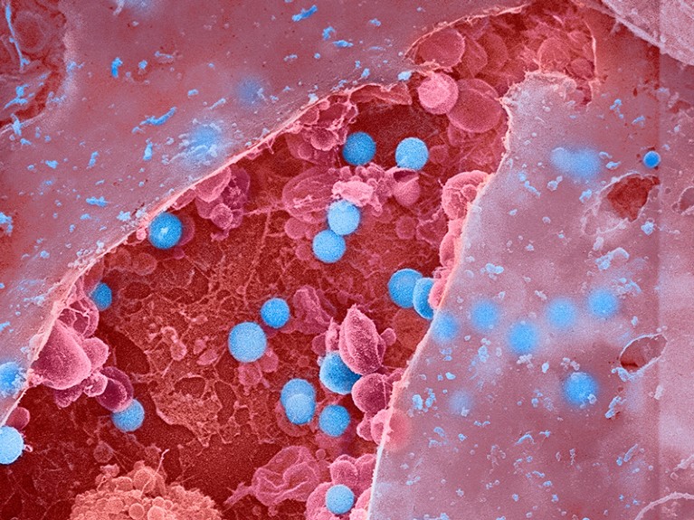
Coloured scanning electron micrograph (SEM) of influenza (flu) viruses (blue) budding from a burst epithelial cell.
In this false-colour scanning electron microscope image, influenza virus particles (blue) stand ready to release from a burst epithelial cell (red).

An analysis of about 450,000 electronic health records has found a link between infections with influenza and other common viruses and an elevated risk of having a neurodegenerative condition such as Alzheimer’s or Parkinson’s disease later in life. But researchers caution that the data show only a possible connection, and that it’s still unclear how or whether the infections trigger disease onset.

The analysis, published in Neuron on 19 January1, found at least 22 links between viral infections and neurodegenerative diseases. Some of the viral exposures were associated with an increased risk of brain disease up to 15 years after infection.

“It’s startling how widespread these associations seem to be, both for the number of viruses and number of neurodegenerative diseases involved,” says Matthew Miller, a viral immunologist at McMaster University in Hamilton, Canada.

Mining health records

This isn’t the first time viruses have been linked to neurodegenerative disease. Infection with a type of herpes virus has been associated with the development of Alzheimer’s2, for instance. And a landmark study published in Science3 last year found the strongest evidence yet that Epstein–Barr virus is tied to multiple sclerosis. But many of these past studies examined only a single virus and a specific brain disease.The quest to prevent MS — and understand other post-viral diseases

To understand whether viruses are linked to brain diseases more broadly, Kristin Levine, a biomedical data scientist at the US National Institutes of Health’s Center for Alzheimer’s Related Dementias in Bethesda, Maryland, and her colleagues analysed hundreds of thousands of medical records to look for instances in which a person had both a viral infection and a brain disease on file.

First, the team examined records from about 35,000 people with brain diseases and about 310,000 people without, sourced from FinnGen, a large Finnish database that includes health information. The team found 45 significant links between infections and brain diseases, and then tested those against more than 100,000 records from another database, the UK Biobank. After this analysis, they were left with 22 significant pairings.

One of the strongest associations was between viral encephalitis, a rare inflammation of the brain that can be caused by multiple types of virus, and Alzheimer’s. People with encephalitis were about 31 times more likely to develop Alzheimer’s later in life than were people who did not have encephalitis. Most other associations were more modest: people who had a bout of flu that led to pneumonia were four times more likely to develop Alzheimer’s than were people who didn’t develop the flu with pneumonia. There were no pairings that suggested a protective link between viral infection and brain disease.

“I’m very excited they’re expanding this research broader than what other studies have looked at,” says Kristen Funk, a neuroimmunologist at the University of North Carolina, Charlotte, who studies the link between herpesviruses and Alzheimer’s.

Data shortcomings

Kjetil Bjornevik, an epidemiologist at the Harvard T.H. Chan School of Public Health in Boston, Massachusetts, and an author of the Epstein–Barr paper in Science, applauds Levine and her colleagues for bringing more attention to the role of viral infections in brain diseases. But he warns that their approach of using medical records “could be problematic” because they analysed only infections that were severe enough to warrant a trip to a health practitioner. Taking milder infections into account might weaken the associations, he says.Are infections seeding some cases of Alzheimer’s disease?

The data are also sourced almost exclusively from people of European ancestry, which means that the findings might not be applicable to the larger global population, Funk says. Furthermore, she adds, outside Europe, “certain viruses are more prevalent”, such as Zika or West Nile virus, so the analysis might have missed links between those pathogens and brain disease. Levine acknowledges the limitations of the analysis; the team worked with the data that were available, she says.

These limitations also underscore the difficulty of untangling whether a viral infection leads to neurodegenerative disease, or whether the disease makes a person more susceptible to infection, Bjornevik says. To make it even more tricky, the authors found that the more time that elapsed between the infection and the diagnosis of brain disease, the weaker the link was. The body is known to begin changing years before symptoms of brain disease develop and a diagnosis is made4, so it’s tough to determine which is causing which, he adds. Another plausible theory is that these viral infections might be accelerating molecular changes in the body that were already ongoing, says Cornelia van Duijn, a genetic epidemiologist at the University of Oxford, UK.Could long COVID be linked to herpes viruses? Early data offer a hint

If future studies add more weight to the connection between viral infection and brain disease, it could offer health officials a tangible way to delay the onset of neurodegenerative disease. Vaccines exist for many of these viruses, van Dujin says. Because multiple types of dementia are diagnosed late in life — close to the average life expectancy — if clinicians could postpone disease onset by even a couple of years, that could mean that many people might never develop the disease, she adds.

“It’s not very clear that the infections are causing brain disease,” she says. But viral infections aren’t pleasant, and if there’s any link to brain disease, “I think we owe it to people to prevent them.”

More Thankful, Less Stressed?


Summary: The state of gratitude lowers systolic blood pressure response through stress-testing experiences, demonstrating that gratitude has a unique buffering effect against both reactions to and recovery from psychological stress.

Source: BIAL Foundation

Researchers from Irish universities carried out a study with 68 adults and found that gratitude has a unique stress-buffering effect on both reactions to and recovery from acute psychological stress, which can contribute to the improvement of cardiovascular health.

Knowing that stress affects human beings and has an impact on their health and well-being, namely causing high blood pressure and increasing cardiovascular morbidity and coronary heart disease, it is important to know our reactions towards stress and find out if there are any factors that can play key stress-buffering roles.

In the article “Gratitude, affect balance, and stress buffering: A growth curve examination of cardiovascular responses to a laboratory stress task”, published in January in the Journal of Psychophysiology, Brian Leavy, Brenda H. O’Connell and Deirdre O’Shea propose that, although previous research suggest that gratitude and affect-balance play key stress-buffering roles, to date little has been known about the impact of these variables on cardiovascular recovery from acute psychological stress.

That was the focus of the study by the researchers from the Universities of Maynooth and Limerick in Ireland, who also sought to find out whether affect balance moderates the relationship between gratitude and cardiovascular reactions to acute psychological stress.

The research carried out at the Irish University of Maynooth involved 68 undergraduate students (24 male and 44 female), aged between 18 and 57 years. This study used a within-subjects experimental design with lab tasks in which stress was induced to participants and then cardiovascular reactivity and recovery in response to this was measured.

This shows a happy woman in a corn flower field
It was also found that affect balance amplifies the effects of state gratitude.

The results showed that state gratitude predicted lower systolic blood pressure responses throughout the stress-testing period, which means that the state of gratitude has a unique stress-buffering effect on both reactions to and recovery from acute psychological stress. It was also found that affect balance amplifies the effects of state gratitude.

These findings have clinical utility as there are several low-cost gratitude interventions which can contribute to well-being (Wood et al., 2010). For example, previous research has shown how cardiac patients who make use of gratitude journals have better cardiovascular outcomes than those who do not (Redwine et al., 2016).

Combined with the results of this study and previous work, gratitude may thus constitute a useful point of intervention for the improvement of our cardiovascular health.

Abstract

Gratitude, affect balance, and stress buffering: A growth curve examination of cardiovascular responses to a laboratory stress task

Previous research has indicated that gratitude and affect-balance play key stress-buffering roles. However, to date there is limited research on the impact of gratitude and affect balance on cardiovascular recovery from acute psychological stress, and whether affect balance moderates the relationship between gratitude and cardiovascular reactions to acute psychological stress.

In this study, 68 adults completed measures of state gratitude, positive and negative affect, and completed a laboratory-based cardiovascular stress-testing protocol. This incorporated a 20-minute acclimatization period, a 10-minute baseline, a 6-minute arithmetic stress task, and an 8-minute recovery period.

Mixed-effects growth curve models were fit and the results indicated that state gratitude predicted lower systolic blood pressure responses throughout the stress-testing period. Affect balance was found to moderate the association between state gratitude and diastolic blood pressure responses to stress, amplifying the effects of state gratitude.

These findings suggest that state gratitude has a unique stress-buffering effect on both reactions to and recovery from acute psychological stress.

Non-invasive Neurotechnology Reduces Symptoms of Insomnia and Improves Autonomic Nervous System Function


Summary: A new closed-loop, acoustic stimulation technique significantly improves sleep quality and autonomic immune system function, and reduces symptoms of insomnia.

Source: Wake Forest University

A good night’s sleep is crucial to health and well-being. Numerous research studies have shown that insomnia can increase the risk of cardiovascular events, obesity, diabetes and other illnesses. 

Now, a new study from researchers at Wake Forest University School of Medicine shows significant improvements in not only sleep quality, but also in improved autonomic nervous system function using a closed-loop, acoustic stimulation neurotechnology.

The study is published online in Global Advances in Integrative Medicine and Health.

Cereset ResearchTM with Standard Operating Procedures (CR-SOP) is the evolution of HIRREM®, or high-resolution, relational, resonance-based electroencephalic mirroring, a noninvasive, closed-loop technology that uses scalp sensors to monitor brainwaves and software algorithms to translate specific frequencies into audible tones of varying pitch.

These tones linked to brainwaves are echoed back in real time via earbuds. This allows the brain a chance to listen to itself, to look at itself in an acoustic mirror.

“CR-SOP allows the brain to reset from stress patterns that contribute to insomnia,” said Charles H. Tegeler, M.D., chair of neurology at Wake Forest University School of Medicine. “During the intervention, the brain continuously updates with respect to its own activity patterns, resulting in auto-calibration or self-optimization.”

While still echoing brainwaves, as with legacy HIRREM, CR-SOP uses an updated platform with faster computers, new sensors and hardware, and computer management during the protocols. This results in faster echoing of brainwaves, shorter sessions and reduced dependence on technologist expertise.

In this randomized and controlled study of 22 adults, researchers compared changes on the Insomnia Severity Index (ISI), a self-report instrument to assess insomnia symptoms. About half of the participants received 10 sessions of CR-SOP linked to brainwaves while the control group received 10 sessions of randomly generated auditory tones. Sessions were received over a mean of 15.3 days. Researchers also recorded heart rate and blood pressure to assess autonomic cardiovascular regulation. 

After completion of the sessions and at follow-up visits up to six weeks later, subjects in the CR-SOP group reported reduced insomnia symptoms. They also showed statistically and clinically significant improvements in autonomic function across multiple measures such as heart rate variability (HRV) and baroreflex sensitivity (BRS) compared to those who received random tones.

HRV is a powerful biometric that reflects the health of the autonomic nervous system, and BRS measures blood pressure regulation. HRV is correlated with a host of important health and well-being outcomes.

This shows a test subject undergoing the treatment
Cereset Research with Standard Operating Procedures (CR-SOP) is the evolution of HIRREM, or high-resolution, relational, resonance-based electroencephalic mirroring, a noninvasive, closed-loop technology that uses scalp sensors to monitor brainwaves and software algorithms to translate specific frequencies into audible tones of varying pitch.

These findings are in line with previous HIRREM research that showed a reduction in insomnia symptoms.

According to Tegeler, the study also used standard operating procedures so that all subjects received the same sequence of protocols. Taken together, this greatly increases the scalability of this approach so that more people might have access, more quickly, he said.

“Closed-loop acoustic stimulation can improve sleep as well as autonomic function in those who suffer from insomnia,” Tegeler said. “This pilot study demonstrates these benefits with CR-SOP from sessions received over a short period. This is also an important step in showing the intervention’s potential scalability for treating more people.”

Ongoing clinical trials are focused on stress and anxiety in health care workers as well as caregivers. 

HIRREM and Cereset Research are registered trademarks of Brain State Technologies based in Scottsdale, Arizona and have been licensed to Wake Forest University School of Medicine for collaborative research since 2011.

Conclusions

This pilot study compared use of a standardized, allostatic, acoustic neurotechnology intervention with a sham, active control condition. The magnitude of change in insomnia severity was clinically relevant and similar to the findings in a prior, fully powered trial, but the differential improvement observed was not statistically significant. Significant improvements were demonstrated in sleep quality and some autonomic function measures.

Cerebral Alveolar Echinococcosis


A 17-year-old boy presented to the emergency department with a 3-week history of dizziness, headache, and weakness of the right leg. Neurologic examination was notable for 4/5 strength in the right lower leg. Computed tomography (CT) of the head revealed ring-enhancing lesions in the frontal lobe and basal ganglia on the left side. Subsequent magnetic resonance imaging of the head also showed the lesions (Panel A; sagittal view, T1-weighted) as well as surrounding edema and midline shift (Panel B; axial view, T2-weighted). Testing for Echinococcus multilocularis and E. granulosus IgG antibodies was negative. Surgical excision of the lesions was performed. Histopathological testing showed necrotic tissue with surrounding granulomatous inflammation and an echinococcal laminated membrane (Panel C, arrow) as well an intact cyst (Panel D, arrow). A reverse-transcriptase–polymerase-chain-reaction assay identified E. multilocularis. CT imaging of the chest and abdomen did not show any other sites of disease. A diagnosis of primary cerebral alveolar echinococcosis was made. Alveolar echinococcosis is an invasive type of echinococcal infection that leads to tissue inflammation and destruction. The causative tapeworm is acquired by means of close contact with infected animals, which this patient had not reported. Postoperatively, a long course of albendazole was prescribed. At 2 months of follow-up, the patient’s symptoms had resolved.

Source: NEJM


Pregnant Acetaminophen Users Risk ADHD in Offspring


Pregnant Acetaminophen Users Risk ADHD in Offspring

Acetaminophen (the active ingredient in Tylenol and Excedrin) is the most common over-the-counter pain medication. A new, long-term study found that pregnant women who take the drug may be placing their unborn children at risk of developing attention deficit disorders.

In the report, “Acetaminophen Use During Pregnancy, Behavioral Problems, and Hyperkinetic Disorders,” published in JAMA Pediatrics, researchers found a compelling pattern of pregnant mothers taking acetaminophen and offspring who are later diagnosed with attention deficit hyperactivity disorder (ADHD), exhibiting ADHD-like behaviors, or taking ADHD medication.

Researchers looked at data on 64,322 children and mothers who were enrolled in a Danish study tracking pregnancies and children, from 1996 to 2002.

A series of computer-assisted telephone interviews taken throughout each pregnancy found that more than one-half of all the mothers reported using acetaminophen. Researchers discovered that children born to mothers taking the medication had up to a 37 percent higher risk of ADHD by age seven.

Mothers who continued to take acetaminophen into the second and third trimesters had the strongest associations with ADHD. According to the report, risks were elevated by 50 percent or more when the mothers used the painkiller for more than 20 weeks into their pregnancy.

According to the National Institutes of Health, ADHD is one of the most common childhood disorders. Symptoms include difficulty staying focused and uncontrollable hyperactive behavior.

The jury is still out about what causes ADHD—and why there are now so many cases. Some doctors insist that the epidemic is merely the result of better diagnosing, while others point to various environmental and genetic factors.

According Dr. Beate Ritz, professor and vice chair of the Epidemiology Department at the University of California, Los Angeles’ (UCLA) School of Public Health and one of the senior authors of the report, the impetus for the study was the drug’s emerging reputation as a hormonal disrupter.

“We know ADHD has to do with the balance of certain brain circuitry and systems that are interacting with each other,” she said. “If we disturb that, we could see how that would lead to some kind of neurodevelopmental disorder or shift.”

The preponderance of ADHD in boys, and general fetal sensitivity to hormonal changes, got researchers thinking about how a widely used hormonal disrupter might affect a developing fetus. Ritz’s colleague, Dr. Jørn Olsen, another senior author and chair of the UCLA Epidemiology Department, has seen previous patterns of fetal hormonal disturbance, with pregnant women and acetaminophen use showing a greater risk for children with undescended testicles.

Safety Reputation

In the 1950s, acetaminophen was a new over-the-counter alternative to remedy pain and headaches, but over the years it has come to dominate the market—not only due to its effectiveness but also its reputation for safety. Since the 1970s, doctors have favored the well-tolerated acetaminophen over aspirin or other painkillers, particularly for pregnant women.

While it may be the safest analgesic in the modern medical arsenal, acetaminophen still carries significant risks that some say require more caution.

In September 2013, ProPublica and NPR collaborated on a report on acetaminophen and found that for more than 30 years, the U.S. Food and Drug Administration (FDA) had either “delayed or failed to adopt measures designed to reduce deaths and injuries” related to the drug.

In that report, the big issue was dosage, which the FDA found to be a “persistent, important public health problem.” While relatively safe at recommended doses, even slight overdoses have been shown to cause liver damage, resulting in about 150 deaths a year, according to the Centers for Disease Control.

Fetal Vulnerability

The FDA recently announced that it will review its over-the-counter drug policy. However, the safety issue becomes even more complicated in regard to acetaminophen as a hormonal disrupter—where even relatively small doses may have a profound effect on the body’s endocrine system, especially in the midst of development.

“We know that during development the fetus actually prepares itself for the environment he or she may encounter after being born,” said Ritz. “That’s very true with nutrition, but there are also certain chemicals, stress hormones that the mother has in her body that can program the brain.”

Instead of traditional measures of toxicity where a high enough dose threshold triggers damage to cells, endocrine disruptive chemicals can interfere in delicate hormonal processes with doses previously considered benign. As a young life builds fine details of its neurological system toward the later stages of fetal development, the body can be particularly vulnerable to a chemical influence disrupting the process, according to Ritz.

“That can happen at much lower doses than what we would normally expect for toxicity,” said Ritz.

UCLA researchers want to see more tests with variations in the study design to see if the pattern holds up. But until that’s available in another decade or so, Ritz urges caution to pregnant mothers.

“I would want to be careful,” she said, adopting a pregnant mother’s perspective. “‘I had to deal with my headache in a different way, but at least I didn’t put my fetus at risk.’”

Surprising Side Effects of Tylenol


Extra Strength Tylenol is displayed in a drugstore in Washington, on July 5, 2006. (Brendan Smialowski/Getty Images)

Extra Strength Tylenol is displayed in a drugstore in Washington, on July 5, 2006.

You know Tylenol can relieve a headache, but are you aware it can cause other side effects, such as increased risk of death or heart toxicity?

Have you ever popped a Tylenol to push through that annoying headache and get on with your day? Most of us have. Each week, millions of Americans take one of the 600 medicines that contain acetaminophen, Tylenol’s active ingredient, for various aches and pains. Acetaminophen is the most common drug ingredient in the US, but this ‘harmless drug’ is linked to over 110,000 injuries and deaths per year.

How can Tylenol, which is doled out like candy, be bad for you? Amazingly, no one really knows how acetaminophen works, but people do know that this drug gets to your brain. Tylenol in your brain is concerning because it depletes glutathione, an antioxidant that is especially necessary for brain health. Our bodies depend on antioxidants to balance oxidative damage and inflammation. If you’re popping a Tylenol with your morning antioxidant-rich smoothie, Tylenol may rob you of the smoothie’s benefits!

(Niloo/Shutterstock)

Beyond Liver Damage

Most people have heard that Tylenol can damage the liver (has anyone ever drunkenly warned you to take a Motrin, not a Tylenol, to prevent a hangover?). But since everything in our bodies is connected, it’s not surprising that Tylenol can do damage beyond your liver. A recent study showed that people who took Tylenol had increased risks of death, heart toxicity, gastrointestinal bleeding, and kidney damage. Importantly, people who took more Tylenol suffered more damage.

It’s also scary how Tylenol affects mood. After swallowing 1000 mg of Tylenol, people exhibited less empathy and blunted positive emotions. For reference, 1000 mg is two extra-strength Tylenol tablets, and the ‘safe’ range is 3000 mg per day. This means that popping two Tylenols can affect you physically and emotionally!

If you’re pregnant or looking to become pregnant, please be especially careful about taking Tylenol. Research has shown that children exposed to acetaminophen in the womb had behavioral, communication, and motor skill problems. Another study linked prenatal acetaminophen exposure with increased ADHD-like behaviors and medication use.

What About Other Painkillers?

Hopefully you’re convinced to think twice before taking a Tylenol, but what about other pain-relievers like Motrin, Aleve, or Advil? These non-steroidal anti-inflammatory drugs (NSAIDs) must be safe, since thirty million people take them every day! Not so fast…

Women who recognize the importance of hormonal balance should be wary. NSAIDs can mess with ovulation, especially progesterone levels, after only 10 days of use. Additionally, NSAIDs injure the small intestine; in one study, 71% of NSAID users showed small intestinal damage, compared to 10% of non-users [5]. Damaged intestines can lead to intestinal permeability, or “leaky gut.” Leaky gut has been linked to depression, ADHD, and allergies.  NSAIDs can cause leaky gut and harm your microbiome, the trillions of beneficial bacteria that live in and on us. Disrupting our bacteria can do more damage than we realize!

How Can I Relieve My Headache?!

Now that you know the surprising dangers of Tylenol and other NSAIDs, what should you take for headaches and other aches and pains? Turmeric, the yellow root found in curry powder, contains a powerful anti-inflammatory and pain reliever called curcumin. This has been used in Ayurvedic and Chinese medicine as a treatment for pain, digestive disorders, and wound healing for centuries. Many studies show the beneficial effects of curcumin; curcumin works as well as ibuprofen to alleviate pain from knee osteoarthritis and PMS. Next time you have a headache, try 1-2 grams of curcumin – or a turmeric latte!

A Drug That Increases Dopamine Can Reverse the Effects of Inflammation on the Brain in Depression


Summary: Levodopa, a drug commonly prescribed for the treatment of Parkinson’s disease that increases dopamine in the brain was found to reverse the effects of neuroinflammation on the reward system and improve symptoms associated with depression.

Source: Emory University

An Emory University study published in Molecular Psychiatry shows levodopa, a drug that increases dopamine in the brain, has potential to reverse the effects of inflammation on brain reward circuitry, ultimately improving symptoms of depression.

Numerous labs across the world have shown that inflammation causes reduced motivation and anhedonia, a core symptom of depression, by affecting the brain’s reward pathways.

Past research conducted by the Department of Psychiatry and Behavioral Sciences at Emory University School of Medicine has linked the effects of inflammation on the brain to decreased release of dopamine, a chemical neurotransmitter that regulates motivation and motor activity, in the ventral striatum.

In the study, researchers demonstrated that levodopa reversed the effects of inflammation on the brain’s functional connectivity in reward circuitry and anhedonia (inability to feel pleasure) in depressed individuals with higher C-reactive protein (CRP), a blood biomarker produced and released by the liver in response to inflammation.

Levels of inflammation can be easily measured by simple blood tests, like CRP, readily available in clinics and hospitals throughout the U.S.

The study included 40 depressed patients with a range of CRP levels from high to low who underwent functional brain scans on two visits after receiving in random order either placebo or levodopa, a drug often prescribed for disorders like Parkinson’s disease.

Levodopa improved functional connectivity in a classic ventral striatum to ventromedial prefrontal cortex reward circuit but only in patients with higher levels of CRP. This improvement in reward circuitry in depressed individuals with higher CRP also correlated with reduced symptoms of anhedonia after levodopa.

This shows a young man crying
Levels of inflammation can be easily measured by simple blood tests, like CRP, readily available in clinics and hospitals throughout the U.S.

“This research demonstrates the translational potential for use of inflammation-related deficits in functional connectivity and could have important implications for the future investigations of precision therapies for psychiatric patients with high inflammation,” says principal investigator and senior author Jennifer C. Felger, Ph.D., associate professor of psychiatry and behavioral sciences, Emory School of Medicine.

Felger says the study findings are critical for two reasons. First, they suggest depressed patients with high inflammation may specifically respond to drugs that increase dopamine.

Second, Felger says these findings also provide additional evidence that functional connectivity in reward circuitry may serve as a reliable brain biomarker for the effects of inflammation on the brain.

“Moreover, as the effect of levodopa was specific to depressed patients with higher inflammation, this functional connectivity may be used to assess the responsiveness of the brain to novel treatments that might be targeted to this subtype of depressed patients in future studies and clinical trials,” says Felger.

Functional connectivity in reward circuitry and symptoms of anhedonia as therapeutic targets in depression with high inflammation: evidence from a dopamine challenge study

Increased inflammation in major depressive disorder (MDD) has been associated with low functional connectivity (FC) in corticostriatal reward circuits and symptoms of anhedonia, relationships which may involve the impact of inflammation on synthesis and release of dopamine.

To test this hypothesis while establishing a platform to examine target engagement of potential therapies in patients with increased inflammation, medically stable unmedicated adult MDD outpatients enrolled to have a range of inflammation (as indexed by plasma C-reactive protein [CRP] levels) were studied at two visits involving acute challenge with the dopamine precursor levodopa (L-DOPA; 250 mg) and placebo (double-blind, randomized order ~1-week apart).

The primary outcome of resting-state (rs)FC in a classic ventral striatum to ventromedial prefrontal cortex reward circuit was calculated using a targeted, a priori approach.

Data available both pre- and post-challenge (n = 31/40) established stability of rsFC across visits and determined CRP > 2 mg/L as a cut-point for patients exhibiting positive FC responses (post minus pre) to L-DOPA versus placebo (p < 0.01).

Higher post-L-DOPA FC in patients with CRP > 2 mg/L was confirmed in all patients (n = 40) where rsFC data were available post-challenge (B = 0.15, p = 0.006), and in those with task-based (tb)FC during reward anticipation (B = 0.15, p = 0.013).

While effort-based motivation outside the scanner positively correlated with rsFC independent of treatment or CRP, change in anhedonia scores negatively correlated with rsFC after L-DOPA only in patients with CRP > 2 mg/L (r = -0.56, p = 0.012).

FC in reward circuitry should be further validated in larger samples as a biomarker of target engagement for potential treatments including dopaminergic agents in MDD patients with increased inflammation.

New Technology May Help Inform Brain Stimulation


Summary: A new technique that uses ultrafast fMRI is able to capture brain activity at sub-second levels. The technique allows for real-time monitoring of the brain under stimulation conditions.

Source: University of Queensland

Brain stimulation, such as Deep brain stimulation (DBS), is a powerful way to treat neurological and psychiatric disorders. While it has provided therapeutic benefit for sufferers of Parkinson’s, Alzheimer’s, and addiction for more than a decade, its underlying neural mechanism is not yet fully understood.

Researchers at the Queensland Brain Institute (QBI) are now one step closer to unravelling the mystery of brain activity to better understand this mechanism and potentially predict DBS outcomes.

The brain is a highly complex network of circuits organised hierarchically with wide-ranging connections. Connections go in different directions, forwards and backwards, and between neurons that are either excitatory – the accelerators of a response – or inhibitory – the brakes modifying a response.

“Say you want to move your hand – once that signal is initiated, we expect that the activity that follows depends on the brain’s neural networks,” Associate Professor Kai-Hsiang Chuang said.

“What we don’t fully understand is how or when these structural and functional components of the brain interact to eventually lead to the outcome of moving your hand.”

Functional MRI (fMRI) is the most popular technique used to study brain networks. fMRI tracks blood flow and oxygenation changes following neural activity, thereby indirectly measuring the functional connections being formed, and giving us an indication of where brain activity is propagating.

Brain activity, however, isn’t as simple as a signal travelling from area to area.

The team at the Chuang laboratory have developed a new ultrafast fMRI technique with a vastly increased temporal resolution, enabling them to capture the dynamics of brain activity at a sub-second level.

Associate Professor Chuang said the new technique had led to more comprehensive understanding of how and when the brain’s structural and functional connections interact.

“The first new discovery we made is that brain activity not only propagates through structural wiring but follows certain preferential circuits depending on their excitatory and inhibitory neuronal distribution,” he said.

“Communication between brain regions of similar cell types becomes more fluent, and the brain activity stronger.”

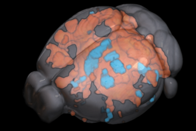
This shows a brain map with the stimulated areas highlighted orange and blue
Mouse brain activity under optogenetic stimulation detected by ultrafast fMRI technique. Red shows a positive response (reflecting excitatory activity) and blue shows a negative response (reflecting inhibitory activity).

The Chuang group tracked the brain activity of mice both while stimulated and at rest using their ultrafast fMRI technique. When the brain was stimulated, activity followed the structural wiring in the forward direction — from A to B and then B to C. When the brain was at rest, activity was more dependent on cell type organisation and less on structural wiring, propagating between C and B but not with A, if that’s where the preferential circuit was.

This means that how information is processed is actually dependent on your state, where it was previously thought that brain activity functioned in the same way whether at rest or busy doing a task.

“The second discovery we made was that the blood signal detected by fMRI could reflect the network organisation and cell type distribution,” Associate Professor Chuang said.

“These findings have significant implications for how brain structure shapes function, and how to predict activity based on the knowledge of this structure. More practically, what we now know will impact the design of DBS and other brain stimulation techniques.

“The next steps are to work with clinicians versed in brain stimulation to determine how we can utilise this knowledge combined with human data to help improve our understanding of DBS.”

This more comprehensive understanding could enable us to better predict DBS results and potentially improve its design for better therapeutic outcomes.

Abstract

Hemodynamic transient and functional connectivity follow structural connectivity and cell type over the brain hierarchy

The neural circuit of the brain is organized as a hierarchy of functional units with wide-ranging connections that support information flow and functional connectivity. Studies using MRI indicate a moderate coupling between structural and functional connectivity at the system level.

However, how do connections of different directions (feedforward and feedback) and regions with different excitatory and inhibitory (E/I) neurons shape the hemodynamic activity and functional connectivity over the hierarchy are unknown.

Here, we used functional MRI to detect optogenetic-evoked and resting-state activities over a somatosensory pathway in the mouse brain in relation to axonal projection and E/I distribution.

Using a highly sensitive ultrafast imaging, we identified extensive activation in regions up to the third order of axonal projections following optogenetic excitation of the ventral posteriomedial nucleus of the thalamus.

The evoked response and functional connectivity correlated with feedforward projections more than feedback projections and weakened with the hierarchy.

The hemodynamic response exhibited regional and hierarchical differences, with slower and more variable responses in high-order areas and bipolar response predominantly in the contralateral cortex.

Electrophysiological recordings suggest that these reflect differences in neural activity rather than neurovascular coupling. Importantly, the positive and negative parts of the hemodynamic response correlated with E/I neuronal densities, respectively. Furthermore, resting-state functional connectivity was more associated with E/I distribution, whereas stimulus-evoked effective connectivity followed structural wiring.

These findings indicate that the structure–function relationship is projection-, cell-type- and hierarchy-dependent. Hemodynamic transients could reflect E/I activity and the increased complexity of hierarchical processing.

Fresh Questions About Oxytocin as the ‘Love Hormone’ Behind Pair Bonding


Summary: The “love hormone” oxytocin may not play as critical a role in bonding as previously believed. Removing the oxytocin receptor in animal models still resulted in monogamous mating, attachment, and parental bonding behaviors, although females without the receptor produced milk in smaller quantities. Findings reveal parenting and bonding aren’t purely dictated by oxytocin receptors.

Source: UCSF

Turning a decades-old dogma on its head, new research from scientists at UC San Francisco and Stanford Medicine shows that the receptor for oxytocin, a hormone considered essential to forming social bonds, may not play the critical role that scientists have assigned to it for the past 30 years. 

In the study, appearing Jan. 27, 2023 in Neuron, the team found that prairie voles bred without receptors for oxytocin and showed the same monogamous mating, attachment, and parenting behaviors as regular voles. In addition, females without oxytocin receptors gave birth and produced milk, though in smaller quantities, than ordinary female voles.  

The results indicate that the biology underlying pair bonding and parenting isn’t purely dictated by the receptors for oxytocin, sometimes referred to as the “love hormone.” 

“While oxytocin has been considered ‘Love Potion #9,’ it seems that potions 1 through 8 might be sufficient,” said psychiatrist Devanand Manoli, MD, PhD, a senior author of the paper and member of the UCSF Weill Institute for Neurosciences. “This study tells us that oxytocin is likely just one part of a much more complex genetic program.”  

CRISPR Voles Pack a Surprise 

Because prairie voles are one of the few mammalian species known to form lifelong monogamous relationships, researchers study them to better understand the biology of social bonding. 

Studies in the 1990s using drugs that prevent oxytocin from binding to its receptor found that voles were unable to pair bond, giving rise to the idea that the hormone is essential to forming such attachments.  

The current project emerged from shared interests between Manoli and co-senior author and neurobiologist Nirao Shah, MD, PhD, then at UCSF and now at Stanford Medicine. Shah had been interested in the biology of oxytocin and social attachment in prairie voles since teaching about the oxytocin studies decades earlier. Manoli, who wanted to investigate the neurobiology of social bonding, joined Shah’s lab in 2007 as a postdoctoral scholar.  

For this study, 15 years in the making, the two applied new genetic technologies to confirm if oxytocin binding to its receptor was indeed the factor behind pair bonding. They used CRISPR to generate prairie voles that lack functional oxytocin receptors. Then, they tested the mutant voles to see whether they could form enduring partnerships with other voles.  

To the researchers’ surprise, the mutant voles formed pair bonds just as readily as normal voles.  

“The patterns were indistinguishable,” said Manoli. “The major behavioral traits that were thought to be dependent on oxytocin – sexual partners huddling together and rejecting other potential partners as well as parenting by mothers and fathers – appear to be completely intact in the absence of its receptor.” 

Labor and Lactation 

Even more surprising for Manoli and Shah than the pair bonding was the fact that a significant percentage of the female voles were able to give birth and provide milk for their pups.
 
Oxytocin is likely to have a role in both birth and lactation, but one that is more nuanced than previously thought, Manoli said. Female voles without receptors proved perfectly capable of giving birth, on the same timeframe and in the same way as the regular animals, even though labor has been thought to rely on oxytocin. 

The results help to clear up some of the mystery surrounding the hormone’s role in childbirth: Oxytocin is commonly used to induce labor but blocking its activity in mothers who experience premature labor isn’t better than other approaches for halting contractions.  

When it came to producing milk and feeding pups, however, the researchers were taken aback. Oxytocin binding to its receptor has been considered essential for milk ejection and parental care for many decades, but half of the mutant females were able to nurse and wean their pups successfully, indicating that oxytocin signaling plays a role, but it is less vital than previously thought.  

“This overturns conventional wisdom about lactation and oxytocin that’s existed for a much longer time than the pair bonding association,” said Shah. “It’s a standard in medical textbooks that the milk letdown reflex is mediated by the hormone, and here we are saying, ‘Wait a second, there’s more to it than that.’” 

Hope for Social Connection  

Manoli and Shah focused on understanding the neurobiology and molecular mechanisms of pair bonding because it is thought to hold the key to unlocking better treatments for psychiatric conditions, such as autism and schizophrenia, that interfere with a person’s ability to form or maintain social bonds.  

This shows heart shaped wind chimes
The results indicate that the biology underlying pair bonding and parenting isn’t purely dictated by the receptors for oxytocin, sometimes referred to as the “love hormone.”

Over the past decade, much hope was pinned on clinical trials using oxytocin to address those conditions. But those results were mixed, and none has illuminated a clear path to improvement.  

The researchers said their study strongly suggests that the current model – a single pathway or molecule being responsible for social attachment –is oversimplified. This conclusion makes sense from an evolutionary perspective, they said, given the importance of attachment to the perpetuation of many social species.  

“These behaviors are too important to survival to hinge on this single point of potential failure,” said Manoli. “There are likely other pathways or other genetic wiring to allow for that behavior. Oxytocin receptor signaling could be one part of that program, but it’s not the be-all end-all.” 

The discovery points the researchers down new paths to improving the lives of people struggling to find social connection.  

“If we can find the key pathway that mediates attachment and bonding behavior,” Shah said, “We’ll have an eminently druggable target for alleviating symptoms in autism, schizophrenia, many other psychiatric disorders.” 
 
 Authors: Additional authors include: Ruchira Sharma, Rose Larios, Nastacia Goodwin, Michael Sherman and Isidero Espineda of UCSF, Maricruz Alvarado Mandujano, YiChao Wei, Srinivas Parthasarthy and Joseph Knoedler of Stanford, and Forrest Rogers, Trenton Simmons, Adele Seelke, Jessica Bond, and Karen Bales of UC Davis, and Annaliese Beery of UC Berkeley. 
 
Funding: This work was supported by NIH grants R01MH123513, R01MH108319, DP1MH099900 and R25MH060482, NSF grant, 1556974, and philanthropy. For details, see the study.  

Abstract

Oxytocin receptor is not required for social attachment in prairie voles

Highlights

  • Prairie voles lacking oxytocin receptor (Oxtr) generated with CRISPR targeting
  • Oxtr−/− voles form pair bonds or social attachments
  • Oxtr−/− voles show parental behavior
  • Oxtr−/− females nurse many of their pups to weaning

Summary

Prairie voles are among a small group of mammals that display long-term social attachment between mating partners. Many pharmacological studies show that signaling via the oxytocin receptor (Oxtr) is critical for the display of social monogamy in these animals. We used CRISPR mutagenesis to generate three different Oxtr-null mutant prairie vole lines. 

Oxtr mutants displayed social attachment such that males and females showed a behavioral preference for their mating partners over a stranger of the opposite sex, even when assayed using different experimental setups.

Mothers lacking Oxtr delivered viable pups, and parents displayed care for their young and raised them to the weanling stage.

Together, our studies unexpectedly reveal that social attachment, parturition, and parental behavior can occur in the absence of Oxtr signaling in prairie voles.