Scientists Link Selfies To Narcissism, Addiction & Mental Illness


Scientists Link Selfies To Narcissism, Addiction & Mental Illness
The growing trend of taking smartphone selfies is linked to mental health conditions that focus on a person’s obsession with looks.

According to psychiatrist Dr David Veal: “Two out of three of all the patients who come to see me with Body Dysmorphic Disorder since the rise of camera phones have a compulsion to repeatedly take and post selfies on social media sites.”

“Cognitive behavioural therapy is used to help a patient to recognise the reasons for his or her compulsive behaviour and then to learn how to moderate it,” he told the Sunday Mirror.

A British male teenager tried to commit suicide after he failed to take the perfect selfie. Danny Bowman became so obsessed with capturing the perfect shot that he spent 10 hours a day taking up to 200 selfies. The 19-year-old lost nearly 30 pounds, dropped out of school and did not leave the house for six months in his quest to get the right picture. He would take 10 pictures immediately after waking up. Frustrated at his attempts to take the one image he wanted, Bowman eventually tried to take his own life by overdosing, but was saved by his mom.

“I was constantly in search of taking the perfect selfie and when I realized I couldn’t, I wanted to die. I lost my friends, my education, my health and almost my life,” he told The Mirror.

The teenager is believed to be the UK’s first selfie addict and has had therapy to treat his technology addiction as well as OCD and Body Dysmorphic Disorder.
Part of his treatment at the Maudsley Hospital in London included taking away his iPhone for intervals of 10 minutes, which increased to 30 minutes and then an hour.

“It was excruciating to begin with but I knew I had to do it if I wanted to go on living,” he told the Sunday Mirror.

Public health officials in the UK announced that addiction to social media such as Facebook and Twitter is an illness and more than 100 patients sought treatment every year.

“Selfies frequently trigger perceptions of self-indulgence or attention-seeking social dependence that raises the damned-if-you-do and damned-if-you-don’t spectre of either narcissism or very low self-esteem,” said Pamela Rutledge in Psychology Today.

The big problem with the rise of digital narcissism is that it puts enormous pressure on people to achieve unfeasible goals, without making them hungrier. Wanting to be Beyoncé, Jay Z or a model is hard enough already, but when you are not prepared to work hard to achieve it, you are better off just lowering your aspirations. Few things are more self-destructive than a combination of high entitlement and a lazy work ethic. Ultimately, online manifestations of narcissism may be little more than a self-presentational strategy to compensate for a very low and fragile self-esteem. Yet when these efforts are reinforced and rewarded by others, they perpetuate the distortion of reality and consolidate narcissistic delusions.

The addiction to selfies has also alarmed health professionals in Thailand. “To pay close attention to published photos, controlling who sees or who likes or comments them, hoping to reach the greatest number of likes is a symptom that ‘selfies’ are causing problems,” said Panpimol Wipulakorn, of the Thai Mental Health Department.

The doctor believed that behaviours could generate brain problems in the future, especially those related to lack of confidence.

The word “selfie” was elected “Word of the Year 2013″ by the Oxford English Dictionary. It is defined as “a photograph that one has taken of oneself, typically with a smartphone or webcam and uploaded to a social media website”.

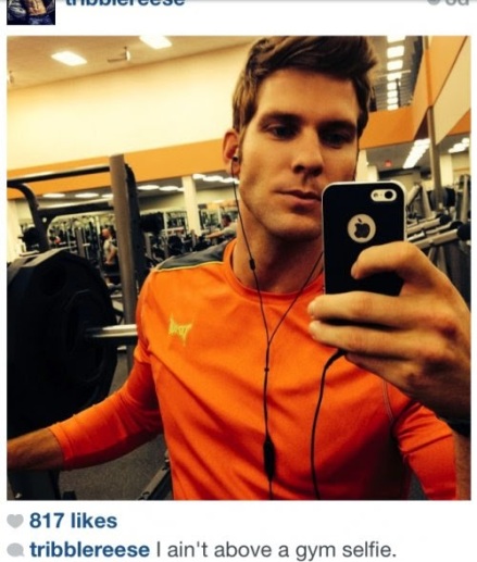
1. The Gym Selfie (Because the checkin isn’t enough.)
Scientists Link Selfies To Narcissism, Addiction & Mental Illness - The Gym Selfie (Because the checkin isn’t enough.)

2. The Pet Selfie (If you want to post a picture of your pet, post a picture of your pet.)
Unless this happens, then it’s ok:
Scientists Link Selfies To Narcissism, Addiction & Mental Illness - The Pet Selfie (If you want to post a picture of your pet, post a picture of your pet.)

3. The Car Selfie AKA The Seatbelt Selfie (You LITERALLY got in the car and thought, “I look so good today, I better let everyone know before I put this thing in drive and head to my shift at the Olive Garden.”)
Scientists Link Selfies To Narcissism, Addiction & Mental Illness - The Car Selfie AKA The Seatbelt Selfie (You LITERALLY got in the car and thought, “I look so good today, I better let everyone know before I put this thing in drive and head to my shift at the Olive Garden

If you can combine the Seatbelt Selfie with the beloved Shirtless Selfie like this unattractive fella below, you..are…GOLD.
Scientists Link Selfies To Narcissism, Addiction & Mental Illness - If you can combine the Seatbelt Selfie with the beloved Shirtless Selfie like this unattractive fella below, you..are…GOLD.

4. The Blurry Selfie (Why?)
Scientists Link Selfies To Narcissism, Addiction & Mental Illness - The Blurry Selfie (Why?)

5. The Just Woke Up Selfie
Scientists Link Selfies To Narcissism, Addiction & Mental Illness - The Just Woke Up Selfie
Yeah right you just woke up.

6. Or even worse, the Pretending to Be Asleep Selfie. (We know you’re not asleep, asshole. You took the damn picture.)
Scientists Link Selfies To Narcissism, Addiction & Mental Illness - Or even worse, the Pretending to Be Asleep Selfie. (We know you’re not asleep, asshole. You took the damn picture.)
Scientists Link Selfies To Narcissism, Addiction & Mental Illness

7. The Add a Kid Selfie (Extra points for a C-section scar.)
Scientists Link Selfies To Narcissism, Addiction & Mental Illness - The Add a Kid Selfie (Extra points for a C-section scar.)

8. The Hospital Selfie (A rare gem. The more tubes you have hooked up to you, the better.)
Scientists Link Selfies To Narcissism, Addiction & Mental Illness - The Hospital Selfie (A rare gem. The more tubes you have hooked up to you, the better.)

9. The “I’m On Drugs” Selfie (This looker below also qualifies as theLook At My New Haircut Selfie.)
Scientists Link Selfies To Narcissism, Addiction & Mental Illness - The “I’m On Drugs” Selfie (This looker below also qualifies as theLook At My New Haircut Selfie.)

10. The Duck Face Selfie (Hey girls. This doesn’t make you prettier. It makes you look stupid and desperate. If that’s what you’re going for, carry on.)
Scientists Link Selfies To Narcissism, Addiction & Mental Illness - The Duck Face Selfie (Hey girls. This doesn’t make you prettier. It makes you look stupid and desperate. If that’s what you’re going for, carry on.)

11. The Pregnant Belly Selfie (Send this to your family and friends, not the entire Internet.)
Scientists Link Selfies To Narcissism, Addiction & Mental Illness - The Pregnant Belly Selfie (Send this to your family and friends, not the entire Internet.)
And yes, that’s a pregnant belly duck face selfie. It’s the unicorn of awful selfies.

12. The “I’m a Gigantic Whore” Selfie
Scientists Link Selfies To Narcissism, Addiction & Mental Illness - The “I’m a Gigantic Whore” Selfie
Nice phone case, by the way.

13. The “I Have Enough Money to Fly On an Airplane” Selfie (AND I own earbuds.)
Scientists Link Selfies To Narcissism, Addiction & Mental Illness - The “I Have Enough Money to Fly On an Airplane” Selfie (AND I own earbuds.)

14. The 3D Selfie. (It takes talent…along with class.)
Scientists Link Selfies To Narcissism, Addiction & Mental Illness - The 3D Selfie. (It takes talent…along with class.)

15. The Say Something That Has Nothing To Do With Anything Selfie(You had a great night? Oh.)
Scientists Link Selfies To Narcissism, Addiction & Mental Illness - The Say Something That Has Nothing To Do With Anything Selfie(You had a great night? Oh.)

16. The “I Live In Filth” Selfie (We all make messes, but if you’re going to post your living quarters on the World Wide Web, pick up your damn room.)
Scientists Link Selfies To Narcissism, Addiction & Mental Illness - The “I Live In Filth” Selfie (We all make messes, but if you’re going to post your living quarters on the World Wide Web, pick up your damn room.)
Source: Disclose.tv via Why Don’t You Try This

The Dangers of Raw Milk: Unpasteurized Milk Can Pose a Serious Health Risk


Milk and milk products provide a wealth of nutrition benefits. But raw milk can harbor dangerous microorganisms that can pose serious health risks to you and your family. According to an analysis by the Centers for Disease Control and Prevention (CDC), between 1993 and 2006 more than 1500 people in the United States became sick from drinking raw milk or eating cheese made from raw milk. In addition, CDC reported that unpasteurized milk is 150 times more likely to cause foodborne illness and results in 13 times more hospitalizations than illnesses involving pasteurized dairy products.

cows and a glass of milk

Raw milk is milk from cows, sheep, or goats that has not been pasteurized to kill harmful bacteria. This raw, unpasteurized milk can carry dangerous bacteria such as Salmonella, E. coli, and Listeria, which are responsible for causing numerous foodborne illnesses.

These harmful bacteria can seriously affect the health of anyone who drinks raw milk, or eats foods made from raw milk. However, the bacteria in raw milk can be especially dangerous to people with weakened immune systems, older adults, pregnant women, and children. In fact, the CDC analysis found that foodborne illness from raw milk especially affected children and teenagers.

“Pasteurized Milk” Explained

Pasteurization is a process that kills harmful bacteria by heating milk to a specific temperature for a set period of time. First developed by Louis Pasteur in 1864, pasteurization kills harmful organisms responsible for such diseases as listeriosis, typhoid fever, tuberculosis, diphtheria, and brucellosis.

Research shows no meaningful difference in the nutritional values of pasteurized and unpasteurized milk. Pasteurized milk contains low levels of the type of nonpathogenic bacteria that can cause food spoilage, so storing your pasteurized milk in the refrigerator is still important.

Raw Milk & Pasteurization: Debunking Milk Myths

While pasteurization has helped provide safe, nutrient-rich milk and cheese for over 120 years, some people continue to believe that pasteurization harms milk and that raw milk is a safe healthier alternative.

Here are some common myths and proven facts about milk and pasteurization:

  • Pasteurizing milk DOES NOT cause lactose intolerance and allergic reactions. Both raw milk and pasteurized milk can cause allergic reactions in people sensitive to milk proteins.
  • Raw milk DOES NOT kill dangerous pathogens by itself.
  • Pasteurization DOES NOT reduce milk’s nutritional value.
  • Pasteurization DOES NOT mean that it is safe to leave milk out of the refrigerator for extended time, particularly after it has been opened.
  • Pasteurization DOES kill harmful bacteria.
  • Pasteurization DOES save lives.

Raw Milk and Serious Illness

Symptoms and Advice

Symptoms of foodborne illness include:

  • Vomiting, diarrhea, and abdominal pain
  • Flulike symptoms such as fever, headache, and body ache

While most healthy people will recover from an illness caused by harmful bacteria in raw milk – or in foods made with raw milk – within a short period of time, some can develop symptoms that are chronic, severe, or even life-threatening.

If you or someone you know becomes ill after consuming raw milk or products made from raw milk – or, if you are pregnant and think you could have consumed contaminated raw milk or cheese – see a doctor or healthcare provider immediately.

The Dangers of Listeria and Pregnancy

pregnant womanPregnant women run a serious risk of becoming ill from the bacteria Listeria which can cause miscarriage, fetal death or illness or death of a newborn. If you are pregnant, consuming raw milk – or foods made from raw milk, such as Mexican-style cheese like Queso Blanco or Queso Fresco – can harm your baby even if you don’t feel sick.

Protect Your Family with Wise Food Choices

Most milk and milk products sold commercially in the United States contain pasteurized milk or cream, or the products have been produced in a manner that kills any dangerous bacteria that may be present. But unpasteurized milk and products made from unpasteurized milk are sold and may be harmful to your health. To avoid getting sick from the dangerous bacteria found in raw milk, you should choose your milk and milk products carefully. Consider these guidelines:/p>

Okay to Eat

  • Pasteurized milk or cream
  • Hard cheeses such as cheddar, and extra hard grating cheeses such as Parmesan
  • Soft cheeses, such as Brie, Camembert, blue-veined cheeses,Queso Fresco cheese and Mexican-style soft cheeses such as Queso Fresco, Panela, Asadero, and Queso Blanco made from pasteurized milk
  • Processed cheeses
  • Cream, cottage, and Ricotta cheese made from pasteurized milk
  • Yogurt made from pasteurized milk
  • Pudding made from pasteurized milk
  • Ice cream or frozen yogurt made from pasteurized milk

Unsafe to Eat

  • Unpasteurized milk or cream
  • Soft cheeses, such as Brie and Camembert, and Mexican-style soft cheeses such as Queso Fresco, Panela, Asadero, and Queso Blanco made from unpasteurized milk
  • Yogurt made from unpasteurized milk
  • Pudding made from unpasteurized milk
  • Ice cream or frozen yogurt made from unpasteurized milk

When in Doubt – Ask!

Taking a few moments to make sure milk is pasteurized – or that a product isn’t made from raw milk – can protect you or your loved ones from serious illness.

  • Read the label. Safe milk will have the word “pasteurized” on the label. If the word “pasteurized” does not appear on a product’s label, it may contain raw milk.
  • Don’t hesitate to ask your grocer or store clerk whether milk or cream has been pasteurized, especially milk or milk products sold in refrigerated cases at grocery or health food stores.
  • Don’t buy milk or milk products at farm stands or farmers’ markets unless you can confirm that it has been pasteurized.

Is Your Homemade Ice Cream Safe?

Each year, homemade ice cream causes serious outbreaks of infection from Salmonella. The ingredient responsible? Raw or undercooked eggs. If you choose to make ice cream at home, use a pasteurized egg product, egg substitute, or pasteurized shell eggs in place of the raw eggs in your favorite recipe. There are also numerous egg-free ice cream recipes available.

Source:www.fda.gov

Milk intake and risk of mortality and fractures in women and men: cohort studies


A diet rich in milk products is promoted to reduce the likelihood of osteoporotic fractures. Milk contains 18 of 22 essential nutrients, including calcium, phosphorus, and vitamin D of especial importance for the skeleton. Intestinal uptake of these nutrients is enhanced by the enzymatic capacity to digest lactose into D-glucose and D-galactose by mutation in the lactase gene, a variant common in those with northern European ancestry.1 2 An intake of dairy foods corresponding to three or four glasses of milk a day has been suggested to save at least 20% of healthcare costs related to osteoporosis.3

A high intake of milk might, however, have undesirable effects, because milk is the main dietary source of D-galactose. Experimental evidence in several animal species indicates that chronic exposure to D-galactose is deleterious to health and the addition of D-galactose by injections or in the diet is an established animal model of aging.4 5 6 7 Even a low dose of D-galactose induces changes that resemble natural aging in animals, including shortened life span caused by oxidative stress damage, chronic inflammation, neurodegeneration, decreased immune response, and gene transcriptional changes.5 7 A subcutaneous dose of 100 mg/kg D-galactose accelerates senescence in mice.5 This is equivalent to 6-10 g in humans, corresponding to 1-2 glasses of milk. Based on a concentration of lactose in cow’s milk of approximately 5%, one glass of milk comprises about 5 g of D-galactose. The increase of oxidative stress with aging and chronic low grade inflammation is not only a pathogenetic mechanism of cardiovascular disease and cancer in humans8 9 but also a mechanism of age related bone loss and sarcopenia.9 10 The high amount of lactose and therefore D-galactose in milk with theoretical influences on processes such as oxidative stress and inflammation makes the recommendations to increase milk intake for prevention of fractures a conceivable contradiction.

Because of the high content of lactose in milk, we hypothesised that high consumption of milk may increase oxidative stress, which in turn affects the risk of mortality and fracture. Meta-analyses of cohort studies for the association between dairy and milk intake in relation to mortality11 and fractures12 13 have displayed no clear pattern of risk, and evidence from randomised trials are lacking. Separating milk intake from the consumption of other dairy products may be of importance since a less pronounced induction of oxidative stress and inflammation in humans is expected with cheese and fermented dairy products (for example, soured milk and yogurt) because of their lower or non-existent lactose and galactose content,14 15 possible probiotic antioxidant and anti-inflammatory effects,16 17 18 and effects on gut microbiota.19 20 21 Indeed, a high intake of fermented milk products has been associated with a decreased risk of cardiovascular diseases,18 22 23 24 whereas a high milk intake is related to a tendency of an unfavourable risk profile for the development of diabetes and cardiovascular disease.18 23 24 We therefore assessed the relation between high milk intake with risk of death and fractures in women and men. We also studied biological markers of oxidative stress and inflammation in relation to milk intake in humans.


Abstract

Objective To examine whether high milk consumption is associated with mortality and fractures in women and men.

Design Cohort studies.

Setting Three counties in central Sweden.

Participants Two large Swedish cohorts, one with 61 433 women (39-74 years at baseline 1987-90) and one with 45 339 men (45-79 years at baseline 1997), were administered food frequency questionnaires. The women responded to a second food frequency questionnaire in 1997.

Main outcome measure Multivariable survival models were applied to determine the association between milk consumption and time to mortality or fracture.

Results During a mean follow-up of 20.1 years, 15 541 women died and 17 252 had a fracture, of whom 4259 had a hip fracture. In the male cohort with a mean follow-up of 11.2 years, 10 112 men died and 5066 had a fracture, with 1166 hip fracture cases. In women the adjusted mortality hazard ratio for three or more glasses of milk a day compared with less than one glass a day was 1.93 (95% confidence interval 1.80 to 2.06). For every glass of milk, the adjusted hazard ratio of all cause mortality was 1.15 (1.13 to 1.17) in women and 1.03 (1.01 to 1.04) in men. For every glass of milk in women no reduction was observed in fracture risk with higher milk consumption for any fracture (1.02, 1.00 to 1.04) or for hip fracture (1.09, 1.05 to 1.13). The corresponding adjusted hazard ratios in men were 1.01 (0.99 to 1.03) and 1.03 (0.99 to 1.07). In subsamples of two additional cohorts, one in males and one in females, a positive association was seen between milk intake and both urine 8-iso-PGF2α (a biomarker of oxidative stress) and serum interleukin 6 (a main inflammatory biomarker).

Conclusions High milk intake was associated with higher mortality in one cohort of women and in another cohort of men, and with higher fracture incidence in women. Given the observational study designs with the inherent possibility of residual confounding and reverse causation phenomena, a cautious interpretation of the results is recommended.

Conclusion

A higher consumption of milk in women and men is not accompanied by a lower risk of fracture and instead may be associated with a higher rate of death. Consequently, there may be a link between the lactose and galactose content of milk and risk as suggested in our hypothesis, although causality needs be tested using experimental study designs. Our results may question the validity of recommendations to consume high amounts of milk to prevent fragility fractures.3 71 72 The results should, however, be interpreted cautiously given the observational design of our study. The findings merit independent replication before they can be used for dietary recommendations.

What is already known on this topic

  • A high milk intake is recommended for the prevention of osteoporotic fractures

  • Milk is the major dietary source of galactose intake

  • The addition of galactose by injection or in the diet is an established animal model of aging by induction of oxidative stress and inflammation

  • Results of previous research on the importance of milk intake for the prevention of fractures and the influence on mortality rates are conflicting

What this study adds

  • A high milk intake in both sexes is associated with higher mortality and fracture rates and with higher levels of oxidative stress and inflammatory biomarkers

  • Such a pattern was not observed with high intake of fermented milk products

Source:http://www.bmj.com

Critical step in DNA repair, cellular aging pinpointed


The body’s ability to repair DNA damage declines with age, which causes gradual cell demise, overall bodily degeneration and greater susceptibility to cancer. Now, research reveals a critical step in a molecular chain of events that allows cells to mend their broken DNA.

The body’s ability to repair DNA damage declines with age, which causes gradual cell demise, overall bodily degeneration and greater susceptibility to cancer.

DNA repair is essential for cell vitality, cell survival and cancer prevention, yet cells’ ability to patch up damaged DNA declines with age for reasons not fully understood.

Now, research led by scientists at Harvard Medical School reveals a critical step in a molecular chain of events that allows cells to mend their broken DNA.

The findings, published March 24 in Science, offer a critical insight into how and why the body’s ability to fix DNA dwindles over time and point to a previously unknown role for the signaling molecule NAD as a key regulator of protein-to-protein interactions in DNA repair. NAD, identified a century ago, is already known for its role as a controller of cell-damaging oxidation.

Additionally, experiments conducted in mice show that treatment with the NAD precursor NMN mitigates age-related DNA damage and wards off DNA damage from radiation exposure.

The scientists caution that the effects of many therapeutic substances are often profoundly different in mice and humans owing to critical differences in biology. However, if affirmed in further animal studies and in humans, the findings can help pave the way to therapies that prevent DNA damage associated with aging and with cancer treatments that involve radiation exposure and some types of chemotherapy, which along with killing tumors can cause considerable DNA damage in healthy cells. Human trials with NMN are expected to begin within six months, the researchers said.

“Our results unveil a key mechanism in cellular degeneration and aging but beyond that they point to a therapeutic avenue to halt and reverse age-related and radiation-induced DNA damage,” said senior author David Sinclair, professor in the Department of Genetics at HMS and professor at the University of New South Wales School of Medicine in Sydney, Australia.

A previous study led by Sinclair showed that NMN reversed muscle aging in mice.

A plot with many characters

The investigators started by looking at a cast of proteins and molecules suspected to play a part in the cellular aging process. Some of them were well-known characters, others more enigmatic figures.

The researchers already knew that NAD, which declines steadily with age, boosts the activity of the SIRT1 protein, which delays aging and extends life in yeast, flies and mice. Both SIRT1 and PARP1, a protein known to control DNA repair, consume NAD in their work.

Another protein DBC1, one of the most abundant proteins in humans and found across life forms from bacteria to plants and animals, was a far murkier presence. Because DBC1 was previously shown to inhibit vitality-boosting SIRT1, the researchers suspected DBC1 may also somehow interact with PARP1, given the similar roles PARP1 and SIRT1 play.

“We thought if there is a connection between SIRT1 and DBC1, on one hand, and between SIRT1 and PARP1 on the other, then maybe PARP1 and DBC1 were also engaged in some sort of intracellular game,” said Jun Li, first author on the study and a research fellow in the Department of Genetics at HMS.

They were.

To get a better sense of the chemical relationship among the three proteins, the scientists measured the molecular markers of protein-to-protein interaction inside human kidney cells. DBC1 and PARP1 bound powerfully to each other. However, when NAD levels increased, that bond was disrupted. The more NAD present inside cells, the fewer molecular bonds PARP1 and DBC1 could form. When researchers inhibited NAD, the number of PARP1-DBC1 bonds went up. In other words, when NAD is plentiful, it prevents DBC1 from binding to PARP1 and meddling with its ability to mend damaged DNA.

What this suggests, the researchers said, is that as NAD declines with age, fewer and fewer NAD molecules are around to stop the harmful interaction between DBC1 and PARP1. The result: DNA breaks go unrepaired and, as these breaks accumulate over time, precipitate cell damage, cell mutations, cell death and loss of organ function.

Averting mischief

Next, to understand how exactly NAD prevents DBC1 from binding to PARP1, the team homed in on a region of DBC1 known as NHD, a pocket-like structure found in some 80,000 proteins across life forms and species whose function has eluded scientists. The team’s experiments showed that NHD is an NAD binding site and that in DBC1, NAD blocks this specific region to prevent DBC1 from locking in with PARP1 and interfering with DNA repair.

And, Sinclair added, since NHD is so common across species, the finding suggests that by binding to it, NAD may play a similar role averting harmful protein interactions across many species to control DNA repair and other cell survival processes.

To determine how the proteins interacted beyond the lab dish and in living organisms, the researchers treated young and old mice with the NAD precursor NMN, which makes up half of an NAD molecule. NAD is too large to cross the cell membrane, but NMN can easily slip across it. Once inside the cell, NMN binds to another NMN molecule to form NAD.

As expected, old mice had lower levels of NAD in their livers, lower levels of PARP1 and a greater number of PARP1 with DBC1 stuck to their backs.

However, after receiving NMN with their drinking water for a week, old mice showed marked differences both in NAD levels and PARP1 activity. NAD levels in the livers of old mice shot up to levels similar to those seen in younger mice. The cells of mice treated with NMN also showed increased PARP1 activity and fewer PARP1 and DBC1 molecules binding together. The animals also showed a decline in molecular markers that signal DNA damage.

In a final step, scientists exposed mice to DNA-damaging radiation. Cells of animals pre-treated with NMN showed lower levels of DNA damage. Such mice also didn’t exhibit the typical radiation-induced aberrations in blood counts, such as altered white cell counts and changes in lymphocyte and hemoglobin levels. The protective effect was seen even in mice treated with NMN after radiation exposure.

Taken together, the results shed light on the mechanism behind cellular demise induced by DNA damage. They also suggest that restoring NAD levels by NMN treatment should be explored further as a possible therapy to avert the unwanted side effects of environmental radiation, as well as radiation exposure from cancer treatments.

In December 2016, a collaborative project between the Sinclair Lab and Liberty Biosecurity became a national winner in NASA’s iTech competition for their concept of using NAD-boosting molecules as a potential treatment in cosmic radiation exposure during space missions.


Source:www.sciencedaily.com

A Brief But Very Real and Blood Boiling History of Why Cannabis is Illegal


 Cannabis has been shown to kill cancer cells, save the lives of countless epileptic children, treat PTSD, heal bones, treat brain trauma, and a slew of other uses science is only beginning to understand. And yet, the only thing dangerous about this seemingly miraculous plant is that police will kidnap, cage, or kill you for possessing it.

Bill Murray makes a good point. #EndTheDrugWarThe Free Thought Project.com (Y)

To understand why the state is so adamant about locking people in cages over a plant, we have to look at the history of that plant’s prohibition.

Looking at the legislation, it’s obvious the Southwestern states outlawed marijuana to control an undesired Mexican population. It wasn’t marijuana that legislatures were fighting, it was its users; cheap Mexican labor was a problem. Congressmen rallied around statements such as, “All Mexicans are crazy, and this stuff [marijuana] is what makes them crazy”, and “Give one of these Mexican beet field workers a couple of puffs on a marijuana cigarette and he thinks he is in the bullring at Barcelona.”

Northeastern states had entirely different reasons for the ban. According to a 1919 New York Times editorial, “No one here in New York uses this drug marijuana. We have only just heard about it from down in the Southwest, but we had better prohibit its use before it gets here. Otherwise all the heroin and hard narcotics addicts…and all the alcohol drinkers…will substitute this new and unknown drug marijuana.”

Utah, however, enacted marijuana law for its own reasons. When the Mormon Church decreed polygamy a mistake in 1910, those in disagreement fled to Mexico. Failing to establish settlement, the group returned to Utah in 1914 with marijuana. The Church, opposed to euphoriants of any kind, declared marijuana prohibited and wrote it, with other religious prohibitions, into the state’s criminal law.

With 27 states prohibiting marijuana, it wasn’t long until federal legislation tried to control this “growing problem”. Not yet able to mandate criminal law, a common states’ rights issue of the time, the legislation came in the form of the Marihuana Tax Act of 1937.


The Marihuana Tax Act of 1937 moved through congress very quickly. The Congressional committee hearings lasted one hour each over two days. The hearings featured several testimonies: Harry Anslinger(the newly named Commissioner of the Federal Bureau of Narcotics), industry spokesmen for rope, paint, and birdseed, and medical testimony from Drs. James C. Munch and William C. Woodward.

Each argument can easily be paraphrased. Mr. Anslinger essentially said that marijuana was a “national menace”. The paint and rope spokesmen didn’t care; they could use other resources. The birdseed spokesman claimed they absolutely needed marijuana seeds to produce shiny coats, and to this day possess an exemption to use “denatured seeds.” Dr. Munch conducted an experiment, from which he couldn’t draw a conclusion. Dr. Woodward, a representative of the American Medical Association, stated, “The American Medical Association knows of no evidence that marihuana is a dangerous drug.”

The bill went to the Congressional floor on Aug. 20; it was there for less than two minutes. When asked what the bill concerned, the Speaker replied, “I don’t know. It has something to do with a thing called marihuana. I think it’s a narcotic of some kind.”

When asked if the AMA supported the bill, one member of the committee replied, “They support this bill 100 percent.” This was a lie, but the bill passed anyway. It then cleared the Senate without debate, and President Franklin D. Roosevelt signed it into law.

Afterward, Mr. Anslinger named Dr. Munch his expert witness; a position he held until 1962. During that time, Dr. Munch went on to repeatedly testify, “After two puffs on a marijuana cigarette, I was turned into a bat,” and claimed that he flew around the room for fifteen minutes before finding himself at the bottom of a two-hundred-foot high ink well.

From that point on, when the public perceived an increase in drug use, the answer was new criminal law with harsher penalties in every offense category. When the federal government discovered that organized crime was funded through illegal narcotics, even harsher penalties were enacted. Through repetition of this pattern, drug penalties increased eightfold over 20 years. The war on drugs had begun.

Those who continue this madness are either incredibly foolish or profiting from it. As the Former UN Secretary General said in an op-ed this week, “The war on drugs is a war on people.”

The time to end the drug war is now. Please share this article with your friends and family so that they will know the lunacy and corruption that led to the suppression of this amazing plant.

 Matt Agorist is an honorably discharged veteran of the USMC and former intelligence operator directly tasked by the NSA. This prior experience gives him unique insight into the world of government corruption and the American police state. Agorist has been an independent journalist for over a decade and has been featured on mainstream networks around the world.

WARNING: The conspiratorial stupidity and mania surrounding the prohibition of cannabis are massively infuriating. 

Using the data compiled at DrugLibrary.orgNick Panetta, the public relations director of UGA NORML sums up the history of drug war quite eloquently.

Historically, marijuana drug laws are the product of a lack of knowledge, and what must either be described as propaganda or complete lunacy. Prior to the federal Marihuana Tax Act of 1937, 27 states had passed laws against Marijuana. Those states could be categorized into three groups: Southwestern, Northeastern, and Utah.

Source:http://thefreethoughtproject.com

Meditation’s Calming Effects Pinpointed in Brain


A new mouse study reveals a set of neurons that may point to physiological roots for the calming effects of breathing control.

During yoga pranayama exercises people practice controlling the breath, or prana, to induce a state of calm and focus. Paying attention to breathing and slowing down respiration is a core component of many mindfulness practices. Research suggests the practice has multiple benefits—it induces an overall sense of well-being while reducing anxiety and improved sleep.

But what exactly is going on in the brain during meditation? Imaging studies of humans have shown brain regions involved in mind-wandering, attention and emotion are involved in various stages of mindfulness practice. A new mouse study, published Thursday in Science, shows that neurons in the brain stem may also mediate the link between breathing and inducing a state of meditative calm.

PACING BREATHS

The basis for the new study dates back to 1991, when a group of neuroscientists at the University of California, Los Angeles, (U.C.L.A.) discovered the pre-Bötzinger complex, an area containing neurons that fired rhythmically in time with each breath. “Quite different from the cardiac pacemaker, the breathing pacemaker has a whole variety of different rhythms—for example, a yawn or a sigh or a gasp,” says study co-author Mark Krasnow, a biochemistry professor at Stanford University. Rather than simply providing air to your lungs, these types of breaths are also associated with social and emotional signals.

Recent evidence suggests the pre-Bötzinger complex can control a variety of breathing behaviors. In a study published last year in Nature, Krasnow and his colleagues reported on a subset of neurons within this brain region that is solely responsible for generating sighs. When the researchers stimulated these neurons in mice, they sighed continuously. But when the team removed those nerve cells, the animals kept breathing, never sighing. Now, the team has uncovered a separate group of neurons in this area that appear to have another specific function: regulating states of calm and arousal.

Krasnow’s team genetically engineered mice to remove a specific subset of neurons that contains two genes: cadherin 9 (Cdh9), a gene that is expressed in the pre-Bötzinger complex, and developing brain homeobox protein 1 (Dbx1), which prior studies had demonstrated are necessary for respiration—without it, mice do not breathe.

When the team removed these Cdh9/Dbx1 neurons from mice, the animals still breathed normally with one slight difference: breaths came more slowly than in normal mice. The rodents were also unusually calm—they spent less time exploring their surroundings and more time sitting still. “We were totally surprised,” says study co-author Kevin Yackle, a professor at the University of California, San Francisco, who conducted the study while he was a postdoc at Stanford. “It certainly wasn’t something we expected to find.”

The researchers also discovered these neurons form connections with the locus coeruleus, another area in the brain stem involved in modulating arousal and emotion. “[One] thing that’s interesting about this, and surprising, is that this small group of neurons is not involved in producing the inspiratory rhythm per se,” says Jeffrey Smith, a neuroscientist at the National Institute of Neurological Disorders and Stroke, who was not involved in the study. Smith, along with one of the current study’s co-authors, neurobiologist Jack Feldman at U.C.L.A., discovered the pre-Bötzinger complex. “It’s now becoming apparent that there’s a lot of structural and functional complexity to the pre-Bötzinger complex itself that we hadn’t really anticipated.”

CREATING CALM

Evidence from human research also suggests meditation and respiration are closely connected. In a recent study, for example, Antoine Lutz, a scientist who studies the neurobiology of meditation at the French National Institute of Health and Medical Research, and his colleagues at the University of Wisconsin–Madison discovered long-term meditators develop slower breathing patterns than those who did not practice on a regular basis. The slower breathing in long-term practitioners may “activate this ascending pathway less,” says Lutz, who was not involved in the current study. “Maybe it’s a signature of a different level of stress.”

According to Lutz, the findings from latest Science paper raise the possibility that “any form of practice—from yoga, pranayama to meditation—that is actively manipulating respiration might be using this pathway to regulate some aspects of arousal.” He points out, however, this pathway may not be as relevant for forms of meditation that do not involve directly controlling respiration. For example, in some types of mindfulness training, individuals simply observe their breath rather than control it.

“Breathing is about staying alive on one level, but it’s also connected to emotional life,” says Christopher Del Negro, a neurophysiologist at the College of William & Mary who was not involved in the work. The studies showing that different neural populations in the pre-Bötzinger complex can also control sighing and regulate arousal, “begin to break that next level of not just talking about breathing for physiology, but breathing for emotional well-being,” he adds.

Understanding how the brain controls breathing could also help develop new therapeutic targets to treat conditions such as anxiety, panic disorders and arousal-related sleep disorders. “[Cardiologists] have ways of pharmacologically controlling the heart rhythm,” Yackle says. “But a similar type of pharmacological approach for breathing doesn’t exist, and I think it could be important in multiple fields of medicine.”

Before that happens, however, neuroscientists will first need to uncover how this brain region works in people. Researchers have found a pre-Bötzinger complex in humans, but its anatomy and physiology are much less understood. For now Krasnow, Yackle and their colleagues plan to investigate the other populations of neurons in the breathing pacemaker of rodents to see what other functions they might find. The present study, though, holds promise of eventually furnishing at least a partial window on the physical underpinnings of an ancient practice.

Source:www.scientificamerican.com

Electric Eye: Retina Implant Research Expands in Europe, Seeks FDA Approval in U.S.


Several technologies to restore sight to retina-damaged eyes are making headway–one seeks to begin human trials in the U.S. and another has already hit the market in Europe.

Promising treatments for those blinded by an often-hereditary, retina-damaging disease are expanding throughout Europe and making their way across the pond, offering a ray of hope for the hundreds of thousands of people in the U.S. left in the dark by retinitis pigmentosa. The disease—which affects about one in 4,000 people in the U.S. and about 1.5 million people worldwide—kills the retina’s photoreceptors, the rod and cone cells that convert light into electrical signals, which are transmitted via the optic nerve to the brain’s visual cortex for processing.

There is no effective treatment for the condition, but researchers are making great strides to remedy this through implants that stimulate still-active nerves in the retina, the layer of tissue at the back of the inner eye. In mid-November Retina Implant, AG, got approval to extend the yearlong phase II human clinical trial of its retinal implant outside its native Tübingen, Germany, to five new sites—Oxford, London and Budapest, along with two additional locations in Germany.The company’s implant is a three- by three-millimeter microelectronic chip (0.1-millimeter thick), containing about 1,500 light-sensitive photodiodes, amplifiers and electrodes surgically inserted beneath the fovea (which contains the cone cells) in the retina’s macula region. The fovea enables the clarity of vision that people rely on to read, watch TV and drive. The chip helps generate at least partial vision by stimulating intact nerve cells in the retina. The nervous impulses from these cells are then led via the optic nerve to the visual cortex where they finally lead to impressions of sight.

Thus far, some patients report having a narrow field of vision partially restored, providing them with enough acuity to locate light sources such as windows and lamps as well as detect lighted objects against dark backgrounds. The chip’s power source is positioned under the skin behind the ear and connected via a thin cable.

Window on the world
For those suffering with retinitis pigmentosa, Retina Implant’s technology creates a small black-and-white window on the world, says Eberhart Zrenner, the company’s co-founder and director and chairman of the University of Tübingen’s Institute for Ophthalmic Research in Germany. Retina Implant has successfully placed chips beneath the retina of nine patients since May 2010. A 10th patient experienced a problem when their optic nerve did not forward the information on the chip to the brain.

Looking ahead, Zrenner hopes to widen patients’ field of vision further. “Because our chip has independent miniature photodiodes, we could arrange three of them in a row beneath the retina,” he says. The ability to produce accurate colors via retinal implants, however, is very complicated and may not be possible for years, he adds. Retina Implant has also developed an outpatient treatment for early-stage retinitis pigmentosa called Okuvision, which uses electric stimulation to help preserve retinal cells.

Sights set on the U.S.
The phase II extension expands Retina Implant’s trial to an additional 25 patients beginning early next year and follows a partnership the company struck in March with the Wills Eye Institute in Philadelphia. Wills is looking to become the lead U.S. clinical trial investigator site for Retina Implant’s technology and to help the company through the U.S. Food and Drug Administration’s (FDA) review process.

Cutting-edge technologies such as sub-retinal implants are typically at a disadvantage when seeking FDA approval due to the lack of a track record, but Retina Implant’s work in Europe provides a precedent for the FDA to consider, says Julia Haller, Wills’s ophthalmologist in chief. “There’s information available to U.S. regulators about how patients have responded so far,” she adds.

Commercial implant
Whereas Retina Implant’s technology is just getting started in the U.S., another retinal implant–maker is already in FDA human clinical trials, which are expected to conclude in July 2014. Second Sight Medical Products sells its Argus II Retinal Prosthesis System in Europe—the first commercial implantation of their device took place October 29 in Pisa, Italy (pdf).

Second Sight’s technology is fundamentally different, converting video images captured by a miniature camera—housed in a special pair of glasses worn by the patient—into a series of small electrical pulses transmitted wirelessly to an array of electrodes implanted on the retina’s surface, rather than under it. These pulses are intended to stimulate the retina’s remaining cells and create the perception of patterns of light in the brain. Epiretinal devices (overlying the retina) such as the Argus II preprocess an image before sending it to the retina. Because the camera does not create an exact simulation of normal retinal outputs, patients need time to learn how to process the information that their brain receives.

Although both Retina Implant and Second Sight’s technologies are still relatively unproved, their potential is great. “As somebody who has to tell families that their child is going to lose all vision and not be able to do any of the things they had dreamed he or she would be able to do, I know that every little step you make, from absolute blindness to being able to see shapes to being able to count fingers and read words makes an incredible impact on a person’s life,” says Haller, who, in addition to being familiar with Retina Implant, has experience implanting Second Sight’s retinal prosthetic devices.

Alternative implants
Retina Implant and Second Sight’s technologies may be the furthest along in terms of testing but they are not the only ones working on ways to treat, and even prevent, retinitis pigmentosa.

A sub-retinal implant under development by Optobionics in Glen Ellyn, Ill., most closely resembles the work of Retina Associates. Optobionics’s Artificial Silicon Retina (ASR) microchip is designed as a stand-alone implant placed behind the retina to directly stimulate the remaining viable cells of the retina. Instead of an external power supply, the Optobionics chip has an array of micro-photodiodes that convert light energy to electrical signals, which stimulate retinal cells. Haller implanted several Optobionics sub-retinal chips as part of a study conducted at the Wilmer Eye Institute at Johns Hopkins in Baltimore throughout 2004 and 2005 while she was a surgeon there (pdf). The company’s funding subsequently ran out, however. Only recently were Optobionics’ co-founders able to acquire the rights to the ASR implant technology. They plan to reorganize a new company under the Optobionics name.

Neurotech Pharmaceuticals, Inc. in Lincoln, R.I., is developing a different type of implant. Their intraocular implant consists of human cells genetically modified to secrete a nerve growth factor they say is capable of rescuing and protecting dying photoreceptors. The implant does not replace retinal tissue but rather is a way to resuscitate damaged retinal cells.

At Weill Cornell Medical College of Cornell University in New York City, neuroscientist Sheila Nirenberg is leading a project to develop an artificial retina with the capacity to reproduce normal vision. Rather than increasing the number of electrodes placed in an eye to capture more information and send signals to the brain, Nirenberg’s work focuses on the quality of the artificial signals themselves so as to improve their ability to carry impulses to the brain.

It will take some time to see which approach works best, Haller says, adding, “All of the treatments for retinitis pigmentosa are experimental right now, so there’s no real comparison yet between what works and what doesn’t.”

Source:/www.scientificamerican.com

Was So Resistant to Bacterial Transfer


Let’s take a time-out to review the five-second rule.

Comedian Elayne Boosler touched on a great deal of the human experience thusly: “My mother was so proud of her housecleaning. She always said, ‘You could eat off my floor’. You can eat off my floor, too. There’re thousands of things down there.” Homer Simpson, spotting a piece of pie on the floor, said, “Mmmm, floor pie!” And then there was the episode of Friends where Rachel and Chandler are picking at a slab of cheesecake that’s fallen on the hallway floor when Joey walks in—and sits down, pulls a fork out of his pocket and says, “Alright, what are we having?”

As these three popular culture examples clearly show, people often eat food that has fallen on the floor. Of course, most people try to pick the food up as quickly as possible after it has hit the deck. That practice has been codified as the five-second rule: it’s safe to eat any comestibles retrieved from the floor within five seconds. Actually, I remember it from when I was a kid as the 15-second rule, but we were on a budget.

Back in March 2014, my Scientific American colleague Larry Greenemeier wrote a Web story about research at Aston University in England that appeared to confirm the five-second rule. (The study results were announced by the institution but were not published in any peer-reviewed journal.) “Food retrieved just a few seconds after being dropped is less likely to contain bacteria than if it is left for longer periods of time,” Greenemeier summarized. “The Aston team also noted that the type of surface on which the food has been dropped has an effect, with bacteria least likely to transfer from carpeted surfaces. Bacteria is much more likely to linger if moist foods make contact for more than five seconds with wood laminate or tiled surfaces.”

At this point, I’m reminded of the famous story of the old Jewish man perturbed by the fact that when he dropped a piece of buttered bread, it landed with the buttered side up. Now, the sticky butter avoiding the floor might seem like good luck. But as life is a vale of tears, the man was troubled that the universe did not appear to be functioning in accordance with the Creator’s vast, eternal plan. So he consulted his rabbi. And the rabbi, after days of study and reflection, arrived at a scientific explanation: the bread was buttered on the wrong side.

Again, the Aston University work, which found contamination by Escherichia coli and Staphylococcus aureus bacteria to be a function of food’s time spent on the floor, was interpreted as supportive of the five-second rule. But not so another study that came out online in September 2016 in the journal Applied and Environmental Microbiology.

That work, by scientists at Rutgers University, tracked Enterobacter aerogenes transfer from various surfaces to different foods. And the researchers state: “Although we show that longer contact times result in more transfer, we also show that other factors, including the nature of the food and the surface, are of equal or greater importance. Some transfer takes place ‘instantaneously’ at times [less than one second], disproving the ‘five second rule.’”

And that’s how the Rutgers study was reported by numerous news outlets—as the debunking of the five-second rule. But what is fascinating to this observer is that both studies basically found the same thing: the degree of bacterial contamination is dependent on contact time, surface type and what we’ll call food Elmeritude, or glueyness. The coverage echoed the different ways the two studies’ conclusions were couched. (Don’t drop food on the couch.)

But what’s truly bothering me is, When did the five-second rule come to pertain to bacterial transfer? Unless I’m misremembering my misspent youth, the key factor in edibility of fallen food was whether it had schmutz all over it. If you picked it up and it was free of dust bunnies or cat hair, bombs away for your stomach acid and immune system to deal with. Anyway, that’s the side my bread is buttered on.

Source:/www.scientificamerican.com

World Cup to Debut Mind-Controlled Robotic Suit


“The beautiful game” will have a robotic addition at this year’s World Cup kickoff.
Clad in a robotic body suit and a cap adorned with electrodes that will detect brain signals and cue leg movement, a paralyzed Brazilian will take to the pitch and move with the assistance of a specially designed exoskeleton during the opening ceremony of the World Cup on June 12.

Legions of fans tuning in to watch the event will see the debut of a technology that, according to its creators, will one day use people’s brain waves to control robotic limbs and effectively make wheelchairs obsolete. This initial demonstration is merely an early prototype, but Duke University neuroengineer Miguel Nicolelis, the man behind the project, envisions a future in which the brain–machine interface will allow individuals who have lost mobility from accidents or disease to get back on their feet—even if a robotic suit is needed to make that happen.

The technology hinges on sensors that listen to a barrage of electrical signals in the brain, reading and translating them into digital commands that, in turn, spark an artificial device to act on the brain’s prompts.

Nicolelis wrote about his plans for this World Cup demonstration in the September 2012 Scientific American (pdf). To learn more about the current state of the technology and exactly what to expect on and off the field, SA spoke with Nicolelis just a week before kickoff.

Several years ago you were already hoping your technology would be ready for a debut at the World Cup opening ceremonies. Can you bring us up to speed on what the exoskeleton will be able to do on June 12?
The eight patients we have worked with in the last few months, who are [mostly] in their late 20s, are able to walk in the lab with the exoskeleton and kick the ball. But they also got a sensation they were walking, which is one of the key objectives here with this project—to give them a feeling that this is not a machine carrying them but that they are actually walking. This is already happening, because not only are they controlling the movements with brain activity but they are getting feedback from the device delivered to their arms where they still have sensation. They all have this phantom sensation—like a phantom limb sensation—and that is very new. We didn’t know that would happen but that is a very important find.

For the World Cup demo we have only a very limited time so it’s more of a symbolic gesture that science can provide the kind of hope that millions of patients around the world would like to have to one day walk again. It’s actually a kickoff for our project. In 16 months we went from zero to full project—we were able to deliver this on time and show that the brain–machine interface has a really big future.

So, what will the selected individual actually be doing that day? Walking out onto the field and kicking the ball as you wrote about in 2012?
I cannot tell you what we are going to be doing. I would not tell my mother, let alone Scientific American—sorry. It’s a surprise for a billion people.

It’s certainly technologically complex to fine-tune communication between the brain and robotic circuitry. How much of the action of the exoskeleton is being controlled by the person and how much is actually automated?
Without the person there is no movement. We are using a noninvasive technique that is an EEG- [electroencephalography-] based interface. So the person has to imagine what kind of movement he or she wants to make and that decision triggers what the exoskeleton does—when it stops, when it kicks the ball. In 2002 I published a paper where I discussed this concept of what we call “share control” in which the higher order decisions are taken by the brain and lower level mechanics are taken by the robot. In real time the person is processing the feedback from the exoskeleton so there is symbiosis. I cannot tell you what percentage is from each.

Right now, is the brain sending that initial “go” signal but the actual steps and kick are preprogrammed into the suit’s computer?
We have a couple patients who modulate the EEG from step to step. We discovered some patients are actually capable of doing that and can control the velocity of the device. This is just coming out in the last few weeks. We have many things that we are going to publish throughout the next few months showing that we have actually extended the abilities of this noninvasive interface. We are basically creating a mental language for the patients to have a variety of actions that they can control and we are just in the beginning of this. The potential is pretty big.

Earlier on you thought you would have to implant electrodes directly into the brain to manipulate the robotic limb, but you did not actually end up doing that. These young adults are wearing a cap with external sensors, right?
The implantable technology is not ready. It needs to show benefits that outweigh the potential dangers of a neurosurgical procedure. I’m a very strong proponent of this technology but it’s not ready for prime time yet. It has to be miniaturized and improved. Our eight patients are very happy with the results and I don’t think they would be happy with implantable technologies that cannot deliver more benefits for locomotion than we have for external sensors.
With new implants we designed—discussed in the journal Nature Methods this month—we can now simultaneously record about 512 channels wirelessly and simultaneously, which no one has done before. These animals [monkeys] were able to control wheelchairs—they drove the wheelchairs around and did all sorts of tasks just by brain control showing that this is possible and has a very nice future, but it’s just not ready for patients right now. [Scientific American is part of Nature Publishing Group.]

The signal comes from EEG but my understanding is EEG is prone to noise and contamination, especially from the muscles and from eye movements. Have you done anything to prove that the signals are coming from the brain and not these other sources?
We are recording EEG simultaneously so we can see if there is any effect.

In theory, could the exoskeleton be controlled equally well by sound or finger movement or something like that?
I don’t know. We haven’t tried.

How did you find the Brazilian patients you’ve been working with for the last few months?
We did it with health authorities in Brazil. We partnered with the largest spinal cord hospital in the country—they have 65,000 patients—and selected the eight we decided were best for this study.

Did you want young adults because they were relatively lightweight?
They are from 22 to 38 or so, so they are in a different range of weights, and some of them are para-athletes so they are healthy young individuals.

Which technologies are novel here—the EEG cap, the suit, the software that processes signal?
I haven’t seen any exoskeleton that is controlled by voluntary brain activity and provides feedback to the patent simultaneously. That doesn’t exist—or at least it hasn’t been published in the literature.

How far is this technology from being usable by paralyzed people around the world?
It’s the same question people asked when people went to the moon, and we already know the answer. This is how science progresses. You make big advances and then you discover what can be done and then you try to apply this by developing new versions that can be used by everybody that needs it.

If things don’t go as planned on the big day and the suit does not operate according to specifications or the patient gets nervous, is there some sort of backup switch that can operate the system?
We have hours and hours of video footage of these patients walking using this device that we will make public and available, and we are hoping that the suit will work.

Is there any particular obstacle you are concerned about for that day like cell phones in the audience or anything like that?
My obstacle right now is just journalists. There are too many pessimists who cannot see the big picture of what this means for science in a developing country and who only look at scientists as a bunch of people that create bombs and things to kill people. We need to portray science as a good endeavor that can improve mankind. That’s what we are trying to do.

Why did you choose the World Cup to debut this technology?
Because the World Cup is Brazil. Brazil is the World Cup. There’s no other country that embodies the beauty of football like Brazil. It makes total sense. We have plans for the Paralympic Games, too. That’s in Brazil also. Don’t worry, we’ll be there.

Source:www.scientificamerican.com

New Limb Regeneration Insight Surprises Scientists


Reactivating a dormant gene enhances mice’s healing abilities.

Limb regeneration remains the stuff of science fiction for humans, but an accidental discovery provides a new window into what it would take for people to grow lost limbs with newtlike flair.

The finding emerged from research into a gene that can turn back the clock on human cells. Young animals are able to recover from tissue damage much better than adults and can even regenerate tissues in the womb. In recent years researchers have eyed a gene called Lin28a, which is active early in life but silenced in most mature tissues. It can reprogram human somatic (nonreproductive) cells, rewinding them back to an embryoniclike state. The work led researchers to stumble upon another potential role for this gene, which enhances the healing power of mice when reactivated.

In the course of his cancer research George Daley of Children’s Hospital Boston and Harvard Medical School was trying to clip holes in the ears of genetically engineered mice so he could tell them apart when, surprisingly, the wounds kept healing. Then he tried a backup identification technique—clipping off the tips of their toes—but the toes regrew. Daley and his colleagues also waxed the backs of the mice and were shocked to find that the fur rapidly grew back. These lab mice had been genetically engineered so that Lin28a remained switched on rather than shutting down after birth, apparently giving the mice supergrowth abilities. “We knew [Lin28a] could reprogram cells back to embryoniclike stem cells but we made this other discovery largely by accident,” says Daley, whose team’s findings were published in the November 7 issue of Cell. The team found they could replicate the healing abilities of the engineered mice by giving nongenetically altered ones drugs that help activate certain metabolic processes—the same pathway Lin28astimulates—revving up and energizing cells as if they were much younger.

The findings reveal that at least part of the reason that most animals cannot regenerate lost limbs lies in their metabolism. When Lin28a turns on and expresses a protein in the body, it boosts the metabolism, apparently fooling the body into thinking that it is younger and spurring a complex cascade of chemical reactions that generate energy. The research shows how the same mechanisms that ordinarily provide cellular energy can also drive more exotic processes such as wound healing.

The power of Lin28a appeared to only extend so far. When mice were no longer babies—at five weeks—the scientists were not able to regenerate their limbs, even if the gene was stimulated. And mice with Lin28aactivation were never able to repair damage to the heart, suggesting that the protein is not equally effective everywhere in the body. One factor that may limit the regeneration is the size of the organs involved, says Yui Suzuki, a developmental biologist at Wellesley College who was not involved with the work. Perhaps the mice can regenerate small organs, such as immature toes, but not larger ones, such as full-size digits or the heart, but the jury is still out.

Scientists have long pursued the goal of human limb regeneration, but uncovering how to kick-start the necessary biological processes or identify the needed pathway for humans to regenerate body parts the way salamanders or starfish do has remained elusive.

Humans do have some regenerative capacities—for example, regrowing fingertips if a sizable portion of the fingernail remains. That process depends on the presence of stem cells tucked in the epithelium underneath the nail, which is a luxury not available throughout the body. The new research, however, could potentially open a way to expand our regenerative playbook by manipulating the activity of genes such as Lin28a or mimicking their effects.

The regrowth process in mice with switched-on Lin28a is beautifully intricate. One of the gene’s molecular targets, for example, is a microRNA (a small noncoding RNA molecule) called let7, which in turn regulates hundreds of other genes, so the effects of Lin28a can set off a complex array of regulatory interactions. The team initially assumed that much of the enhanced wound-healing ability stemmed from Lin28a shutting off that target, let7. But employing a genetic trick, they used antibiotics to block let7 and discovered that simply obstructing the microRNA was not enough—fingering Lin28a as the healing agent. Daley’s next steps will focus on reactivating the Lin28a pathway to stimulate wound healing in internal organs.

Lin28a has already been linked to the timing of puberty in mice and a predisposition to diabetes. It is also a prime regulator of cellular metabolism and division in organisms as diverse as worms and humans. “This is a gene that has now stimulated tremendous interest, and that is a testimony to its central role in many areas of biology,” Daley says. But spurring human regenerative abilities with the gene remains a long way off—no drugs are known to effectively turn Lin28a on in humans. “This is exciting and illuminating research on the principle of regeneration,” Daley says. “I hope it will stimulate other research that would have clinical implications.”

Source:www.scientificamerican.com