XX chromosomes put women’s hearts at risk.


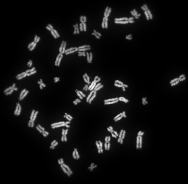
New research at the University of Kentucky has confirmed that the presence of XX sex chromosomes increases the amount of fat circulating in the blood, which leads to narrowing of the arteries and ultimately a higher risk of heart attacks and coronary artery disease.

The research was published in Nature Communications. The leading cause of death in women is coronary artery disease (CAD), but women develop CAD almost 10 years later than men. For many years, scientists attributed this decade-long delay in disease development to the protective effects of sex hormones. There is a lot of evidence that hormones like estrogen and progesterone protect the heart, but scientists had little data on the influence of the genetic component—the X chromosome—on the heart.

man and woman in medical laboratory

A team led by Lisa Cassis, a researcher in the UK College of Medicine’s pharmacology and nutritional sciences department, set out to understand the role of sex chromosomes in the cardiovascular system. Cassis’s team studies chromosome effects in mice, and for this most recent discovery they were able to zero in on XX chromosomes by removing hormones.

According to Yasir Al-Siraj, a postdoctoral scholar and the paper’s first author, if the levels of circulating lipids transported by the blood are too high, they will start to accumulate in and on the artery wall, leading to plaque buildup. These plaques harden and narrow the artery, reducing blood flow to the vital organs.

The team looked at lipids absorbed from the diet and made in the liver. Cassis, who also serves as the UK vice president for research, said, “We looked at how our X sex chromosomes were influencing the levels of lipids in the blood and in the arteries.” What they found is that an XX sex chromosome combination promotes efficient use of fat.

human chromosomes

Women need fat to bear and feed babies, Cassis explained: “We’re set up, potentially through our XX sex chromosomes, so that we can effectively absorb that lipid from the diet and put it into our fat cells and maybe even make it in the liver.” Everything is fine until women hit menopause and the protective effects of hormones disappear, leaving  women with what Cassis calls “that XX thrifty, fat-absorbing kind of genotype.”

The team is looking at genes that are changed in the liver and in the intestine to find novel targets for drug development. If they can find target genes that influence atherosclerosis, scientists can then explore the effects of existing drugs or develop new ones.

Al-Siraj said his next step is to study the role of the number of X chromosomes in atherosclerosis. “We don’t know if our findings are due to the presence of two X chromosomes or due to the absence of the Y chromosome,” he said. These findings may also drive choice of diet for post-menopausal women. Cassis said, “For example, if they’re very effective fat absorbers, obviously, once they get post-menopausal, they need to be careful about the fat content.”

Bone Loss May Indicate Poor Heart Health in Dialysis Patients


High parathyroid hormone levels and bone loss may predict progression of coronary artery calcification (CAC) in patients receiving dialysis, according to a study published online April 2 in the Journal of the American Society of Nephrology.

“We discovered that high parathyroid hormone and the consequential bone loss are major risk factors for progression of vascular calcifications,” Hartmut H. Malluche, MD, from the Division of Nephrology, Bone and Mineral Metabolism, University of Kentucky, Lexington, commented in a news release.

“These two factors were heretofore not appreciated and were independent from traditional known risk factors,” he added.
Elevated parathyroid hormone levels cause the release of calcium from bone, leading to bone loss and thinning. Most patients receiving dialysis for chronic kidney disease have CAC. CAC increases the risk for cardiovascular events, which in turn cause the majority of deaths in patients with CKD, the authors note.

Therefore, Dr Malluche and colleagues recommend monitoring bone loss with measurements of parathyroid hormone or bone mineral density (BMD) as a way to predict progression of CAC in patients receiving dialysis.

Between August 2009 and April 2013, the researchers enrolled 213 participants from 38 dialysis centers in Kentucky. Participants underwent measurement of routine laboratory tests, serum markers of bone metabolism, and CAC at baseline and 1 year. The researchers also evaluated BMD at both points using dual-energy X-ray absorptiometry scans and quantitative computed tomography. They assessed CAC using multislide computed tomography of the heart and CAC square root of coronary artery calcification volume, an analytic technique that accounts for variability in scanning.

About 80% of participants had CAC at baseline, and almost 50% of these had measurements suggesting high risk for cardiovascular events. One third of participants had osteoporosis.

Independent positive predictors of baseline CAC included coronary artery disease, diabetes, length of time receiving dialysis, age, and concentration of fibroblast growth factor 23, which regulates serum phosphate levels and helps maintain bone strength. In contrast, BMD of the spine inversely predicted baseline CAC.
CAC progression at 1 year occurred among three quarters of the 122 patients who completed the study. Independent risk factors for CAC progression included age, osteoporosis (β = 4.6; 95% confidence interval, 1.8 – 7.5; P = .002), and baseline total or whole parathyroid hormone more than nine times the normal value, after adjusting for age (β = 6.9; 95% confidence interval, 2.4 – 11.4; P = .003).

The researchers note several limitations for the study, including exclusion of about 20% of screened patients because of severe comorbidities or impaired mental status. In addition, the prospective, short-term nature of the study precluded determination of disease mechanisms and long-term relationships.

Dr Malluche noted in the press release that important links may exist between the level of calcification in bones and calcifications in blood vessels.

“Studies need to be done to find out whether prevention of bone loss will reduce progression of vascular calcifications,” he emphasized.