Brain Scans Reveal How Drinking Turns People Into Raging Assholes


We all have that friend who gets a little out of hand when they start drinking alcohol. Maybe he gets loud, or maybe she starts fights with strangers for looking at her funny. Alcohol seems to induce aggression, changing the brain in a way that makes a drunk person more likely to see minor social cues as threats, but how it does so has always been a bit of biological mystery.

Scientists found that alcohol-induced aggression was correlated to decreased activity in the prefrontal cortex.

But in a paper published in the journal Cognitive, Affective, & Behavioral Neuroscience, a team of researchers led by Thomas Denson, Ph.D., of the University of New South Wales School of Psychology use brain scans to show that alcohol changes activity in certain key parts of the brain related to aggression and emotion.

Using functional magnetic resonance imaging (fMRI), a technique that tracks changes in blood flow in the brain, the team looked at the brains of 50 young men after they consumed either two alcoholic drinks or two non-alcoholic placebo drinks. These volunteers engaged in a task that gauged their level of aggression in the face of provocation, which revealed the parts of the brain that become more active in such situations.

These scans show how alcohol-induced aggression was related to decreased activity in the prefrontal cortex, caudate, and ventral striatum, but increased activity in the hippocampus.

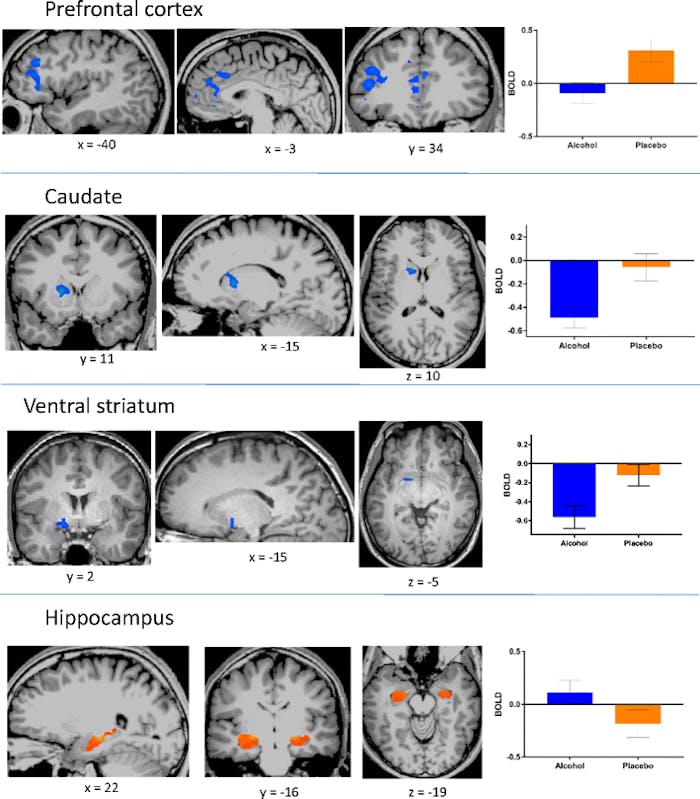
The researchers found that alcohol-induced aggression was correlated with decreased activity in prefrontal cortex, caudate, and ventral striatum, but increased activity in the hippocampus. These parts of the brain all control key factors in aggression: The prefrontal cortex is associated with thoughtful action and social behavior, the caudate is linked to the brain’s reward system and inhibitory control, and the ventral striatum is a part of the reward system that makes you feel good when you do something good. The hippocampus, meanwhile, is associated with emotion and memory.

These results support previous hypotheses that prefrontal cortex dysfunction is associated with alcohol-induced aggression. Taking all these brain areas together, the researchers say their findings suggest that intoxicated people have trouble processing information through their working memory. In short, they suspect that alcohol focuses a person’s attention on the cues that could instigate aggression while taking attention away from their knowledge of social norms that say violence is not acceptable.

Along similar lines, they also suspect that alcohol could make relatively minor cues seem aggressive or violent, which can cause a drunk person to overreact to a minor incident, like someone looking at them funny or accidentally bumping into them at the bar. Denson’s previous research on the angry brain found a lot of overlap in the way the prefrontal cortex behaves when someone is drunk and angry versus when they’re simply ruminating on their anger while sober.

This research proposes some possible brain biomarkers for alcohol-induced aggression, which is a significant public health issue. According to the Centers for Disease Control and Prevention, in the United States, alcohol-related violence — including homicide, child abuse, suicide, and firearm injuries — was responsible for more than 16,000 deaths between 2006 and 2010, the most recent years the agency reported figures.

While the new study doesn’t propose a solution per se, it does build on our body of knowledge around an age-old question: Why do some people become assholes when they get drunk?

Abstract: Alcohol intoxication is implicated in approximately half of all violent crimes. Over the past several decades, numerous theories have been proposed to account for the influence of alcohol on aggression. Nearly all of these theories imply that altered functioning in the prefrontal cortex is a proximal cause. In the present functional magnetic resonance imaging (fMRI) experiment, 50 healthy young men consumed either a low dose of alcohol or a placebo and completed an aggression paradigm against provocative and nonprovocative opponents. Provocation did not affect neural responses. However, relative to sober participants, during acts of aggression, intoxicated participants showed decreased activity in the prefrontal cortex, caudate, and ventral striatum, but heightened activation in the hippocampus. Among intoxicated participants, but not among sober participants, aggressive behavior was positively correlated with activation in the medial and dorsolateral prefrontal cortex. These results support theories that posit a role for prefrontal cortical dysfunction as an important factor in intoxicated aggression.

Infants use prefrontal cortex in learning.


Researchers have long thought that the region of the brain involved in some of the highest forms of cognition and reasoning — the prefrontal cortex (PFC) — was too underdeveloped in young children, especially infants, to participate in complex cognitive tasks. A new study suggests otherwise. Given the task of learning simple hierarchical rules, babies appeared to employ much the same circuits as adults doing a similar task.

A baby wears a near infrared spectroscopy-sensing headband in the lab of Associate Professor Dima Amso.

Researchers have long thought that the region of the brain involved in some of the highest forms of cognition and reasoning — the prefrontal cortex (PFC) — was too underdeveloped in young children, especially infants, to participate in complex cognitive tasks. A new study in the Journal of Neuroscience suggests otherwise. Given the task of learning simple hierarchical rules, babies appeared to employ much the same circuits as adults doing a similar task.

The findings suggest that even at the age of 8 months, a baby’s PFC appears properly adapted to the kinds of tasks important to an infant of that age, said study senior author Dima Amso, associate professor of cognitive, linguistic and psychological sciences (CLPS) at Brown University.

“The wow factor isn’t ‘Look the PFC works,'” Amso said. “It’s that what seems to be happening is that its function is a really good fit for what these babies need to be mastering at that moment in their development.”

Of course babies aren’t yet equipped for writing essays or planning the day’s errands, Amso said, but their brains are properly adapted for learning essential elements in their world and how best to organize them. The PFC is not offline. Instead it’s appropriately mature for the goals of babyhood.

An example from bilingualism

To make this discovery, Amso, lead author Denise Werchan, fellow CLPS professor Michael Frank and then postdoctoral researcher Anne Collins, who is now assistant professor at the University of California at Berkeley, devised a task initially developed by Collins and Frank to test PFC function in adults. The infant version was made to parallel the circumstance of growing up in a bilingual family. Maybe Mom and her side of the family speak English, while Dad and his family speak Spanish. The babies must learn that different groups of people use different words for the same things.

To scientists, this association of some people with one language and other people with another is an example of a “hierarchical rule set.” The person speaking is the higher-level context that determines what language will be used. Babies must learn that Mom and her brother will say “cat” when Dad and his sister will say “gato” to refer to the same family pet.

The team wanted to determine how baby brains handle this task. They recruited 37 babies to learn a simple, abstracted version of the bilingual scenario while their brain activity and behavior were gently monitored.

On screens before them, babies were shown a face and then an image of a toy. At the same time they’d hear a particular nonsense word in a voice associated with the face, as if the depicted person — call her “person 1” — was calling the depicted toy by that word. Then they’d see a different face with a different associated voice call the same toy by a new word (i.e. as if “person 2” was speaking a different language). Over several rounds, switching back and forth, the babies were exposed to these associations of person 1 with one vocabulary and person 2 with a distinct vocabulary.

After that phase the babies were then introduced to person 3 on the screen who used the same words as person 1, but also introduced a few new ones (in the bilingual family metaphor, think of person 3 as Dad’s sister, if person 1 is Dad). If the babies were learning the rules, they’d associate person 3’s new words with person 1, because they were otherwise speaking the same rule set or “language.”

In the blink of an eye

The researchers tested whether the learning was occurring by next presenting the babies with persons 1 and 2 saying some of the new vocabulary of person 3. Babies who’d been learning should react differently to each instance. They should look longer at the unexpected case of person 2 using a word from the vocabulary of person 3. In fact, that’s what the babies did. On average they gazed a couple of seconds longer at that surprising situation of person 2 using an inconsistent language than they did at the expected case of person 1 speaking like person 3.

Meanwhile, the researchers were tracking brain activity by means of a Near Infrared Spectroscopy (NIRS) machine provided, along with technical support, by TechEn, Inc. NIRS safely records brain activity over the scalp and is therefore becoming important to infant research, Amso said. Babies wear a special headband that holds near-infrared sensors over the scalp area of interest. The sensors detect how much infrared light is absorbed by hemoglobin in the blood and therefore report where brain activity is greatest (because that’s where the blood goes).

The researchers also tracked the babies’ eye blinks because recent research has found that eye blinks reflect the degree of involvement of the neurotransmitter dopamine. When adults learn hierarchical rules, Frank and Collins have combined experimental data with computer models of brain function to suggest, that the key circuit involved is a connection between the PFC and another region called the striatum. That connection is mediated and reinforced by dopamine.

The results of the infrared recording and the eye blink tracking both supported the hypothesis that as the babies were learning they were actively employing the PFC, similarly to adults. Both PFC activity — specifically in the right dorsolateral PFC — and eye blinks were significantly elevated when babies were asked to switch from one “language” to another, which is the most cognitively demanding moment for the PFC during the task.

“Once you learn these hierarchical structures, each time you need to access or use one of them you need to update the structure into working memory,” Werchan said. “When the task switches you need to update information into PFC.”

Moreover, the degree of the babies’ elevated PFC and eye blink activity predicted how distinctly they responded to the unexpected situation of a person speaking inconsistently with their language — a measure of how well the babies learned the rule structures.

Developing a new view of development

Amso said the findings suggest that early neurodevelopment should be viewed differently than before. Rather than regarding young brains as immature and less functional, a better perspective may be to regard them as constantly adapting to meet the key challenges they face. When healthy, they are as sophisticated as they need to be.

“Atypical development, then, might reflect an inability to adapt to an environmental challenge, or an earlier adaptation because of a negative environment. Amso said. “We and others are probing with these ideas as relevant to PFC development.”

Autism May Begin Before Birth, Autopsy Study Reveals


Disorganized neurons in the prefrontal cortex suggest that brain abnormalities in children with autism may begin before birth, a detailed postmortem study shows.

The analysis revealed the presence of patches of disorganized neurons in areas that mediate functions that are disturbed in autism, including social, emotional, communication, and language function, according to the study authors, led by Rich Stoner, PhD, Autism Center of Excellence and the Department of Neuroscience, University of California at San Diego.

The patches suggest that brain cell activity has been disrupted during pregnancy, which possibly points more to a genetic than an environmental trigger for autism.

“Such abnormalities may represent a common set of developmental neuropathological features that underlie autism and probably result from dysregulation of layer formation and layer-specific neuronal differentiation at prenatal developmental stages,” the authors write.

The study was published March 27 in the New England Journal of Medicine.

New Insight

Researchers obtained 42 fresh-frozen postmortem cortical tissue blocks from the superior or middle frontal gyrus of the dorsolateral prefrontal cortex, posterior superior temporal cortex, or occipital cortex of boys and girls aged 2 to 15 years with and without autism. The tissue blocks were selected for their high RNA integrity, thereby ensuring the quality of the samples.

The investigators used a large panel of highly selective markers for specific cell subtypes and a subset of 25 autism candidate genes. These included biomarkers for brain cell types in different layers of the cortex and genes implicated in autism.

They detected what they described as “pathological patches of abnormal laminar cytoarchitecture and disorganization” in samples of the prefrontal and temporal cortex samples, but not the occipital cortex. The patches were found in 91% (10 of 11) of the children with autism (cases) and 9% (1 of 11) of control individuals.

The patches had fewer cells expressing layer- or cell-type-specific markers than normally present in fully differentiated cortical neurons and decreased expression of certain autism candidate genes.

The focal patches of abnormal gene expression measured 5 to 7 mm in length and were located in areas adjacent to apparently unaffected areas of the cortex. They spanned multiple contiguous neocortical layers; the clearest evidence of abnormal expression was found in layers 4 and 5.

“This defect indicates that the crucial early developmental step of creating 6 distinct layers with specific types of brain cells ― something that begins in prenatal life ― had been disrupted,” study investigator Eric Courchesne, PhD, Autism Center of Excellence and the Department of Neuroscience, University of California at San Diego, said in a release.

“The finding that these defects occur in patches rather than across the entirety of cortex gives hope as well as insight about the nature of autism,” he added.

Patches Widespread

The fact that the researchers sampled only small portions of cortex but observed focal patches in nearly every case sample suggests that “pathological patches are widespread across prefrontal and temporal cortex in children with autism,” according to investigators.

The patches were present both in boys and girls, in high- and low-functioning children, and regardless of the cause of death or postmortem interval.

Presentation of the patches varied across cases, which, said the authors, was unexpected given the phenotypic heterogeneity of autism.

“However, the features that we describe here may explain some of the heterogeneity of autism: disorganized patches in different locations could disrupt disparate functional systems in the prefrontal and temporal cortexes and potentially influence symptom expression, response to treatment, and clinical outcome,” the researchers write.

Although the data suggest a novel pathologic mechanism in autism, the exact biological process is unknown.

“The identified laminar disorganization could result from migration defects resulting in the failure of cells to reach their targeted destination and the accumulation of such cells in nearby regions,” the authors write.

Or, they added, the patches could reflect de novo changes early in neurodevelopmental processes, which yield regions of affected progenitor cells adjacent to regions of unaffected progenitor cells.

In any case, the data are consistent with an early prenatal origin of autism, or at least prenatal processes, that may confer a predisposition to autism, according to investigators.

Although the sample size was small compared with postmortem studies of adult diseases, it is as large ― or larger ― than most previous such studies of autism, the authors note.

Key Advance

Commenting on the study for Medscape Medical News, Thomas Frazier, PhD, director, Cleveland Clinic Children’s Center for Autism, in Ohio, said it indicates that in autism, abnormalities in brain cell development occur prior to birth and that this leads to dysfunctional regions of the brain.

“The key advance from this research is that it suggests that very early developmental processes lead to autism,” said Dr. Frazier. “The findings support genetic disruptions leading to brain disorganization or possibly very early interactions between genes and the prenatal environment.”

Dr. Frazier predicted that some experts may find the results controversial because the findings “suggest that brain abnormalities begin before birth, making it less likely that a purely environmental insult causes autism.”

The Lasting Impacts of Poverty on the Brain.


Poverty shapes people in some hard-wired ways that we’re only now beginning to understand. Back in August, we wrote about some provocative new research that found that poverty imposes a kind of tax on the brain. It sucks up so much mental bandwidth – capacity spent wrestling with financial trade-offs, scarce resources, the gap between bills and income – that the poor have fewer cognitive resources left over to succeed at parenting, education, or work. Experiencing poverty is like knocking 13 points off your IQ as you try to navigate everything else. That’s like living, perpetually, on a missed night of sleep.

That finding offered a glimpse of what poverty does to a person during a moment in time. Picture a mother trying to accomplish a single task (making dinner) while preoccupied with another (paying the rent on time). But scientists also suspect that poverty’s disadvantages – and these moments – accumulate across time. Live in poverty for years, or even generations, and its effects grow more insidious. Live in poverty as a child, and it affects you as an adult, too.

Some new research about the long-term arc of poverty, particularly on the brain, was recently published in theProceedings of the National Academy of Sciences, and these findings offer a useful complement to the earlier study. In this new paper, researchers from the University of Illinois at Chicago, Cornell, the University of Michigan, and the University of Denver followed children from the age of 9 through their early 20s.

The Lasting Impacts of Poverty on the Brain

Those who grew up poor later had impaired brain function as adults—a disadvantage researchers could literally see in the activity of the amygdala and prefrontal cortex on an fMRI scan. Children who were poor at age 9 had greater activity in the amygdala and less activity in the prefrontal cortex at age 24 during an experiment when they were asked to manage their emotions while looking at a series of negative photos. This is significant because the two regions of the brain play a critical role in how we detect threats and manage stress and emotions.

Poor children, in effect, had more problems regulating their emotions as adults (regardless of what their income status was at 24). These same patterns of “dysregulation” in the brain have been observed in people with depression, anxiety disorders, aggression and post-traumatic stress disorders.

Over the course of the longitudinal study – which included 49 rural, white children of varying incomes – these same poor children were also exposed to chronic sources of stress like violence and family turmoil, or crowded and low-quality housing. Those kinds of stressors, the researchers theorize, may help explain the link between income status in childhood and how well the brain functions later on. That theory, they write, is consistent with the idea that “early experiences of poverty become embedded within the organism, setting individuals on lifelong trajectories.”

To add some of these findings together: Poverty taxes the ability of parents to do all kinds of things, including care for their children. And the developmental challenges that children face in a home full of stressed adults may well influence the adults that they, themselves, become.

Magic Mushrooms Repair Brain Damage From Extreme Trauma.


A new study by The University of South Florida has found that low doses of the active ingredient in magic mushrooms repairs brain damage caused by extreme trauma, offering renewed hope to millions of sufferers of PTSD (Post-Traumatic Stress Disorder).

The study confirms previous research by Imperial College London, that psilocybin, a naturally occurring compound present in “shrooms”, stimulates new brain cell growth and erases frightening memories. Mice conditioned to fear electric shock when hearing a noise associated with the shock “simply lost their fear”, says Dr. Juan Sanchez-Ramos, who co-authored the study. A low dose of psilocybin led them to overcome “fear conditioning” and the freeze response associated with it faster than the group of mice on Ketanserin (a drug that counteracts the receptor that binds psilocybin in the brain) and a control group on saline.

shroomery
An estimated 5 percent of Americans – more than 13 million people – have PTSD at any given time, according to the PTSD Alliance. The condition more often associated with combat veterans, is twice as likely to develop in women because they tend to experience interpersonal violence (such as domestic violence, rape and abuse) more often than men.

PTSD is not just psychological

Common symptoms, such as hyper-vigilance, memory fragmentation, flashbacks, dissociation, nightmares and fight or flight responses to ‘triggers’, are generally thought to be psychological and therefore treatable by learning to change thought processes. But new research suggests that they may in fact be the result of long term physiological mutations to the brain.

In the South Florida University study, the mice treated with low doses of psilocybin grew healthy new brain cells and their overactive medial prefrontal cortex regions (common in PTSD sufferers) were restored to normal functionality.

Further independent studies (http://www.thedoctorwillseeyounow.com ) have shown that the hippocampus part of the brain is damaged by extreme stress and that this is specific to PTSD and not associated with anxiety or panic disorders.

Dr. Sanchez-Ramos acknowledged that there was no way of knowing whether the mice in the experiment experienced altered states of consciousness or hallucinations – commonly experienced with magic mushrooms, but he believed the doses were too low to cause psychoactive effects.

Magic Mushrooms Repair Brain Damage From Extreme Trauma.


A new study by The University of South Florida has found that low doses of the active ingredient in magic mushrooms repairs brain damage caused by extreme trauma, offering renewed hope to millions of sufferers of PTSD (Post-Traumatic Stress Disorder).

The study confirms previous research by Imperial College London, that psilocybin, a naturally occurring compound present in “shrooms”, stimulates new brain cell growth and erases frightening memories. Mice conditioned to fear electric shock when hearing a noise associated with the shock “simply lost their fear”, says Dr. Juan Sanchez-Ramos, who co-authored the study. A low dose of psilocybin led them to overcome “fear conditioning” and the freeze response associated with it faster than the group of mice on Ketanserin (a drug that counteracts the receptor that binds psilocybin in the brain) and a control group on saline.

An estimated 5 percent of Americans – more than 13 million people – have PTSD at any given time, according to the PTSD Alliance. The condition more often associated with combat veterans, is twice as likely to develop in women because they tend to experience interpersonal violence (such as domestic violence, rape and abuse) more often than men.

PTSD is not just psychological
Common symptoms, such as hyper-vigilance, memory fragmentation, flashbacks, dissociation, nightmares and fight or flight responses to ‘triggers’, are generally thought to be psychological and therefore treatable by learning to change thought processes. But new research suggests that they may in fact be the result of long term physiological mutations to the brain.

In the South Florida University study, the mice treated with low doses of psilocybin grew healthy new brain cells and their overactive medial prefrontal cortex regions (common in PTSD sufferers) were restored to normal functionality.

Further independent studies  have shown that the hippocampus part of the brain is damaged by extreme stress and that this is specific to PTSD and not associated with anxiety or panic disorders.

Dr. Sanchez-Ramos acknowledged that there was no way of knowing whether the mice in the experiment experienced altered states of consciousness or hallucinations – commonly experienced with magic mushrooms, but he believed the doses were too low to cause psychoactive effects.

Decriminalisation of psilocybin could help millions
Previous studies have shown that low doses of psilocybin produce no consciousness state altering effects. Administered in the correct amount, psilocybin could therefore be assumed to safely treat PTSD with minimal risk of adverse side effects. Magic mushrooms could help millions recover from the debilitating cycles of fight and flight and other conditioned biological responses caused by extreme trauma, if only they weren’t listed as a dangerous Schedule 1 drug with no medical benefits.

Meanwhile, doctors are authorised to dispense powerful, side-effect laden pharmaceutical drugs to army vets and others suffering from the symptoms of PTSD without any evidence that these treatments actually work, according to a major review by the committee of the Institute of Medicine on the topic.

The situation is so bad that an average of 18 American veterans commits suicide every day linked to the sharp rise in prescription drugs, depression, and other psychological conditions. Safe, natural alternatives to pharmaceuticals such as homeopathic and herbal remedies have been found to alleviate symptoms. Meditation has also been shown to reduce high activity levels in the amygdala (the brain’s emotional centre) experienced in PTSD sufferers as anxiety, stress and phobias.

The Neuroscience of Everybody’s Favourite Topic.


Why do people spend so much time talking about themselves?

Human beings are social animals. We spendlarge portions of our waking hours communicating with others, and the possibilities for conversation are seemingly endless—we can make plans and crack jokes; reminisce about the past and dream about the future; share ideas and spread information. This ability to communicate—with almost anyone, about almost anything—has played a central role in our species’ ability to not just survive, but flourish.

the-neuroscience-of-everybody-favorite-topic-themselves_1

How do you choose to use this immensely powerful tool—communication? Do your conversations serve as doorways to new ideas and experiences? Do they serve as tools for solving the problems of disease and famine?

Or do you mostly just like to talk about yourself?

If you’re like most people, your own thoughts and experiences may be your favorite topic of conversation.  On average, people spend 60 percent of conversationstalking about themselves—and this figure jumps to 80 percent when communicating via social media platforms such as Twitter or Facebook.

Why, in a world full of ideas to discover, develop, and discuss, do people spend the majority of their time talking about themselves? Recent research suggests a simple explanation: because it feels good.

In order to investigate the possibility that self-disclosure is intrinsically rewarding, researchers from the Harvard University Social Cognitive and Affective Neuroscience Lab utilized functional magnetic resonance imaging (fMRI). This research tool highlights relative levels of activity in various neural regions by tracking changes in blood flow; by pairing fMRI output with behavioral data, researchers can gain insight into the relationships between behavior and neural activity. In this case, they were interested in whether talking about the self would correspond with increased neural activity in areas of the brain associated with motivation and reward.

In an initial fMRI experiment, the researchers asked 195 participants to discuss both their own opinions and personality traits and the opinions and traits of others, then looked for differences in neural activation between self-focused and other-focused answers. Because the same participants discussed the same topics in relation to both themselves and others, researchers were able to use the resulting data to directly compare neural activation during self-disclosure to activation during other-focused communication.

Three neural regions stood out. Unsurprisingly, and in line with previous research, self-disclosure resulted in relatively higher levels of activation in areas of the medial prefrontal cortex (MPFC) generally associated with self-related thought. The two remaining regions identified by this experiment, however, had never before been associated with thinking about the self: the nucleus accumbens (NAcc) and the ventral tegmental area (VTA), both parts of the mesolimbic dopamine system.

These newly implicated areas of the brain are generally associated with reward, and have been linked to the pleasurable feelings and motivational states associated with stimuli such as sexcocaine, and good food. Activation of this system when discussing the self suggests that self-disclosure, like other more traditionally recognized stimuli, may be inherently pleasurable—and that people may be motivated to talk about themselves more than other topics (no matter how interesting or important these non-self topics may be).

This experiment left at least one question unanswered, however. Although participants were revealing information about themselves, it was unclear whether or not anyone was paying attention; they were essentially talking without knowing who (if anyone) was on the other end of the line. Thus, the reward- and motivation-related neural responses ostensibly produced by self-disclosure could be produced by the act of disclosure—of revealing information about the self to someone else—but they could also be a result of focusing on the self more generally—whether or not anyone was listening.

In order to distinguish between these two possibilities, the researchers conducted a follow-up experiment. In this experiment, participants were asked to bring a friend or relative of their choosing to the lab with them; these companions were asked to wait in an adjoining room while participants answered questions in a fMRI machine. As in the first study, participants responded to questions about either their own opinions and attitudes or the opinions and attitudes of someone else; unlike in the first study, these participants were explicitly told whether their responses would be “shared” or “private”; shared responses were relayed in real time to each participant’s companion and private responses were never seen by anyone, including the researchers.

In this study, answering questions about the self always resulted in greater activation of neural regions associated with motivation and reward (i.e., NAcc, VTA) than answering questions about others, and answering questions publicly always resulted in greater activation of these areas than answering questions privately.  Importantly, these effects were additive; both talking about the self and talking to someone else were associated with reward, and doing both produced greater activation in reward-related neural regions than doing either separately.

These results suggest that self-disclosure—revealing personal information to others—produces the highest level of activation in neural regions associated with motivation and reward, but that introspection—thinking or talking about the self, in the absence of an audience—also produces a noticeable surge of neural activity in these regions. Talking about the self is intrinsically rewarding, even if no one is listening.

Talking about the self is not at odds with the adaptive functions of communication. Disclosing private information to others can increase interpersonal liking and aid in the formation of new social bonds—outcomes that influence everything from physical survival to subjective happiness. Talking about one’s own thoughts and self-perceptions can lead to personal growth via external feedback. And sharing information gained through personal experiences can lead to performance advantages by enabling teamwork and shared responsibility for memory. Self-disclosure can have positive effects on everything from the most basic of needs—physical survival—to personal growth through enhanced self-knowledge; self-disclosure, like other forms of communication, seems to be adaptive.

You may like to talk about yourself simply because it feels good—because self-disclosure produces a burst of activity in neural regions associated with pleasure, motivation, and reward.  But, in this case, feeling good may be no more than a means to an end—it may be the immediate reward that jump-starts a cycle of self-sharing, ultimately leading to wide varieties of long-term benefits.

Source: Scientific American


Brain scans of rappers shed light on creativity.


Functional magnetic resonance imaging shows what happens in the brain during improvisation.

Rappers making up rhymes on the fly while in a brain scanner have provided an insight into the creative process.

Freestyle rapping — in which a performer improvises a song by stringing together unrehearsed lyrics — is a highly prized skill in hip hop. But instead of watching a performance in a club, Siyuan Liu and Allen Braun, neuroscientists at the US National Institute on Deafness and Other Communication Disorders in Bethesda, Maryland, and their colleagues had 12 rappers freestyle in a functional magnetic resonance imaging (fMRI) machine.

The artists also recited a set of memorized lyrics chosen by the researchers. By comparing the brain scans from rappers taken during freestyling to those taken during the rote recitation, they were able to see which areas of the brain are used during improvisation. The study is published today in Scientific Reports1.

The results parallel previous imaging studies in which Braun and Charles Limb, a doctor and musician at Johns Hopkins University in Baltimore, Maryland, looked at fMRI scans from jazz musicians2. Both sets of artists showed lower activity in part of their frontal lobes called the dorsolateral prefrontal cortex during improvisation, and increased activity in another area, called the medial prefrontal cortex. The areas that were found to be ‘deactivated’ are associated with regulating other brain functions.

“We think what we see is a relaxation of ‘executive functions’ to allow more natural de-focused attention and uncensored processes to occur that might be the hallmark of creativity,” says Braun.

He adds that this suggestion is “a little bit controversial in the literature”, because some studies have found activation of the dorsolateral prefrontal cortex in creative behaviour. He suggests that the discrepancy might have to do with the tasks chosen to represent creativity. In studies that found activation, the activities — such as those that require recall — may actually be less creative.

“We try to stick with more natural creative processing, and when we do that we see this decrease in the dorsal lateral regions,” says Braun.

Pump down the volume

Rex Jung, a clinical neuropsychologist at the University of New Mexico in Albuquerque, has also studied the link between brain structures and creativity, finding an inverse relationship between the volume of some frontal lobe structures and creativity3. “Some of our results imply this downregulation of the frontal lobes in service of creative cognition. [The latest paper] really appears to pull it all together,” he says. “I’m excited about the findings.”

Jung says that this downregulation is likely to apply in other, non-musical areas of creativity — including science.

The findings also suggest an explanation for why new music might seem to the artist to be created of its own accord. With less involvement by the lateral prefrontal regions of the brain, the performance could seem to its creator to have “occurred outside of conscious awareness”, the authors write.

Michael Eagle, a study co-author who raps under the name Open Mike Eagle, agrees: “That’s kind of the nature of that type of improvisation. Even as people who do it, we’re not 100% sure of where we’re getting improvisation from.”

Liu says that the researchers are now working on problems they were unable to explore with freestylers — such as what happens after the initial burst of creative inspiration.

“We think that the creative process may be divided into two phases,” he says. “The first is the spontaneous improvisatory phase. In this phase you can generate novel ideas. We think there is a second phase, some kind of creative processing [in] revision.”

The researchers would also like to look at how creativity differs between experts and amateurs of a similar artistic ilk to freestylers: poets and storytellers.

Watch the video on youtube.URL: http://www.youtube.com/watch?v=LmreCiyV-Kc&feature=player_embedded

Source: Nature