New Alzheimer’s treatment accelerates removal of plaque from the brain in clinical trials


West Virginia University researchers used ultrasound technology to increase drug delivery to the brain

FOX News medical contributor Dr. Marc Siegel explains a new blood test that could lead to early detection of Alzheimer’s disease.

A new Alzheimer’s therapy has shown potential in the first human trials.

Researchers at the West Virginia University Rockefeller Neuroscience Institute (RNI) found that by pairing focused ultrasound in combination with antibody therapies, they were able to accelerate the removal of amyloid-beta plaques from the brains of patients with Alzheimer’s disease.

The study findings were published in The New England Journal of Medicine on Jan. 11.

An abnormal buildup of amyloid-beta proteins is one of the hallmarks of Alzheimer’s, as these proteins clump together to form plaques that interfere with neurons in the brain. 

Anti-amyloid-beta monoclonal antibody treatments, such as aducanumab and lecanemab, have proven to be effective in clearing these plaques and slowing disease progression.

Ultrasound MRI

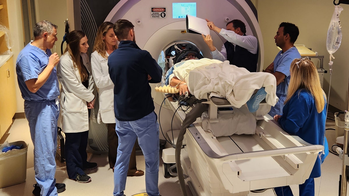
An Alzheimer’s patient undergoes focused ultrasound treatment with the WVU RNI team. (Rockefeller Neuroscience Institute (RNI) at West Virginia University (WVU))

But until now the drugs have been limited by the blood-brain barrier (BBB), which is designed to keep harmful substances from reaching the brain, according to a press release from RNI.

“A study like this is important because it demonstrates that there may be safe ways to increase drug delivery to the brain without any serious adverse effects.”

More than 98% of drugs are blocked by the barrier, which means patients require higher doses and more frequent therapies, the researchers noted.

AY

In this study, scientists used a focused ultrasound (FUS) system to temporarily open the blood-brain barrier, which allowed the antibodies to have greater access to areas of the brain with high amyloid-beta plaques.

After six months of antibody treatment, the study participants had an average of 32% more reduction in amyloid-beta plaques in areas where the BBB was opened compared to areas where the drug was used without the ultrasound, the release stated.

Focused ultrasound

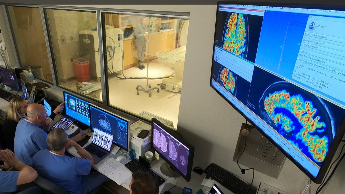
The WVU RNI team, shown in the MRI suite’s control area, plans ultrasound blood-brain barrier treatment. (Rockefeller Neuroscience Institute (RNI) at West Virginia University (WVU))

“This was a first in human safety and feasibility study in three participants demonstrating that the BBB opening can accelerate clearance of beta amyloid plaques,” study lead Dr. Ali Rezai, director of the Rockefeller Neuroscience Institute (RNI) at WVU, told Fox News Digital. 

“Non-invasive focused ultrasound is an outpatient procedure that allows for targeted delivery of therapeutics to the brain that can potentially accelerate the benefit of the antibody treatment in Alzheimer’s disease,” he added.

The three patients, between the ages of 59 and 77, all had mild Alzheimer’s disease.

During the study, they received six monthly infusions of the aducanumab antibody. 

After each treatment, the focused ultrasound was used to open the BBB at the sites of the highest plaque buildup.

While there are some potential risks associated with ultrasound use, such as brain swelling and hemorrhage, Rezai said those effects were not observed in this study.

Focused ultrasound MRI

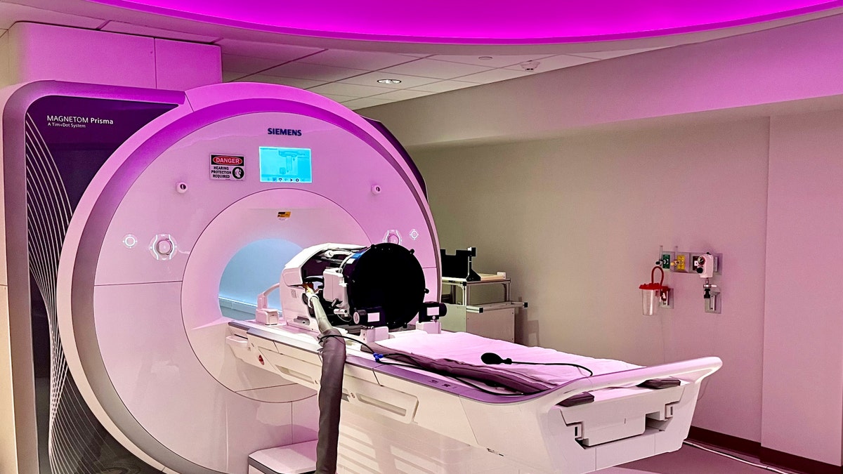
The focused ultrasound helmet unit with 1,024 ultrasound transducers attaches to the MRI table for MRI-guided treatment. (Rockefeller Neuroscience Institute (RNI) at West Virginia University (WVU))

“We verified with MRI scans that the BBB opening was temporary and it closed 24 to 48 hours after the FUS procedure,” he told Fox News Digital.

The reductions in amyloid plaques were verified in PET scans.

This was the first step toward larger studies; in those, researchers will be able to evaluate more patients and larger areas of the brain, Rezai noted.

In the next phase of the clinical trial, the ultrasound therapy will be paired with lecanemab, another anti-beta amyloid antibody.

Ultrasound targeting

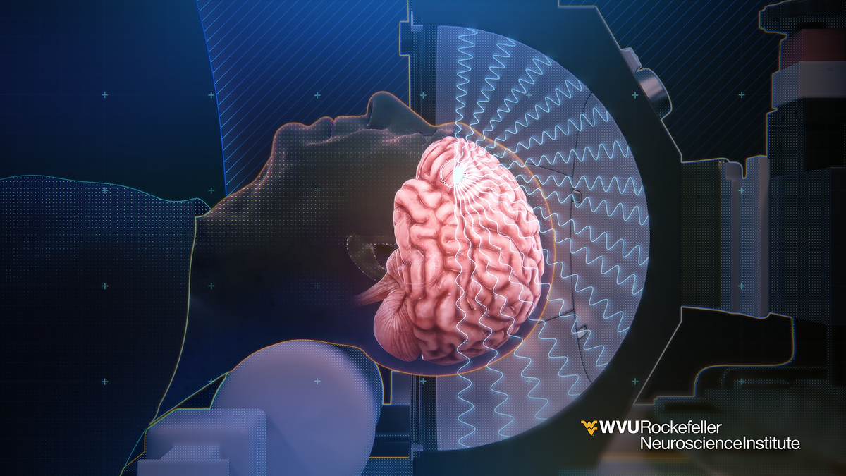
This 3-D illustration shows how ultrasound waves from inside the helmet converge on a focal point on the brain used for blood-brain barrier opening.  (Rockefeller Neuroscience Institute (RNI) at West Virginia University (WVU))

Dr. James Galvin, director of the Comprehensive Center for Brain Health at UHealth, the University of Miami Health System, was not involved in the WVU research but shared his reaction.

“A study like this is important because it demonstrates that there may be safe ways to increase drug delivery to the brain without any serious adverse effects,” he told Fox News Digital. 

“Focused ultrasound has been used in other treatment paradigms for brain diseases such as Parkinson’s disease and brain tumors,” Galvin went on. 

Galvin also cautioned that this research was conducted with only three patients and was not a placebo-controlled study. 

Alzheimer's awareness

As of 2023, an estimated 6.7 million Americans age 65 and older were living with Alzheimer’s. (iStock)

“It was also designed as a safety study and not appropriately powered to detect significant clinical changes,” he added. “It is still too early to make any specific recommendations, but I am excited to see if there are planned follow-up studies with a larger number of patients.”

Rebecca M. Edelmayer, PhD, senior director of scientific engagement at the Alzheimer’s Association, was also not involved in the study but called the results “very intriguing,” albeit preliminary.

“The blood brain barrier, in its healthy form, protects the brain from harmful agents that could reach it via the bloodstream,” she told Fox News Digital via email.

“Getting therapeutics across this barrier — from the bloodstream into the brain tissue — is a challenge for any drug used to treat brain diseases, including drugs to treat Alzheimer’s disease.”

Edelmayer added that while this was a “very small study of relatively short length,” it was a worthwhile way to test a “cutting-edge idea” for improving the effectiveness of Alzheimer’s medications.

Focused ultrasound technology

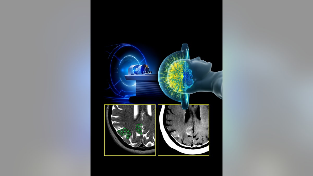
This illustration of the MRI-guided focused ultrasound system includes a representation of treatment delivery, the target region, and the corresponding opening of the blood-brain barrier demonstrated by contrast enhancement. (Rockefeller Neuroscience Institute (RNI) at West Virginia University (WVU))

Focused ultrasound-induced blood-brain barrier opening has also been shown to improve drug delivery to treat brain tumors, Edelmayer pointed out. 

“This is a great example of how learnings from research in other diseases might be repurposed for Alzheimer’s disease and other dementia.”

Are Mental Disorders the Result of Neuroinflammation?


Are Mental Disorders the Result of Neuroinflammation?

New research is increasingly finding that not only are many cognitive disorders such as Alzheimer’s and other forms of dementia related to inflammation among brain tissues, but classic mental diseases such as schizophrenia and depression may also be connected to brain inflammation.

Recent research from Japan’s Kyushu University Medical School and Saga University have been revealing that many mental disorders are produced by inflammation involving the microglia cells of the brain.

What are microglia?

The microglia are immune cells categorized as macophages, but they are focused upon the health of the central nervous system – particularly the brain and spine. Microglia roam the neurons of these areas in search for toxins, intruders and possible infections.

Particularly at issue in cognitive issues such as dementia and Alzheimer’s disease is the build up of amyloid plaque among brain cells. The microglia are the immune cells that prevent and clean up plaque build up among brain cells.

When the microglia populations are damaged or otherwise altered, the brain and CNS becomes increasingly susceptible to mental disorders such as dementia, schizophrenia, depression and mental fatigue.

Neuroinflammation and microglia

Neuroinflammation is the result of damage among brain cells. Healthy microglia populations are focused upon preventing inflammation among neurons, in turn preventing damage to brain cells.

However, when brain cells are damaged, microglia work harder to repair the damage by producing a variety of inflammatory factors. The damage to neurons, evidenced by these inflammatory factors, hamper the brain’s function.

Like other types of macrophages, the microglia are formed within the bone marrow. Once they migrate to the brain, they differentiate into particular responsibilities and different regions. Some microglia are focused on infections, others are focused upon toxins or damaged cells. Others stimulate the repair of brain tissues.

Plaque and neuroinflammation

The build-up of plaque among brain cells – connected to dementia – stimulates the microglia as they work to try to remove the damage. Damage from oxidative stress and glycation byproducts have been linked to this build up of plaque among brain cells.

Research by Dr. Akira Monji and associates has connected mental disorders such as schizophrenia, depressive states and cognitive issues to these increases in microglia inflammatory factors such as nitric oxide and cytokines. When the microglia have rapidly expanded in the face of damage to brain cells, they produce these inflammatory factors. Dr. Monji’s research has shown that the brain tissues of schizophrenia, depression and dementia patients have increased levels of these microglia inflammatory factors.

Furthermore, their research has found that one of the central mechanisms of psychiatric drugs is that they reduce levels of these inflammatory factors – temporarily.

Like most pharmaceuticals, this temporary reduction of inflammatory factors does little to prevent or reduce the cause of the inflammation. Furthermore, by blocking inflammatory factors, the drugs work to interfere with the damage repair that is taking place, driven by the microglia immune cells. This is often the scenario for drugs that are focused upon the symptoms rather than the causes of a condition.

What causes neuroinflammation?

The cause of neuroinflammation, as shown in numerous dementia studies, relates to oxidative damage. Oxidation is produced through an imbalance between toxins that form oxidative radicals and those antioxidants that neutralize those radicals. When the system is not balanced, oxidation takes place, not only among tissues among the cardiovascular system, but also among brain tissues.

This of course is why recent research, such as a new French and Finnish INSERM study, has linked cognitive decline to increased obesity, diabetes and heart disease.

The study, led by Dr. Mika Kivimaki and associates from the French government’s investigative body, INSERM, with support from the U.S. National Institutes of Medicine, studied 6,401 adults between 39 and 63 years old. They found that people who were obese and suffered from metabolic disorder (cardiovascular disease and/or diabetes) had more than a 22% greater cognitive decline than those who were of normal weight with no metabolic disorder.

This study has been confirmed by others that have related cognitive decline to cardiovascular disease, sedentary lifestyles, obesity and increased levels of toxins.

These issues have all been connected to higher levels of inflammation in the body.

The bridge to these relationships has been provided by the Japanese research, finding that neuroinflammation is one of the key factors affecting mental disorders of many types.

Antioxidants and mental disorders

Numerous studies have shown that antioxidants neutralize oxidative radicals that produce inflammation. The very term “antioxidant” is founded upon research showing that particular phyto-chemicals directly neutralize the oxidative effects of radicals formed by toxins.

This is supported by the research.

For example, in another large French study published this March in the Journal of Nutrition, researchers found that a healthy diet with greater antioxidant intake was associated with reduced risk of cognitive decline after the researchers removed factors relating to exercise, alcohol intake, calories, gender, age, education and obesity.

This is of course despite the fact that other research has found that smoking and lack of exercise both increase the rate of cognitive decline, as illustrated in a study by some of the same INSERM researchers that linked obesity and cognitive decline.

What this all means is that mental disorders are no longer conditions that necessarily fall within the abstract domain of behavioral psychology and psychiatry, where treatments such as lobotomy, electric shock and psychotropic drugs have produced a myriad of adverse mental and physical effects.

Rather, the research is conclusive: Many mental disorders are in fact produced by poor diets and poor lifestyles, and thus are to a great degree preventable.

This research also brings mental disorders within the realm of natural health and nutrition. What is now known is that a person with a healthy diet containing plenty of antioxidants, together with an active lifestyle, has a significantly reduced risk of having a mental disorder.

More importantly, a person with a healthier, antioxidant-rich diet will also stand a better chance of remaining alert into their elderly years.