Candida auris was first identified in 2009 in Japan and is resistant to many anti-fungal drugs.
Authorities in Washington state confirmed an outbreak of a deadly fungal infection that has been on the rise across the United States in recent years.
Officials in King County, Washington, said in a statement on Tuesday that an outbreak involving three patients infected by Candida auris, or C. auris, was reported at Kindred Hospital in Seattle beginning in mid-January. Another case was detected on Jan. 26 at a nursing home in nearby Snohomish County, officials said.
“These patients had previously tested negative for C. auris when they were first admitted,” it said. “This is the first known outbreak of C. auris in Washington state.”
The health department said it will “work together” with Kindred Hospital to limit the transmission of C. auris, including keeping those who test positive for the fungal infection to stay “away from other patients.”
Kindred Hospital will also notify other facilities that “received patients who were previously at Kindred and will notify facilities that may receive any patient who screens positive for C. auris,” the county’s statement added.
“As is the case with many multi-drug resistant organisms, it can be difficult to identify the initial source of the infection, and while the investigation is ongoing, the original source of C. auris in this situation may never be identified,” the statement continued. “However, with the early identification of these cases, there is a greater opportunity to reduce the risk of further spread.
The cluster of cases comes as the fungal infection continues to spread across the United States, according to data provided by the U.S. Centers for Disease Control and Prevention (CDC). Since 2016, cases of C. auris have risen each year.
First Detection in the US
Between 2019 and 2021, the CDC said, 17 states identified their first cases, with clinical cases rising nationwide from 476 in 2019 to 1,471 in 2021.
“Screening cases tripled from 2020 to 2021, for a total of 4,041,” the CDC said.
The reason for the increase, the agency added, is due to a multitude of factors such as poor practices around general infection prevention and control in health care facilities, and also possibly due to an increase in reporting and detecting cases.
S. auris was first identified in 2009 in Japan, officials said. The CDC classifies the infections as an urgent public health issue because the fungus is “often resistant to multiple antifungal drugs, spreads easily in healthcare facilities, and can cause severe infections with high death rates.”
“In general, C. auris is not a threat to healthy people,” the CDC said. “People who are very sick, have invasive medical devices, or have long or frequent stays in healthcare facilities are at increased risk for acquiring C. auris.”
It added that the mortality rate of a severe infection is between 30 and 60 percent, but because it generally affects people with compromised immune systems, the CDC considers it rare.
People can get the infection through direct contact with an infected individual or by touching objects or surfaces that are contaminated. Researchers have previously said that the fungus can live for at least two weeks on surfaces and objects.
John Lynch, with the University of Washington’s Harborview Medical Center in Seattle, told KIRO-7 TV that the fungus is described as “an emerging pathogen so a newer infectious disease,” adding that it is still “very difficult to know if someone has Candida auris infection” and that “people can have it on their skin or parts of their body without causing any disease.”
The only way to know if someone is infected, he added, is through laboratory testing.
“The reason why we’re concerned about Candida auris is because it spreads so easily, especially in health care environments, especially among patients who have underlying conditions and have devices in their bodies,” Meagan Kay, a health official with King County and Seattle, told the outlet. “This is really not a big concern for the public, we’re reporting about it for transparency and for people to understand the work we’re doing to keep people safe.”
Symptoms
Infection with C. auris can lead to a range of symptoms, officials say. The fungus can infect the bloodstream, ears, wounds, urinary tracts, and other parts of the body, according to the Cleveland Clinic.
General symptoms include fever, low blood pressure, lethargy and fatigue, low body temperature, chills, high heart rate, or pain or pressure in the ear if the fungus infects the ear, it says.
“Since many people who get C. auris infections are already seriously ill, symptoms of C. auris may not be noticeable. Many people carrying C. auris don’t have symptoms but can still pass it to someone else,” the clinic says.
Individuals who are at risk of infection include those with diabetes or blood cancer, had surgery recently, been on antibiotics for a long time or use them often, or have been in a health care facility or hospital for a lengthy period of time, it says. Those using a central venous line, breathing tube, catheter, or a similar device are also at risk.
Since 2021, there has been an increase in cholera cases and their geographical distribution globally. In 2021, 23 countries reported cholera outbreaks, mainly in the WHO Regions of Africa and the Eastern Mediterranean. This trend has continued into 2022 with over 29 countries (Figure 1) reporting cholera cases or outbreaks. As of 30 November 2022, 16 of these have been reporting protracted outbreaks. Many of those countries reported higher case numbers and case fatality ratio (CFR) than in previous years. The average cholera CFR reported globally in 2021 was 1.9% (2.9% in Africa), well above acceptable (<1%) and the highest recorded in over a decade.
This year the number of cholera cases and cholera-associated deaths have surged globally following years of decline. Of particular concern are the outbreaks in 13 countries, which did not report cholera cases in 2021. Of these, some had not reported any cholera outbreaks for many years (between three and 30), and several are not considered cholera-endemic countries.i,ii The current situation represents a resurgence of the ongoing seventh pandemic of cholera which began in 1961.
The simultaneous progression of several cholera outbreaks, compounded in countries facing complex humanitarian crises with fragile health systems and aggravated by climate change, poses challenges to outbreak response and risks further spreading to other countries. The overall capacity to respond to the multiple and simultaneous outbreaks is strained due to the global lack of resources, including the oral cholera vaccine, as well as overstretched public health and medical personnel, who are often dealing with multiple disease outbreaks at the same time.
Epidemiology
Cholera is an acute diarrheal infection characterized, in its severe form, by extreme watery diarrhea and potentially fatal dehydration. It is caused by the ingestion of food or water contaminated with the bacterium Vibrio cholerae. It has a short incubation period, ranging between two hours and five days. Most people will develop no or only mild symptoms; less than 20% of ill persons develop acute watery diarrhoea with moderate or severe dehydration and are at risk for rapid loss of body fluids, dehydration, and death. Despite being easily treatable with rehydration solution, cholera remains a global threat due to its high morbidity and mortality in vulnerable populations with a lack of access to adequate health care.
Seven distinct pandemics of cholera have been recorded during the past two centuries. The seventh pandemic, which is still ongoing today, is considered to have occurred principally between 1961 to 1974. During this period, following (re)introduction, many countries transitioned to becoming cholera-endemic. While global incidence greatly decreased in the late 1990s, cholera remained prevalent in parts of Africa and Asia.
The global burden of cholera is largely unknown because the majority of cases are not reported, however, previous studies estimate 2.9 million cases, and 95,000 deaths occur annually.
Figure-1: Incidence of cholera casesiii(including estimated cases of acute watery diarrhoea (AWD)iv) per 100,000 population reported to WHO from 1 January to 30 November 2022vNote: countries in white did not report any cholera cases in 2022
Figure-2:Cholera cases* reported to WHO by year and continent, global CFR, 1989-2021**
* In 2017 and 2019, Yemen accounted for 84% and 93% of all cholera cases respectively (Weekly Epidemiological Report 2018, 2020).
**The data for 2022 is not included in the epidemic curve due to (i) incompleteness (data available until 30 November 2022), (ii) provisional estimates. Official reporting of case counts per country to WHO is expected at the end of the year.
To note: data on cholera are often incomplete and underreporting is common. Several countries do not have reporting systems for cholera. This is why complete lists of countries with outbreaks, nor accurate case and death counts cannot be provided.
DRIVERS OF THE CURRENT OUTBREAKS AND CHALLENGES IMPACTING THE RESPONSE
The main drivers and challenges for controlling and containing the current cholera outbreaks are outlined below. Yet, addressing the need for, and lack of funding to prevent outbreaks is critical. Large-scale investments in water and sanitation infrastructure have largely led to the elimination of cholera in Europe and the Americas. Significant investments should support water, sanitation, and hygiene (WASH) interventions for cholera prevention and control. Such interventions should consider the social context and be supported by the best available evidence and updated models of cholera transmission.1
Climate change – widespread floods and drought
Of the countries that have reported cholera outbreaks in 2022, many are experiencing natural disasters such as cyclones (Mozambique, Malawi), flooding (Pakistan, Nigeria), and drought (countries in the Horn of Africa). Major flooding and above-normal hurricane seasons increase outbreak severity and the propensity for regional spread.The upcoming rainy/cyclone season, which is predicted to be severe, has the potential to spread the disease across Southern Africa. The above-normal hurricane season in the Americas is affecting several countries in the Caribbean and Central America causing major flooding. Post-monsoon season (and post-floods) is usually associated with a cholera peak in South Asia. Additionally, many countries experienced droughts leading to cholera2 due to poor access to water, marginalization of refugees and nomadic populations, and expansion of informal urban settlements.
Humanitarian crises, political instability, and conflict
Increasing humanitarian crises due to conflict, political instability, and a lack of development are leaving an increasing number of people at risk for cholera across all WHO regions. Of the countries that have reported outbreaks, nine are experiencing conflict or political violence in affected areas (Afghanistan, Cameroon, the Democratic Republic of Congo, Haiti, the Islamic Republic of Iran, Nigeria, Somalia, the Syrian Arab Republic, and Yemen). In two of these countries (Ethiopia and Cameroon), the current outbreak is not affecting conflict areas, but there is a high risk of spreading into areas of ongoing conflict, which would complicate the response.
Multiple ongoing emergencies
Several countries with cholera outbreaks are also responding to multiple other disease outbreaks including mpox (monkeypox), dengue, chikungunya, measles, and the ongoing COVID-19 pandemic. This also strains the overall response capacity to cholera, particularly in countries with limited resources.
Sub-optimal / delayed surveillance.
The lack of data hinders response. There are several country- and surveillance-specific reasons for the lack of data: (1) Countries with inadequate surveillance systems overall; (2) Countries with robust surveillance systems, which only report cholera from sentinel sites or do not include cholera at all; (3) Lack of data sharing; (4) Breakdown of surveillance systems during humanitarian crises and political instability; (5) Insufficient capacity for lab confirmation and use of heterogenous case definitions (eg. cholera versus acute watery diarrhea).
Medical commodities supply chain
At the time of this report, the global supply of cholera kits is depleted, and suppliers are struggling to meet demand. Delays or shortages of medical supplies can lead to preventable and avoidable deaths. WHO is facilitating global coordination and alternate sources of supply are being sought, but these will not be available immediately.
Limited availability of healthcare resources
The number of outbreaks and geographic scope has stretched the capacity of healthcare services to implement a comprehensive multisectoral response. Parallel large-scale, high-risk outbreaks and other public health and humanitarian crises are further stretching resources and limiting the capacity to respond. In addition, the emigration of skilled medical personnel during a humanitarian crisis, interruption in routine health services such as vaccination leading to (re)-emergence of vaccine-preventable diseases, destruction or inaccessibility of healthcare infrastructure, and violence against health workers have hindered outbreak response activities.
Availability of oral cholera vaccine
The global stockpile of Oral Cholera Vaccine (OCV) is currently insufficient to meet all requests for two doses of preventive vaccination. As a result, on 20 October 2022, the International Coordinating Group (ICG) members (IFRC, MSF, UNICEF, and WHO) took the unprecedented decision to temporarily limit all reactive OCV campaigns to one single dose. The production of OCV is a continuous process with around 2.5 million doses produced monthly. As vaccine manufacturers are producing at their maximum current capacity, there is no short-term solution to increase production. While using a single dose instead of two doses will allow more people to be protected in the short term, this strategy has its limitations, and it is unclear how long immunity will last. To solve the problem in the long term there needs to be an increase in global vaccine production. Since the creation of the global stockpile in 2013, more than 50 million doses of OCV have been successfully used in various settings through mass campaigns.3
REGIONAL OVERVIEW
In the table below, some countries under monitoring are described. These include countries with recently reported outbreaks of cholera, countries where we have observed a continuous rise in cases with challenges to control the outbreak, countries with protracted outbreaks with challenges to control, countries with repeated outbreaks in 2022, countries with large vulnerable populations, and countries where insecurity and conflict hinder the response.
Public health response
WHO is working with partners at global, regional & country level to support Member States in the following cholera outbreak response activities:
Coordination
Providing a forum for technical expertise exchange through the Global Task Force on Cholera Control (GTFCC) coordination, and cooperation on cholera-related activities to strengthen the country’s capacity to prevent and control cholera.
Providing technical support to all ongoing outbreaks (laboratory, case management, OCV, WASH).
Collaborating with key partners (UNICEF, MSF) to coordinate supply and optimal access to supplies.
Leveraging resources to support global monitoring of the cholera pandemic, provide technical support to countries, enhance data collection and reporting, strengthen advocacy, and provide medical and non-medical items to countries in need, especially for case management and diagnosis.
Supporting the deployment of experts through GAVI, GOARN, and standby partners.
Surveillance
Strengthening surveillance including strengthening diagnostic algorithms, use of rapid diagnostic tests, collecting and transporting of samples, and strengthening laboratory capacity to culture V. cholerae.
Vaccine
Providing guidance to identify target populations for vaccination and requesting vaccine through the ICG mechanism, in the context of acutely limited supply.
Supporting advocacy to increase OCV production and engage new vaccine manufacturers.
Working with countries to identify the areas/hotspots where vaccination is most needed.6
Case management
Strengthening case management and improving access to treatment for patients by setting-up dedicated healthcare facilities (Cholera Treatment Centres (CTCs) and Cholera Treatment Units (CTUs)) and training health workers and provision of technical guidance
Infection Prevention and Control (IPC)
Conducting advocacy and resource mobilization activities to support cholera prevention and control at national, regional, and global levels.
Risk communication and community engagement (RCCE)
Working closely with Member States and partners tostrengthen risk communication and community engagement plans and strategies, adapted to local beliefs and contexts, to encourage behavioural change and adoption of appropriate protective measures such as vaccination, and ensuring safe food, water, and hygiene practices.
Providing support to increasing risk perception and knowledge among communities about the disease, its symptoms, associated risks, precautions to take, and when to seek treatment when symptoms appear.
Water, Sanitation, and Hygiene (WASH)
Working closely with Member States and partners to strengthen water, hygiene, and sanitation systems through multi-sectoral mechanisms, including IPC and guidance on water quality monitoring.
Supporting countries for the implementation of effective cholera control strategies and monitoring of progress.
Operations, Support, and Logistics (OSL)
Working closely with suppliers to secure Cholera Kits, sourcing other WASH supplies, and establishing a pipeline for bulk items.
WHO risk assessment
On 26 October 2022, WHO assessed the risk of cholera at the global level as very high, remaining a global threat to public health and an indicator of inequity and lack of social development. There has been an increase in global reported cholera outbreaks with 29 countries, mainly in the WHO African and Eastern Mediterranean Regions, reporting outbreaks to WHO in 2022 with many of those reporting higher case numbers and case fatality ratios (CFR) than in previous years.
Several countries are in the midst of complex humanitarian crises with fragile health systems, inadequate access to clean water and sanitation, and insufficient capacity to respond to these outbreaks. Climate change and lack of development also contribute to outbreaks, and cross-border population movements. The latter, along with increased global travel following the COVID-19 pandemic, further increase the risk of international spread.
The number of outbreaks occurring simultaneously across all WHO Regions is straining the overall epidemic response capacity. Protracted outbreaks of cholera are draining public health response personnel and depleting resources.
Due to the global shortage of OCV, the ICG recently made the unprecedented decision to temporarily suspend the second dose strategy for the outbreak response. There are also significant delays and shortages of medical supplies that can lead to preventable and avoidable deaths.
WHO advice
WHO recommends improving access to proper and timely case management of cholera cases, improving access to safe drinking water and sanitation infrastructure, as well as improving infection prevention and control in healthcare facilities. These measures along with the promotion of preventive hygiene practices and food safety in affected communities are the most effective means of controlling cholera. Effective risk communication and community engagement strategies are needed to encourage behavioral change and adoption of appropriate preventive measures.
The OCV should be used in conjunction with improvements in water and sanitation to control cholera outbreaks and for prevention in targeted areas known to be at high risk for cholera.
WHO recommends Member States to strengthen and maintain surveillance for cholera, especially at the community level, for the early detection of suspected cases and to provide adequate treatment and prevent its spread. Early and adequate treatment limits the CFR of patients to less than 1%.
WHO does not recommend any travel or trade restrictions on Member States based on the currently available information. However, as the outbreak also affects border areas where there is a significant cross-border movement, WHO encourages Member States to ensure cooperation and regular information sharing across all levels of the organization so that any spread across the border is quickly assessed and contained.
D’Mello-Guyett, L., Gallandat, K., Van den Bergh, R., Taylor, D., Bulit, G., Legros, D., Maes, P., Checchi, F., Cumming, O., 2020. Prevention and control of cholera with household and community water, sanitation and hygiene (WASH) interventions: A scoping review of current international guidelines. PloS One 15, e0226549. https://doi.org/10.1371/journal.pone.0226549
About the International Coordinating Group (ICG) on Vaccine Provision [WWW Document], n.d. URL https://www.who.int/groups/icg/about (accessed 12.12.22).
i A cholera-endemic area is one where confirmed cholera cases were detected during the last three years with evidence of local transmission (i.e., the cases are not imported from elsewhere). A cholera outbreak/epidemic can occur in both endemic countries and in countries where cholera does not regularly occur.
ii Lebanon and Syria were not identified as endemic. Source: Global Task Force on Cholera Control (GTFCC) Ending Cholera: a global roadmap to 2030 strategy.
iii This is provisional estimates for 2022
iv AWD: Acute watery diarrhoea is an illness characterized by three or more loose or watery (non-bloody) stools within a 24-hour period (GTFCC).
v WHO receives data from sentinel sites for Bangladesh. This data only specifies acute water diarrhoea (AWD) cases. True estimates are used for data from India.
A really loud whomp, whomp could probably be heard emanating from the headquarters of the U.S. Centers for Disease Control and Prevention (CDC) after the agency admitted that Wuhan coronavirus (Covid-19) “vaccines” never provided any real protection against the so-called “Omicron,” or Moronic, variant of the Fauci Flu.
After many months of lying to the American public about how Moronic does not stand a chance in the face of a Chinese Virus injection, the CDC now says the shots were not so effective after all.
Nearly half of all Moronic hospitalizations across the United States, we are told, are occurring in people who were “fully vaccinated” and “boosted.” These are the people who should have been just fine, according to the government, because they were triple-or-more injected for Chinese Germs.
Instead, the fully jabbed and boosted are now fully sick and dying. And the CDC is reluctantly admitting that the injections it pushed and pushed and pushed provided no protection – and probably made matters worse. (Related: Earlier in the year, European researchers published a paper revealing that covid injections do nothing against the Moronic variant of the Chinese Virus.)
“When you look at who’s hospitalized, it’s much more likely that they will have been vaccinated because so many people are vaccinated now,” Abraar Karan, an infectious disease specialist at Stanford University, is quoted as saying.
“The real comparison is how many hospitalizations do we have now versus in the past when people were not vaccinated or not up-to-date with boosters.”
Most Americans skipped the “boosters,” suggesting the jig is up
The CDC still insists that the so-called “Delta” variant of the Fauci Flu would have been much worse had people not gotten injected. There is zero proof of this, of course, but when has the CDC ever provided any proof to back its lies?
The latest claim is that immunity from the injections wanes rather quickly post-injection, and that the fully jabbed and boosted have to keep getting boosted – for the rest of their lives – in order to stay out of the hospital and avoid early death.
The unvaccinated, on the other hand, have permanent natural immunity because they did not tamper with their immune systems – which are fully capable, by the way, of developing immunity to diseases without the need for any shots.
Most of the country, we are told – but not by much – got the “main course” of injections. Most did not, however, get boosted, which speaks volumes about the growing awareness among the population that the whole thing was a scam.
Even some of the most diehard injection supporters decided to take a pause after the first two shots, recognizing that their friends and family members are now dying from sudden adult death syndrome (SADS) despite being fully jabbed (and that their unvaccinated friends are fine).
It is going to be a tough sell to convince a critical mass of Americans to get any further injections for the dead horse known as covid. And yet the drug industry and government regulators continue to try.
“The mRNA vaccine MADE Omicron,” wrote a commenter at Natural News about where Moronic actually came from. “Every honest virologist predicted it would happen. With so many people with natural immunity, it is highly unlikely that COVID would ever mutate back to again produce the original killer spike proteins. It just isn’t going to happen.”
“I will continue to maintain healthy Vitamin D levels (50 to 60 ng/ml), because it provides a broadband protection against all variants of COVID past, present, and future mutations.”
This electron microscopic image depicts a monkeypox virion. CDC/ Cynthia S. Goldsmith
Monkeypox appears to have exploded out of nowhere in the past two weeks, spreading across Europe, the Americas, and other regions. But warning signs appear to have gone unheeded.
An unusual and long-running outbreak in Nigeria should have served as notice that it was only a matter of time before this orthopoxvirus pushed its way to the center of the infectious diseases stage, experts say.
After decades without cases, Nigeria experienced a large monkeypox outbreak starting in 2017 that continues to this day. Prior to this year, that outbreak spread beyond Nigeria’s borders eight times, with infected people traveling to the United States, the United Kingdom, Israel, and Singapore.
Chikwe Ihekweazu, the former director general of the Nigeria Centre for Disease Control, said his country sought help to try to decipher what was going on with monkeypox. But the requests didn’t get much traction.
As such, some critical questions about monkeypox — including the true case fatality of the West African clade of the virus, the one circulating now, as well as how many people, on average, each infected person transmits to — remain unclear.
“There wasn’t a lot of interest to support that work until now — sadly,” said Ihekweazu, who was recently named to head the World Health Organization’s new Berlin-based hub for pandemic and epidemic intelligence. “It never really received the interest it needed to answer some of these questions.”
Nigeria has detected 558 suspected cases — 241 of them confirmed — since the current outbreak began in 2017.
Nigerian CDC
“When we saw this emerge suddenly in Nigeria in 2017 out of the blue literally, we were all very surprised,” he told STAT. “In a way, it’s similar to the surprise around the world right now, because it’s a similar scenario. Suddenly from nowhere, we had a lot of cases in the Niger Delta part of Nigeria in the south.”
Further investigation revealed cases around the country, Ihekweazu said. “So very interesting that a virus that we hadn’t seen for about 40 years at the time in Nigeria suddenly appears and appears in multiple places at the same time.”
The Nigerian CDC tried — to date without success — to figure out how people were being infected with the virus. Some small mammals are believed to be the host species of the virus, but efforts to find the virus in the wild have so far failed.
The rest of the world appears to be rapidly catching up with Nigeria. There have been over 300 suspected cases detected since the United Kingdom reported in mid-May that it has diagnosed cases of monkeypox in people who had not traveled to one of the countries in West or Central Africa where the virus is endemic. Of the cases outside of Africa, 219 have been confirmed, the European Centre for Disease Prevention and Control said Wednesday.
Ihekweazu said before the Covid-19 pandemic he tried to raise awareness of the problem monkeypox could pose. In 2019, the London-based think tank Chatham House convened a meeting to discuss the risks, said David Heymann, a professor of infectious diseases epidemiology at the London School of Hygiene and Tropical Medicine, who chaired the meeting. Among them was the possibility of sexual transmission of monkeypox because some people who contracted the virus developed lesions on their genitals or in their genital region.
The current outbreak appears to have taken off when the virus began to transmit among men who have sex with men.
The virus is not transmitted through sex per se; there’s no evidence, for example, that it is passed through semen or vaginal fluids. But the skin-to-skin contact experienced during sex can lead to transmission, if one of the partners has monkeypox lesions.
Anne Rimoin, an infectious diseases epidemiologist at the University of California, Los Angeles, who has studied monkeypox since 2002, agreed with Ihekweazu that people who study poxviruses knew spread of monkeypox was a possibility. The eradication of smallpox in 1980 and the cessation of use of smallpox vaccine — which offers some protection against monkeypox — created an ecological void experts feared another poxvirus might fill.
“There have been a million tabletop exercises and other things, looking at the dissemination of monkeypox, of smallpox, of other poxviruses. This is not a completely unanticipated situation here,” Rimoin said. “We knew all along, as population immunity waned and potentially individual immunity waned for those who were vaccinated, we would potentially see cases of monkeypox or other poxviruses spreading.”
From 2018 onward, there were sporadic instances where travelers infected in Nigeria brought the virus to countries where monkeypox is not found. Ihekweazu said each exportation put recipient countries on high alert to try to prevent domestic spread, with cases treated in high containment facilities while they were infectious. But help to stop the virus from spreading at its source didn’t follow.
“So basically you pull out the army whenever there’s a single case exported. But there’s no interest in working together with the country from which the cases are coming to try and understand it a little bit more,” he said.
He suggested in the aftermath of the Covid pandemic, the world may be more open to understanding the need to nip infectious disease in the bud. “This was all pre-Covid. So … hopefully people’s general sense of these things have changed a little bit that we do pay a little bit more attention.”
Objective To characterise the clinical features of monkeypox infection in humans.
Design Descriptive case series.
Setting A regional high consequences infectious disease centre with associated primary and secondary care referrals, and affiliated sexual health centres in south London between May and July 2022.
Participants 197 patients with polymerase chain reaction confirmed monkeypox infection.
Results The median age of participants was 38 years. All 197 participants were men, and 196 identified as gay, bisexual, or other men who have sex with men. All presented with mucocutaneous lesions, most commonly on the genitals (n=111 participants, 56.3%) or in the perianal area (n=82, 41.6%). 170 (86.3%) participants reported systemic illness. The most common systemic symptoms were fever (n=122, 61.9%), lymphadenopathy (114, 57.9%), and myalgia (n=62, 31.5%). 102/166 (61.5%) developed systemic features before the onset of mucocutaneous manifestations and 64 (38.5%) after (n=4 unknown). 27 (13.7%) presented exclusively with mucocutaneous manifestations without systemic features. 71 (36.0%) reported rectal pain, 33 (16.8%) sore throat, and 31 (15.7%) penile oedema. 27 (13.7%) had oral lesions and 9 (4.6%) had tonsillar signs. 70/195 (35.9%) participants had concomitant HIV infection. 56 (31.5%) of those screened for sexually transmitted infections had a concomitant sexually transmitted infection. Overall, 20 (10.2%) participants were admitted to hospital for the management of symptoms, most commonly rectal pain and penile swelling.
Conclusions These findings confirm the ongoing unprecedented community transmission of monkeypox virus among gay, bisexual, and other men who have sex with men seen in the UK and many other non-endemic countries. A variable temporal association was observed between mucocutaneous and systemic features, suggesting a new clinical course to the disease. New clinical presentations of monkeypox infection were identified, including rectal pain and penile oedema. These presentations should be included in public health messaging to aid early diagnosis and reduce onward transmission.
Introduction
On 6 May 2022, the UK High Consequence Infectious Diseases (HCID) network was alerted to an individual with monkeypox who had recently returned from West Africa. Six further infected individuals were identified the following week, without epidemiological linkage to West Africa. As of 12 July, 1735 people had been identified with monkeypox in the UK, most (96%) occurring in gay, bisexual, or other men who have sex with men, and 79% occurring in London.12 People with monkeypox infection have also been reported in several other non-endemic countries in Europe and the Americas, with the highest reported case loads outside of the UK in Spain and Germany.3
Monkeypox is due to an orthopoxvirus, which rarely causes disease in humans. Although the exact reservoir of the virus is still unknown, rodents are suspected to play a part in transmission. The virus was first identified in 1958, among primates in captivity for research purposes.4 Two genetically distinct viral clades are described: Central African (Congo Basin) and West African.5 The first reports of humans becoming infected were recorded in 1970, when a smallpox-like illness was investigated in areas of the Democratic Republic of Congo thought to be free of variola.67 Monkeypox is endemic in the Congo Basin and West Africa, where outbreaks involving 23 to 88 people have been described.89 Several animal species are susceptible to the infection, and animal to human transmission through handling and ingesting wild game animals has been identified as the primary route of infection in African outbreaks, followed by human to human transmission through close contact with infected individuals.10 Spread of respiratory droplets and direct contact with skin lesions and scabs have been described as the predominant routes of transmission between humans, but transmission can also occur via fomites.11 In 2003, the first monkeypox outbreak in the Western hemisphere was reported in 11 people in the United States who had been in close contact with infected prairie dogs. These animals had been transported alongside a Giant Gambian rat, presumed to be the primary source of the infection.12 Since 2018, travel associated monkeypox infection has been diagnosed in four people in the UK, with onward transmission to three further people.13 Sporadic cases of imported infections have also been reported in the US, Singapore, and Israel.14
The incubation period of monkeypox is currently understood to be about 12 days (range 5-24 days).1112 Classic descriptions of monkeypox infection depict biphasic clinical features, with a prodromal phase characterised by fever, malaise, sweats, lymphadenopathy, and headache, followed by skin eruption 2-4 days later.11 Skin lesions follow a typical pattern of evolution, starting as macules and progressing into papules, vesicles, and pustules, which subsequently crust over and then desquamate.1315 Historically, lesions have appeared simultaneously and progressed sequentially.16 Lesions have predominantly affected the face (95% of infected people), palms and soles (75%), mucous membranes (70%), and, less commonly, genitals.5 Most infections are self-limiting and relatively mild, with symptoms lasting 2-4 weeks. Severe manifestations of infection include encephalitis, secondary skin infection, pneumonia, and ocular disease leading to loss of vision. Higher risk populations include neonates, children, and those with immunodeficiency.17
Monkeypox is designated as a high consequence infectious disease in the UK.18 In the 2022 outbreak, the rapid community spread meant that most infected individuals were managed at home after risk assessment.19 The box shows the current UK Health Security Agency case definition of possible and probable monkeypox infection.20
UK Health Security Agency case definition of possible and probable monkeypox infection as of 16 July 2022
Possible infection
A person with a febrile prodrome* compatible with monkeypox infection where there is known prior contact with a confirmed case in the 21 days before symptom onset.
Or
A person with an illness where the clinician has a high suspicion of monkeypox (for example, this may include prodrome or atypical presentations with exposure histories deemed high risk by the clinician, or classical rash without risk factors).
Probable infection
A person with an unexplained rash on any part of their body plus one or more classical symptom or symptoms of monkeypox infection*† since 15 March 2022 and either:
has an epidemiological link to a confirmed or probable case of monkeypox in the 21 days before symptom onset
reported a travel history to West or Central Africa in the 21 days before symptom onset
is a gay or bisexual man or man who has sex with men
*Consists of fever ≥38°C, chills, headache, exhaustion, muscle aches (myalgia), joint pain (arthralgia), backache, and swollen lymph nodes (lymphadenopathy).
†Acute illness with fever (>38.5°C), intense headaches, myalgia, arthralgia, back pain, lymphadenopathy.
The observed clinical features of monkeypox infection in the 2022 UK outbreak differ from those in historical reports. We describe the characteristics and clinical features of monkeypox infection in people managed through a single south London centre and present a series of novel presentations.
Methods
Setting
We conducted a retrospective observational analysis of people with polymerase chain reaction (PCR) confirmed monkeypox virus, who were tested and managed through a south London HCID centre. The centre is one of five HCID centres in the UK and serves an inner city central and south London population. Swabs for diagnostic sampling were taken from the lesions at affiliated community sexual health and HIV medicine services, on admission to hospital (inpatient ward or emergency department) or on transfer of patients with suspected monkeypox from neighbouring NHS trusts (see supplementary figure 1). Samples were processed at the Rare and Imported Pathogens Laboratory at Porton Down, UK.21 People with suspected and confirmed monkeypox infection were risk stratified according to disease severity, immune status, and their ability to self-isolate, and managed accordingly. As part of routine clinical care, individuals were clinically assessed before testing. All people with a positive PCR test result for monkeypox virus took part in a telephone consultation to be counselled about their result and to conduct a risk assessment.
Inclusion criteria and data collection
All people tested for monkeypox virus between 13 May and 1 July 2022 were identified through routine tracking of samples sent from the centre’s virology laboratory to the Rare and Imported Pathogens Laboratory. Those who tested positive were included for further study.
Clinical data were collected through one of three electronic healthcare systems: Electronic Patient Record iSOFT Clinical Manager 1.6 (iSOFT Group, Falls Church, VA), eNoting Client (an in-house patient records system), and preView (IMS MAXIMS, Milton Keynes, UK). Data were collected on personal characteristics, signs and symptoms reported at presentation, mucocutaneous manifestations (description, number, characteristics, and locations), risk factors as defined by the UK Health Security Agency (travel, contacts, and sexual history), HIV status, and sexual health screen results. Typical lesions were defined as macules, papules, vesicles, pustules, umbilication, crust, or scab.
Statistical analysis
We calculated means and medians for continuous data, and percentages for nominal data. The Clopper-Pearson exact method was used to calculate confidence intervals for symptom prevalence. Kaplan Meier for length of stay analysis was calculated using Graphpad Prism version 9.3.1. All other analysis was calculated using Microsoft Excel version 16.62.
Patient and public involvement
The research question for this study was formed through discussions with patients. Although there was no further direct patient or public involvement in this paper owing to limited resources, we have asked members of the public to read our manuscript after submission and also plan to disseminate key messages through social media and conferences.
Results
Of 295 people tested for monkeypox virus by PCR between 13 May and 1 July 2022, 197 (66.8%) tested positive. Overall, 155 (78.7%) of the participants presented via affiliated sexual health and HIV medicine services, 24 (12.2%) via an emergency department, and 18 (9.1%) after acute admission to a ward.
Description of cohort
Personal characteristics
All 197 infected individuals were men. The median age was 38 years (interquartile range 32-42 years, range 21-67 years).
Clinical presentations
All 197 participants (100%, 95% confidence interval 97.8% to 100%) presented with mucocutaneous manifestations. These had a range of documented descriptions (see supplementary figure 2) and not all lesions progressed through the traditionally recognised evolution of macule to papule to vesicle to pustule to scab. Lesions were most commonly found on the genitals (n=111, 56.4%, 49.1% to 63.4%) and anus or perianal area (n=82, 41.6%, 34.7% to 48.8%). Genital lesions or perianal lesions, or both, occurred in 174 participants (88.3%, 83.0% to 92.4%) (table 1).
Table 1
Characteristics and anatomical location of lesions in participants with monkeypox infection
The median number of lesions at presentation was 5 (interquartile range 3-11). Eight (4.1%) participants had more than 100 lesions. A numerical count of lesions was not documented for 29 participants. Twenty two (11.2%, 7.1% to 16.4%) participants presented with a solitary lesion: 12 involved the genitals and seven the perianal area. Twenty seven (13.7%, 9.2% to 19.3%) participants described mucocutaneous manifestations as being pruritic and 27 (13.7%, 9.2% to 19.3%) reported a concomitant widespread maculopapular rash.
Systemic illness was reported by 170 (86.3%) participants. The most commonly described systemic symptoms were fever (n=122, 61.9%, 54.8% to 68.7%), lymphadenopathy (n=114, 57.9%, 50.6% to 64.9%), and myalgia (n=62, 31.5%, 25.1% to 38.5%) (table 2). In contrast with existing case reports suggesting that prodromal systemic symptoms precede skin lesions, we observed a variable temporal association between mucocutaneous and systemic features. In 102/166 (61.5%, 53.6% to 68.9%) participants, symptoms developed before the onset of mucocutaneous manifestation and in 64 (38.5%, 31.1% to 46.4%) after (n=4, unknown). Twenty seven participants (13.7%, 9.2% to 19.3%) presented with mucocutaneous manifestations without systemic symptoms.
Table 2
Symptoms reported at time of presentation in participants with monkeypox infection
Notably, 71 (36.0%, 29.3% to 43.2%) participants reported rectal pain or pain on defecation, 33 (16.8%, 11.8% to 22.7%) sore throat, and 31 (15.7%, 11.0% to 21.6%) penile oedema. Overall, 31/111 (27.9%, 19.8% to 37.2%) participants with genital lesions had penile oedema and 60/82 (73.2%, 62.2% to 82.4%) participants with perianal lesions had rectal pain.
Exposure to infection
Of the 197 participants, 196 (99.5%) identified as gay, bisexual, or other men who have sex with men.
Forty one of 155 (26.5%) participants reported known close contact with someone who showed symptoms of or had confirmed monkeypox infection (n=42 not recorded).
Fifty four (27.4%) participants had a history of travel abroad within four weeks before symptom onset. Most common destinations were within western Europe: Spain (20), France (8), Belgium (4), Germany (4), and Greece (4). One participant had returned from an endemic area (West Africa).
Overall, 170/177 (96.0%) reported sexual contact with a male partner within 21 days of symptoms developing (n=20 unknown).
HIV and sexual health
Seventy of the 197 (35.5%) participants had HIV-1 co-infection (n=2 unknown). Sixty four (91.4%) of these participants were receiving antiretroviral therapy (n=4 unknown) (table 3). Fifty five (78.6%) had an undetectable HIV-1 viral load (<200 copies/mL) (n=13 unknown). The median CD4 count was 664 cells/μL (interquartile range 522-894 cells/μL) (n=40 unknown).
Table 3
HIV co-infection and immune status of participants with monkeypox infection
Of those tested for concomitant sexually transmitted infections, 34 (21.1%) tested positive for Neisseria gonorrhoeae, 18 (11.2%) for Chlamydia trachomatis, 11 (7.0%) for herpes simplex virus 1 or 2, and 6 (3.7%) for Treponema pallidum (table 4). Overall, 56/178 (31.5%) participants had a concomitant sexually transmitted infection, and 12 of these cases had more than one simultaneous sexually transmitted infection. Nineteen participants were not screened for any sexually transmitted infection at initial review.
Table 4
Concomitant sexually transmitted infections in participants with monkeypox infection
Twenty five (12.7%) participants were admitted to hospital, of whom 20 (10.2% of the total cohort) were admitted for clinical reasons. The remainder were admitted for containment as they were unable to effectively self-isolate at home.
The most common clinical reasons for admission were perianal or rectal pain (8/20 participants) and penile swelling (5/20). Three participants had perianal or groin abscesses. Two participants had tonsillar abscesses. Two participants required ophthalmology review owing to eye involvement. Urinary retention, superimposed bacterial lower respiratory tract infection, and disseminated lesions in the context of immunocompromise occurred in one patient each. Of 20 participants admitted to hospital for clinical reasons, 15 (75.0%) had HIV co-infection. Three (15.0%) of the admitted participants were considered to have immunosuppression due to either HIV or immunosuppressive treatment.
No participants required organ support or died. One participant required patient controlled analgesia with fentanyl for severe rectal pain. Five participants had substantial proctitis confirmed on magnetic resonance imaging (MRI), one participant with rectal pain developed a rectal perforation that was managed conservatively, and one patient developed necrotising secondary bacterial infection. Participants with rectal pain were treated with oral and topical analgesia (paracetamol, ibuprofen, opioids, and lidocaine gel); rectal suppositories containing emollient, mesalazine, or steroid; and oral laxatives. To date no adverse events associated with these treatments have been observed.
Median length of stay for discharged participants was 8 days (interquartile range 3.5 to 10 days) (see supplementary figure 3). One participant remains in hospital, and one additional patient was transferred to a different hospital for capacity reasons.
Test negative participants
Of the 98/295 people who tested negative for monkeypox virus by PCR, the most common clinical presentations were rash (n=46, 47.0%), oral or genital ulcers (n=16, 16.3%), and pustules (n=24, 24.5%). Other presentations included rectal symptoms (n=8, 8.2%), sore throat (n=2, 2.0%), fever (n=1, 1.0%), and hidradenitis suppurativa (n=1, 1.0%).
An alternative diagnosis was identified in 49 participants (50.0%), including T pallidum (n=14, 14.3%), herpes simplex virus (n=13, 13.5%), N gonorrhoeae (n=12, 12.2%), varicella zoster virus (n=7, 7.1%), C trachomatis (n=6, 6.1%), bacterial skin infection (n=3, 3.1%), Mycoplasma genitalium (n=1, 1.0%), skin infestation (n=1, 1.0%), and new HIV-1 infection (n=1, 1.0%).
Novel presentations
We describe presentations of monkeypox infection in the participants that are not commonly reported. Some symptoms were severe and required hospital admission. Images represent both a range of presentations and a series of progression, giving an insight into the clinical course of the disease in an outbreak largely centred on gay, bisexual, and other men and men who have sex with men.
Penile oedema
Of the 31 participants who reported penile oedema, five had documented paraphimosis or phimosis.
One participant, a 34-year-old circumcised man, presented with multiple penile lesions with clinically significant associated oedema. He had a history of Crohn’s disease and was receiving adalimumab. He initially described multiple small, vesicular lesions on the penile shaft, coronal sulcus, and scrotum, which enlarged over the next two days, becoming umbilicated, flesh coloured papules (fig 1). The lesions then became more indurated, and the patient developed fever and cervical lymphadenopathy. On day 5 of symptoms, he developed erythema and swelling that extended from the mid-penile shaft to the glans. Overnight the swelling progressed rapidly, and the patient was admitted to hospital for assessment.
On examination, 14 large, umbilicated lesions were identified along the penile shaft, coronal sulcus, and scrotum. There was associated subcutaneous oedema with no evidence of necrosis, and the skin was not tense or painful. Single pustular lesions on the participant’s arm, back, and hip were also noted, along with inguinal lymphadenopathy. He was able to urinate. Results of a Treponema pallidum particle assay and rectal swab for N gonorrhoeae and C trachomatis nucleic acid amplification tests were negative, respectively. The urology team advised conservative management with cold compression and massage, and analgesia including topical lidocaine gel, ibuprofen, and oral morphine sulphate. Over the next 48 hours the swelling remained unchanged, with bruising extending from the glans towards the penile base. The swelling subsequently subsided gradually, and the patient was discharged on day 13. By day 16 the swelling had largely resolved, and the penile lesions had crusted over.
Secondary bacterial infection
One participant, a 47-year-old man with a history of HIV (viral load <200 copies/mL on antiretroviral therapy, CD4 count 755 cells/μL), was referred for review with extensive genital lesions, penile swelling, and purulent penile discharge.
He attended the emergency department when he first noticed spreading vesicles on his scrotum. A swab taken from the lesion confirmed monkeypox virus. The patient re-presented to the emergency department with progressive scrotal swelling, pain, and worsening penile ulceration and was subsequently admitted to hospital. On examination, extensive purulent lesions were identified on the penis and scrotum, with surrounding oedema (fig 2, also see supplementary figure 4). Vesicles were also noted on the arms and torso. No pain was elicited during digital rectal examination. Although there was no urinary retention or dysuria, the patient was catheterised because of concerns about increasing swelling of the penis. He was treated with co-amoxiclav to cover for a superadded bacterial infection but was switched to meropenem and clindamycin because of clinical suspicion of Fournier’s gangrene. A swab sample taken from the penis grew Staphylococcus aureus and Streptococcus dysgalactiae. Lesions were negative for herpes simplex virus. A computed tomography scan showed extensive penile ulceration, a large hydrocele, and fluid within the scrotum. There was no collection or gas within soft tissue. The participant remains an inpatient at the time of writing.
Fig 2
Secondary bacterial infection of penis due to Staphylococcus aureus and Streptococcus dysgalactiae. Also see supplementary figure 4
Overall, 71 (36.0%) participants reported rectal pain or pain on defecation, and this was a common reason for admission (n=8). Five participants had proctitis confirmed on MRI, with one having a perforated rectum and one a perianal abscess.
One participant, a 46-year-old man with a history of HIV (viral load <200 copies/mL on antiretroviral therapy, CD4 count 1200 cells/μL), presented with severe rectal pain.
Symptoms started with fever, sore throat, and fatigue, followed by severe rectal pain. He was seen in the sexual health service, started on empirical doxycycline for proctitis, and tested for monkeypox virus. Over the next two days the patient developed a papular rash on his upper arms and trunk. A week after symptom onset, the rectal pain became so severe the patient required admission to hospital for pain control.
On examination, a papular rash with white exudates was identified in the oral cavity, along with right sided cervical lymphadenopathy. A cluster of tender, white perianal papules were located at the 3 o’clock position. Digital rectal examination elicited noticeable tenderness in the rectum and anal canal. The patient had ongoing fevers and continued to develop new skin lesions. He was started on tecovirimat 600 mg twice daily for 14 days. Results were negative for N gonorrhoeae and C trachomatis (triple site (throat, rectal, and urethral) sampling). No evidence of concomitant T pallidum infection was found.
MRI on day 12 of symptoms showed active proctitis with evidence of a localised lower rectal wall perforation and associated collection (fig 3). The patient was treated conservatively with intravenous ceftriaxone and metronidazole.
Fig 3
T2 weighted magnetic resonance imaging scan of pelvis showing a 3.5 cm cavity in left mesorectum, adjacent to the rectal wall representing an area of localised perforation (arrow)
In total, 22 (11.2%) participants presented with a solitary cutaneous lesion.
One participant, a 53-year-old man with a history of HIV (viral load <200 copies/mL on antiretroviral therapy), presented with a single skin lesion on his thigh. Initially this was a small papule on the medial right thigh but developed into a painful mass with surrounding erythema. After review by a general practitioner, the patient started flucloxacillin, but with no benefit. He presented to the emergency department because the lesion had increased in size. He had no associated fever or other systemic symptoms.
On examination a 4×2 cm, tender area of induration with a central area of crusting was noted, along with bilateral inguinal lymphadenopathy (fig 4). The patient was admitted to hospital for treatment with intravenous antibiotics and further investigation. Ultrasound imaging showed inflamed subcutaneous tissues within the upper right thigh, with a tract to a further lesion in the upper right outer thigh, and reactive groin lymph nodes (see supplementary figure 5). Samples were negative for Leishmania, Rickettsiae, and T pallidum, and for N gonorrhoeae and C trachomatis (triple site (throat, rectal, and urethral) sampling). The patient was discharged with oral co-amoxiclav. However, because of ongoing symptoms and the increase of monkeypox infection in the UK, he was reassessed and tested for monkeypox virus, with a positive result 13 days after symptom onset. How the patient became infected is unclear and there was no known sexual or other exposure to the virus. The patient was reviewed on day 18 by virtual consultation, at which time the crust on the thigh lesion had fallen off.
Fig 4
Development of solitary lesion on right upper inner thigh, tracking laterally to outer thigh. Also see supplementary figure 5
Seventy (35.5%) participants had cutaneous manifestations at different stages of evolution at a single time point documented in the clinical notes.
One participant, a 48-year-old man, presented with polymorphic skin lesions having first noticed a single erosion on his scrotum, which spread to the penile base and foreskin. On day 3 he developed pustular lesions with an erythematous base on his arms, behind his knee, below his ear, and on the bridge of his nose (fig 5). He attended the sexual health service and emergency department with ulcerated genital lesions and was treated with flucloxacillin. On day 5 he developed systemic symptoms, including fever, myalgia, back pain, headaches, and lethargy. By day 17 the genital lesions had crusted over; however, the patient developed new pustular lesions on his hands. By day 24, the lesions on the hands, legs, and face had crusted over. The previously crusted scrotal and penile lesions became ulcerated, and the patient was treated with co-amoxiclav for a suspected secondary bacterial infection. A swab grew Streptococcus pyogenes. Screening results were negative for herpes simplex virus, T pallidum, N gonorrhoeae, and C trachomatis.
Fig 5
Cutaneous lesions on the nose, hand, and penis over time. On day 17 there were fresh pustular lesions on the hand, a partly scabbed lesion on the face, and fully scabbed lesions on the penis
Twenty seven (13.7%) participants reported an erythematous maculopapular rash of varying distribution and rapid onset, separate to areas of blistering or pustules. One of these participants had positive syphilis serology (n=4 unknown).
One participant, a 36-year-old man with a history of HIV (viral load <200 copies/mL on antiretroviral therapy, CD4 count >400 cells/μL), reported a rapidly progressive maculopapular rash soon after developing perianal vesicles.
The vesicles initially progressed into three pruritic, pustular, perianal lesions. On day 4 the patient presented to the sexual health service with rectal pain, tenesmus, rectal bleeding, and difficulty defecating. He was treated empirically for proctitis with doxycycline 100 mg twice daily and aciclovir 400 mg three times daily. On day 6 the patient awoke to a widespread symmetrical, pruritic maculopapular rash across his torso, back, legs, and buttocks, and reported inguinal lymphadenopathy (fig 6; also see supplementary figure 6). He denied any fever or systemic features. Results for herpes simplex virus, N gonorrhoeae, and C trachomatis (3 in 1 sampling) and T pallidum were negative. By day 8 the perianal lesions had begun to crust over, tenesmus had improved, and the rash had started to diminish.
Fig 6
Symmetrical maculopapular rash of the torso, back, and buttocks. Also see supplementary figure 6
Twenty seven (13.7%) participants had oropharyngeal lesions and nine (4.6%) had tonsillar erythema, pustules, oedema, or abscess.
One participant, a 25-year-old man, presented with a right sided tonsillar abscess.
He described developing right sided neck pain, quickly followed by an erythematous, pruritic rash over his trunk. He subsequently developed fever, progressively worsening right submandibular swelling, and pain, and he reported fatigue. The swelling increased, resulting in dysphagia and difficulty breathing. The patient was referred to his local ear, nose, and throat centre where a right tonsillar abscess was observed.
A single papule was noted on the patient’s right forearm. A swab taken from the papule tested positive for monkeypox virus, and the patient was transferred to the high consequence infectious diseases ward. On examination he had a widespread symmetrical erythematous maculopapular rash over his chest (sparing the midline), back, and upper arms, with areas of confluent erythema (fig 7). Smaller areas of a petechial rash were also noted. The right tonsil was enlarged, with an overlying pustular lesion and yellow-green exudate, with associated right cervical lymphadenopathy (fig 7). A small, crusted lesion was evident on each antecubital fossa. The patient had no genital or anal lesions. He was treated with benzylpenicillin and metronidazole. Tonsillar and skin swabs tested positive for monkeypox virus by PCR. Over the course of hospital admission, the rash subsided and the dysphagia improved. Two repeat throat swabs tested positive for monkeypox virus by PCR. Results for N gonorrhoeae and C trachomatis were negative. Additionally, test results for blood cultures, respiratory viral screen, herpes simplex virus, and varicella zoster virus PCR, and HIV, Epstein Barr virus, cytomegalovirus, and mumps IgM were all negative.
Fig 7
(Left) Symmetrical erythematous maculopapular rash on back and upper arms, with areas of confluent erythema. (Right) Right tonsillar enlargement with an overlying pustular lesion and yellow-green exudate with slight deviation of the uvula
Two participants had soft tissue abscesses identified on ultrasound examination.
One of these participants, a 45-year-old man with a history of HIV (viral load <200 copies/mL on antiretroviral therapy), presented with a left sided groin abscess 10 days after he had shaved the area. The patient attended the emergency department for a left inguinal swelling, which had enlarged over three days, and the patient had associated fever and headache. The swelling had an overlying pustule, which the patient had described as an ingrown hair follicle.
On examination, the swelling, measuring 6×8 cm, was incised and drained by the surgical team. The next evening the patient developed papules and pustules over the mons pubis and face, followed by his neck, wrists, and back (eight lesions in total). Test results for N gonorrhoeae and C trachomatis (triple site (throat, rectal, and urethral) sampling) were negative. About five days later all the lesions had crusted over.
Confluent lesions
One participant, a 40-year-old man with a history of HIV (viral load <200 copies/mL on antiretroviral therapy, CD4 count >500 cells/μL), first presented with vesicular lesions at the base of his penis that he had attributed to shaving. He then developed a fever, cervical lymphadenopathy, headache, fatigue, and loss of appetite. He subsequently developed lesions on his face, hands, torso, thighs, and penile shaft (fig 8). Oral flucloxacillin was started because of the erythema around the lesions. The genital lesions progressed from vesicles to pustules, which in the next five days scabbed over. The scabbed lesions then coalesced and ulcerated, with substantial yellow purulent exudate. On day 8 of symptom onset the patient presented to the emergency department and was discharged owing to no clinical concern. He was admitted to hospital three days later for pain management, wound care, and treatment of presumed secondary bacterial infection. He received intravenous co-amoxiclav, octenisan wash, and fucidin cream, and the appearance of the lesions improved. A wound swab showed heavy mixed growth, including coliforms. Test results for N gonorrhoea and C trachomatis (triple site (throat, rectal, and urethral) testing), herpes simplex virus, and T pallidum were all negative. The patient was discharged after five days with prescribed oral co-amoxiclav.
Fig 8
Progression of penile lesions. Multiple lesions progressed to become confluent, subsequently forming a large ulcer
We describe the clinical characteristics of the first 197 patients with monkeypox infection diagnosed or managed within a south London HCID centre and associated sexual health and HIV services during the 2022 outbreak in London. We identified important differences in clinical manifestations between the current outbreak and previous outbreaks in endemic regions, which colleagues in the wider healthcare setting, including primary care and clinics specialising in genitourinary medicine; ear, nose, and throat conditions; and infectious diseases should be aware of to facilitate early diagnosis of monkeypox infection (table 5).
Table 5
Summary of signs and symptoms of monkeypox infection in a London 2022 cohort compared with previous reports from the Democratic Republic of the Congo in 2007-11 and Nigeria in 2017-18
The characteristics of the cohort we describe differ from those of populations affected in previous outbreaks in endemic regions. In previous outbreaks where a higher proportion of the population had been vaccinated against smallpox, most infections occurred in young children.2324 More recently, outbreaks of the West African and Congo Basin clades have affected both adults and children, with male patients being disproportionately represented in some reports in West and Central Africa.10222526 In contrast with previous reports, the current cohort comprised men only, and most (99.5%) identified as gay, bisexual, or other men who have sex with men. Only one participant had recently travelled to an endemic region; this study therefore further corroborates ongoing autochthonous transmission within the UK.
This cohort identifies relatively common symptoms currently excluded from public health messaging and diagnostic criteria. Fourteen per cent of this cohort did not meet the current UK Health Security Agency definition for a probable case. Although not widely described in literature, penile swelling and rectal pain were common presentations in this cohort and the most frequent indications for hospital admission. Severity of symptoms did not, however, always correlate with a high lesion burden or typical patterns of cutaneous manifestations. Five participants presented with abscesses. These patients had a low lesion burden or atypical rashes and therefore monkeypox infection was not suspected during initial review on surgical wards.
At presentation, almost half (47.2%) of the cohort had exclusively mucocutaneous manifestations or developed systemic symptoms after rather than preceding the onset of lesions. This contradicts the current UK Health Security Agency probable case definition, which requires typical systemic symptoms to be present in addition to cutaneous lesions and epidemiological risk.20 The predilection of lesions to genital, perianal, and perioral or tonsillar areas, and the history of recent sexual contact in 96% of our cohort suggests lesions may initially form at the site of inoculation, followed by the development of systemic symptoms and subsequent dissemination of lesions. However, some of the participants, such as those with solitary lesions, did not develop further dissemination. More than a third (35.5%) of this cohort described a polymorphic rash, a finding that has been recognised in other emerging evidence from this outbreak.27 Lesions appearing at different stages and timepoints could be a consequence of autoinoculation. Widespread maculopapular rashes were also observed that did not become pustular or ulcerated. These patterns represent a change in the clinical presentation of the disease.
Solitary lesions and tonsillar signs were not previously known to be typical features of monkeypox infection. On initial presentation, single lesions could be mistaken for other conditions such as syphilis, lymphogranuloma venereum, and ingrown hair follicles. Throat features included ulcers, pain, secondary bacterial superinfection, and quinsy, which could all be mistaken for bacterial tonsillitis. Infection in patients presenting in such ways may have gone undiagnosed in the community for some time. This could help to explain why the outbreak had become so widespread at the point of detection.
Just under a third (31.5%) of the cohort screened for a sexually transmitted infection had a co-infection. The most common co-infections were N gonorrhoeae and C trachomatis on rectal sampling, which might have increased the severity of rectal symptoms at presentation. In those who tested negative for monkeypox virus, the most common alternative diagnoses were syphilis, herpes simplex virus, varicella zoster virus, N gonorrhoeae, and C trachomatis. It is imperative to screen all people for sexually transmitted infections who present to healthcare settings with suspected monkeypox infection to ensure prompt diagnosis and treatment of co-infections.
Policy implications
This study supports previous findings that monkeypox infection is generally a self-limiting disease with a low fatality rate.17 No deaths were reported in the cohort and no patients required level 2 or 3 care. Many patients do, however, seem to require admission for symptom control, which during a growing outbreak has important implications for the allocation of healthcare resources. Additionally, we have recognised some serious complications of monkeypox infection, including severe penile oedema, tonsillar abscess requiring monitoring for airway patency, imaging confirmed proctitis, and rectal perforation.
Only a quarter of this cohort had known contact with someone with confirmed monkeypox infection, raising the possibility of either asymptomatic or paucisymptomatic transmission. Understanding these findings will have major implications for contact tracing, public health advice, and ongoing infection control and isolation measures.
Strengths and limitations of this study
This cohort captures a spectrum of disease severity, encompassing those presenting to sexual health services, attending emergency departments, and requiring hospital admission, including transfers between hospitals. Limitations of this study, however, are the retrospective design, observational nature, potential variability of clinical record keeping, and single centre geographically limited data. The lack of prospective, prespecified data collection criteria means that some findings might be underestimated if not documented at the time.
Conclusions
These findings confirm the ongoing unprecedented community transmission among gay, bisexual, and other men who have sex with men seen in the UK and many other non-endemic countries. Urgent research is needed to further understand the modes of transmission of monkeypox virus, particularly around sexual contact, and also the possibility of asymptomatic spread. We have highlighted new clinical presentations and shown photographs to assist clinicians in the diagnosis of monkeypox infection.
Rectal pain and penile oedema were the most common presentations requiring hospital admission in this cohort, yet these symptoms are not currently included in public health messaging. We recommend clinicians consider monkeypox infection in those presenting with these symptoms. Those with confirmed monkeypox infection with extensive penile lesions or severe rectal pain should be considered for ongoing review or inpatient management. The variable temporal association between mucocutaneous and systemic features, presence of solitary lesions, and biphasic appearance of lesions represent a variation from the classic features.
The continued growth of this outbreak means that spread to vulnerable populations is possible, including immunocompromised individuals and children, and the implications of this are not yet understood. Nosocomial transmission is an infrequent but avoidable consequence of unrecognised monkeypox infection in patients admitted to hospital.1328 Disseminating awareness of atypical presentations is of vital clinical importance as failure to recognise monkeypox infection as a possible differential could pose a major risk to healthcare professionals and other contacts. Continued research will impact local and national infection control and isolation policies and guide the development of new diagnostics, treatments, and preventive measures. It is vital that as these research efforts continue, the populations that are already affected in endemic regions with higher reported mortality secondary to monkeypox infections are not excluded from the development and implementation of these interventions.
What is already known on this topic
Previous cases of human monkeypox infection in the UK were imported or directly related to imported cases from West Africa, with limited reported human to human transmission
The symptoms included in the current UK Health Security Agency case definitions are based on those documented in previous outbreaks
What this study adds
Common symptoms were identified that are not included in current public health messaging, including rectal pain and penile oedema
Features suggesting a change from the classic presentation of the disease were observed, including a variable temporal association between mucocutaneous and systemic features and a biphasic appearance of lesions
Data characterising the clinical presentations, progress, and management of these cases is urgently needed to help guide both the management of patients with monkeypox infection and the response to the outbreak.
A new and growing Ebola outbreak is hitting the Democratic Republic of Congo, and additional concerns have been raised as three infected people escaped their quarantine hospital, potentially infecting countless others.
The three patients had been quarantined in the northwestern city of Mbandaka, a port city with a population of nearly 1.2 million. Two of the patients have passed away, while a third has been found alive and brought back to the hospital for observation. Medecins Sans Frontieres said that two of the escapees had been brought by their families to a church to pray.
World Health Organization Spokesman Tarik Jasarevic told ABC News that while the incident was very concerning, it isn’t unusual for people to wish to spend their final moments in their homes with loved ones. WHO staff is now redoubling its efforts to track down everyone who might have come into contact with these patients.
The problem is compounded by the fact that Ebola is so easily spread. Exposure to the body, fluids, or even personal items of someone who has died from the disease can spread it easily, something that not everyone there is aware of. The WHO is working with community and religious leaders to get the word out in hopes of keeping infections to a minimum.
Another challenge is the fact that traditional practices in the area don’t match up with health recommendations, particularly when it comes to funeral practices. In addition, some of the rural population does not believe in Ebola in the first place and has no faith in the ability of Western medicine to help.
Get more news like this without being censored: Get the Natural News app for your mobile devices. Enjoy uncensored news, lab test results, videos, podcasts and more. Bypass all the unfair censorship by Google, Facebook, YouTube and Twitter. Get your daily news and videos directly from the source! Download here.
Workers from the WHO and Oxfam are going door to door to let everyone know what hygienic precautions they can take to lower their chances of contracting the deadly disease. They’re also letting them know about symptoms to look out for, which include headache, muscle pain, fatigue, fever, diarrhea, vomiting, rash, and bleeding or bruising.
How far will the current outbreak spread?
Until recently, the current Ebola outbreak had been confined to the country’s rural areas, but it has now made its way to bigger cities like Mbandaka, where it has the potential to spread to many more people. The city’s location along the Congo River and its use as a transit hub is raising fears about just how far the outbreak could spread. The city of Kinshasa, which has a population of 10 million, is just downstream, and across the river is the Republic of the Congo’s capital, Brazzaville.
So far, 58 people have reported hemorrhagic fever symptoms in the country, although it’s likely that there are many more cases going unreported given the general mistrust of doctors in the country. Thirty cases have tested positive for Ebola, 14 are suspected, and 14 are considered probable. Some of the infected include health care workers. Twenty-two people have died so far in what is the country’s ninth outbreak since the deadly virus was first identified in 1976, and the outbreak only started earlier this month.
Experts have said that the outbreak has now reached a critical point, with the next few weeks indicating whether they’ll be able to keep the outbreak under control or if it will hit urban areas in full force. Health workers have a list of more than 600 people who are known to have come into contact with confirmed cases, and they are working hard to keep it from becoming a repeat of past outbreaks. One of the biggest Ebola outbreaks struck Guinea, Sierra Leone, and Liberia between 2013 and 2016, killing more than 11,300 people.
Gastroenteritis outbreaks, 86% of which are caused by norovirus, are common in nursing homes. Norovirus infection is thought to be associated with substantial morbidity and mortality in nursing home residents, but the exact risk is undefined.
In this retrospective cohort study, researchers used linked databases of infection outbreaks and Medicare nursing homes to assess the incidences of hospitalizations and deaths during norovirus outbreaks in 308 nursing homes in Oregon, Wisconsin, and Pennsylvania. Four hundred seven outbreaks were reported during 2009 and 2010, with a median of 26 cases per outbreak. In analyses adjusted for seasonal differences, risk for hospitalization was 9% higher and risk for death was 11% higher during outbreaks than at other times.
Comment: These results put some hard numbers to the trends that are observed clinically: Risks for hospitalization and death rise during norovirus outbreaks in nursing homes. The authors estimate that about 100 excess hospitalizations and 45 excess deaths occurred in these homes during the study period, which translates to 500 to 600 excess deaths in nursing home residents nationwide during 2 years. Norovirus vaccine development (now under way) and more aggressive infection control strategies are warranted.


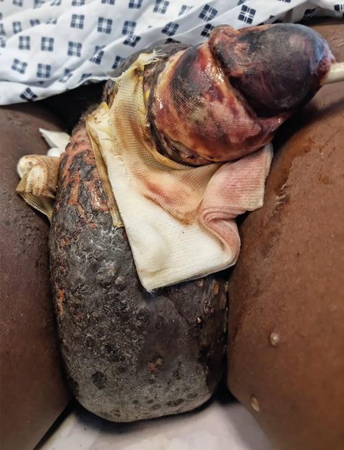