Microplastics found in patients after heart surgery


Everywhere scientists look for microplastics, they’ve found them — food, water, air and some parts of the human body. But examinations of our innermost organs that aren’t directly exposed to the environment are still limited.

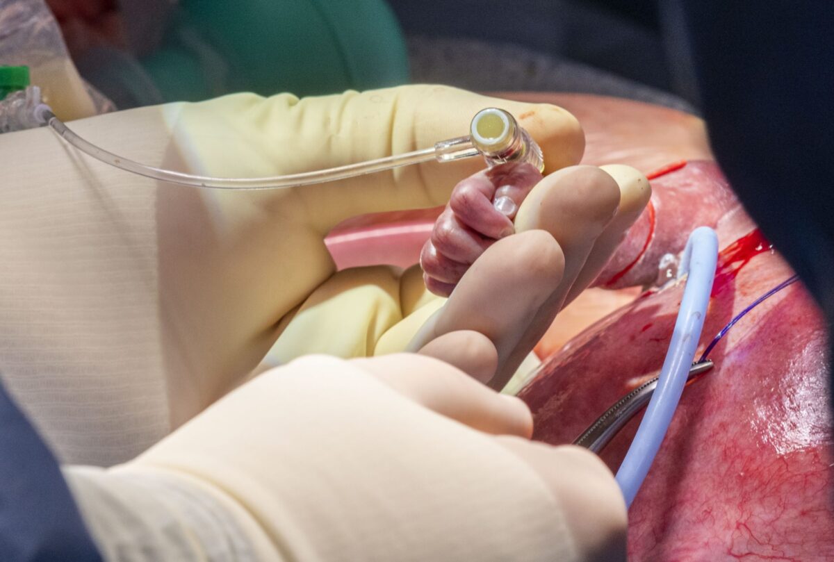
Now, in a pilot study of people who underwent heart surgery, researchers in ACS’ Environmental Science & Technology report that they have found microplastics in many heart tissues. They also report evidence suggesting that microplastics were unexpectedly introduced during the procedures. 

Microplastics are plastic fragments less than 5 millimeters wide, or about the size of a pencil eraser. Research has shown that they can enter the human body through mouths, noses and other body cavities with connections to the outside world. Yet many organs and tissues are fully enclosed inside a person’s body, and scientists lack information on their potential exposure to, and effects from, microplastics. So, Kun Hua, Xiubin Yang and colleagues wanted to investigate whether these particles have entered people’s cardiovascular systems through indirect and direct exposures.

Harmful substances in the soil, water and air also endanger people’s health. In recent years, environmental medicine has been increasingly concerned with the consequences of climate change.

In a pilot experiment, the researchers collected heart tissue samples from 15 people during cardiac surgeries, as well as pre- and post-operation blood specimens from half of the participants. Then the team analyzed the samples with laser direct infrared imaging and identified 20 to 500 micrometer-wide particles made from eight types of plastic, including polyethylene terephthalate, polyvinyl chloride and poly(methyl methacrylate). This technique detected tens to thousands of individual microplastic pieces in most tissue samples, though the amounts and materials varied between participants. All of the blood samples also contained plastic particles, but after surgery their average size decreased, and the particles came from more diverse types of plastics. 

Although the study had a small number of participants, the researchers say they have provided preliminary evidence that various microplastics can accumulate and persist in the heart and its innermost tissues. They add that the findings show how invasive medical procedures are an overlooked route of microplastics exposure, providing direct access to the bloodstream and internal tissues. More studies are needed to fully understand the effects of microplastics on a person’s cardiovascular system and their prognosis after heart surgery, the researchers conclude. 

Unborn Baby Partially Removed from Womb for Heart Surgery, Placed Back, Born 10 Weeks Later


In a rare, delicate, and ultimately lifesaving surgery, an unborn baby was partially removed from his mother’s womb at 26 weeks so that a tumor growing on his heart could be removed. He was then placed back in his mom’s womb for a further 10 weeks before being born at nearly full term.

Doctors at Cleveland Clinic, Ohio, said in a press release the baby is only the second person in the world to undergo this unique surgery, and survive. Today, he is thriving.

Baby Rylan Harrison Drinnon was diagnosed in the spring of 2021 with intrapericardial teratoma with fetal hydrops in utero, an extremely rare condition leading to heart failure if left untreated, according to the statement.

Epoch Times Photo
Dr. Hani Najm, who led the heart surgery team, inserts an IV line in the fetus’s right arm to deliver fluids and medications as needed.

“As far as we know, Cleveland Clinic is the second academic medical center in the world to have performed this fetal surgery successfully with continued pregnancy and delivery,” said Dr. Darrell Cass, director of Cleveland Clinic’s Fetal Surgery and Fetal Care Center.

“In this case, time was of the essence. Shortly after the patient arrived at Cleveland Clinic, imaging tests showed that the tumor kept growing and the fetus’s heart function was deteriorating.”

The malignant mass was compressing the left side of unborn baby Rylan’s heart, cutting off circulation and leading to an accumulation of fluid around his heart and other organs.

Parents Sam and Dave Drinnon of Pittsburgh were referred to Cleveland for their expertise, said Cass. A multidisciplinary team from Cleveland Clinic and Children’s Clinic performed surgery in May 2021 to remove Rylan’s tumor.

After making a “Caesarean section-like incision” to expose the mother’s uterus, the team, led by Dr. Hani Najm, Cleveland Clinic’s chair of pediatric and congenital heart surgery, used ultrasound to locate the placenta and fetus. They opened the uterus and lifted out Rylan’s arms to expose his chest. Najm removed the tumor from the baby’s beating heart before placing him back in the uterus, in a surgery lasting 3 1/2 hours.

“As soon as the tumor was removed, the compression of the left atrium disappeared, and there was a nice blood flow that was almost back to normal,” Najm reported.

Epoch Times Photo
Baby Rylan Harrison Drinnon.

Both mom and baby recovered well, and Rylan was able to remain in the womb until near full term, according to the statement.

Maternal-fetal medicine specialist Dr. Amanda Kalan, who attended the surgery, oversaw Sam’s aftercare and the delivery of her healthy baby boy by C-section on July 13, 10 weeks after the surgery.

Cass expressed pride in his team for their massive success.

Epoch Times Photo

“This tumor was growing rapidly in the exact wrong spot,” Cass explained. “We needed to act quickly and decisively to rescue the fetus … as far as we know, Cleveland Clinic is the second academic medical center in the world to have performed this fetal surgery successfully, with continued pregnancy and delivery.”

Only one previous incidence, said Cass, has ever been documented in the world’s medical literature.

Najm claimed that such innovative fetal surgery “provides hope to other families who may receive a similar devastating diagnosis.”

Looking to the future, Rylan will likely need surgery to reposition his sternum, which did not heal properly in the womb. Doctors will monitor his heart health as he grows to ensure the tumor does not reappear.

The Drinnons are beyond grateful for the lifesaving intervention.

“Now they have this beautiful boy, Rylan, and they think he’s going to be special,” Cass, told Cleveland.com. “He’s going to grow up to be a completely normal kid that just had a really unique odyssey to get to where he is now.”

Blood Test Marker Could Gauge Risks After Heart Surgery


About 2 million adults worldwide undergo heart surgery each year, and checking blood levels of a certain protein could help assess their risk of death within 30 days, a new study shows.

Blood tests to check levels of troponin (a type of protein found in heart muscle) have long been used to evaluate the risk of death and serious complications after heart attack, but the tests are not commonly done after heart surgery.

This new study found that elevated troponin levels were associated with an increased risk of death after heart bypass or open heart surgery.

“This study is a landmark for the health teams taking care of patients after cardiac surgery,” said study co-author André Lamy, a professor of surgery at McMaster University in Canada.

“For the first time, we have a marker that is fast and reliable for the monitoring of these patients after cardiac surgery,” Lamy said in a university news release.

This study included nearly 16,000 adult heart surgery patients, average age 63, in 12 countries. By 30 days after surgery, more than 2% of patients had died, and about 3% had experienced a major vascular complication, such as heart attack, stroke or a life-threatening blood clot.

The patients’ troponin levels were measured before and daily for the first few days after surgery.

“We found that the levels of troponin associated with an increased risk of death within 30 days were substantially higher  200 to 500 times the normal value  than troponin levels that surgical teams are currently told defines the risk of a patient having one of the most common complications after heart surgery  myocardial injury, a heart muscle injury associated with increased deaths,” said lead study author P.J. Devereaux. He’s a senior scientist at McMaster and a cardiologist at Hamilton Health Sciences.

The study was published March 2 in the New England Journal of Medicine.

SOURCE: McMaster University, news release, March 2, 2022

Doctors Perform Heart Surgery On Baby Still Inside Womb.


Children’s Hospital of Philadelphia is world-renowned for its fetal surgery in which an operation is performed on a baby still inside the womb.

Recently, in yet another historic procedure, CHOP doctors undertook a risky and delicate operation on a tiny fetal heart.

Health reporter Stephanie Stahl has the exclusive story of what it took to save an unborn baby named Juan.

After some setbacks, an excited family who learned they were finally going home to South America said goodbye to the team that saved their baby.

Baby Juan and his parents, Cecilia Cella and Pablo Paladino, are headed back to Uruguay where the infant has become a celebrity.

“We receive calls, messages from people we don’t know,” said Pablo Paladino.

Since October, the family has been camped out at CHOP where doctors saved little Juan’s life with an intervention that is largely unheard of in many places.

“It was a hard time, crazy time, but we are extremely happy how everything was solved,” said Paladino.

When Cella was five months pregnant, a routine ultrasound showed a mass on the baby’s heart.

Their doctor in Uruguay sent the images to his friend, Dr. Jack Rychik, who is the director of the Fetal Heart Program at CHOP.

“The minute I saw this I recognized there was a giant tumor sitting on the heart,” said Rychik.

It was a rare pericardial teratoma and the only hope was fetal surgery where doctors would operate on the baby Juan’s heart inside the womb.

“We never heard this before,” said Paladino.

“I started laughing, like what, they do that,” said Cella.

The family raced to Philadelphia because CHOP is the only place where the risky fetal heart operation has been done successfully on just one other occasion.

“We’re operating on two patients here with the single intent,” said Rychik. “Our goal is to get to the tumor and resect the tumor but we also have mother and baby.”

Juan is now the second baby to survive the fetal surgery.

The procedure was performed when his mother was 21 weeks pregnant with the baby.

“His heart at the time of surgery was the size of a peanut. The size of the tumor was three-times the size of the heart,” explained Rychik. “Had we waited an additional day, we probably would have been too late.”

After the fetal surgery, the pregnancy continued as the family waited in Philadelphia.

At 31 weeks, Juan was born on Dec. 11, but the tumor had grown back so there was a second heart surgery.

“There were a lot of chances the baby was going to die,” said Paladino.

And now, with a big scar on his chest, Juan is 3 months old and healthy.


However, his prognosis is unknown.

“There are no other human beings alive today who have had fetal surgery for this removal of this type of tumor that are 30, 40, 50, 60 years old. We could then say what the prognosis is going to be,” says Dr. Rychik.

But for Cella and Paladino, at least their son has a chance after the fetal surgery that still has their heads spinning.

“Crazy, unbelievable, I look at him, I can’t believe what they did here,” said Cella. “It’s awesome.”

“We are grateful that we came here,” added Paladino. “The doctors are amazing.”

The family is now back home in Uruguay where little Juan continues to thrive.

The tumor that was on his heart was benign.

It’s unknown as to what causes this type of tumor, but it is usually a fatal condition.

Source:http://philadelphia.cbslocal.com

Space technology studied for heart surgery.


0918-Feature-Cooling Cap_Blog

A Chicago heart surgeon is trying to determine whether a cooling technology originally developed for astronauts could help patients recover more quickly after open-heart surgery.

The device, called a “Cooling Cap,” uses targeted hypothermia to slow brain activity, potentially limiting or preventing damage by reducing the metabolic demands on the brain.

“You can slow down the metabolism, decrease the pathways and turn off mechanisms that can create adverse outcomes for the patient,” said Paul J. Pearson, M.D., Ph.D., a co-director for NorthShore University HealthSystem’s Cardiovascular Institute in Chicago, who is leading a feasibility study on the device for use during cardiac surgery.

Doctors have used techniques to lowering body temperature during cardiac surgery for decades as a way to minimize the detrimental effects of using the heart-lung machine, a “pump” that temporarily takes over the function of the heart and lungs during surgery.

Originally designed along with other cooling garments used by astronauts to protect them from the extremes of space, the “Cooling Cap” received a grant by the U.S. Department of Defense for use treating traumatic brain injuries on battlefields and gained Food and Drug Administration approval for such use in 2012. The ongoing study is the first to use the technology for heart surgery.

By cooling the brain during surgery, Pearson hopes to speed recovery times by reducing common side effects such as delirium, slow or difficult waking from anesthesia and temporary cognitive disruption. He said studies have shown that patients who experience such side effects following surgery don’t recover as well as those who don’t and may also experience worse survival rates.

“What we’re trying to do is get to a point where we can take very sick and complex patients through heart surgery and have them wake up like a 30-year-old who had a hernia repair,” Pearson said. “This is one more technique that may improve outcomes that are already significantly better than they were 10 years ago.”

Pearson and his colleagues are initially using the technology on 20 patients undergoing heart surgery at NorthShore to see if it could be used in the operating room without disrupting the medical team or other equipment needed for the procedure. Once that study is complete, the technology would undergo a larger study analyzing patient outcomes.