Gene discovery offers new hope for people living with chronic skin disease


Gene discovery offers new hope for people living with chronic skin disease
Dr. Chelisa Cardinez (pictured) says the discovery finally provides an explanation for why patients with psoriasis may go on to develop psoriatic arthritis, leaving them with joint pain, stiffness and swelling.

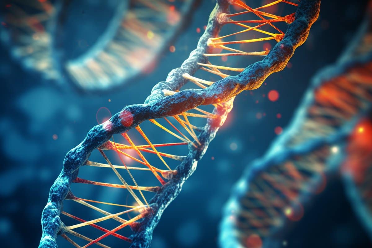
Scientists from The Australian National University (ANU) have discovered a gene mutation is responsible for causing psoriasis—a chronic inflammatory skin disease that causes patients to develop red, scaly and itchy patches across their body. The research is published in Nature Communications.

According to ANU researcher Dr. Chelisa Cardinez, if two copies of this mutated gene (known as IKBKB) are present, patients with psoriasis may go on to develop psoriatic arthritis, leaving them with joint pain, stiffness and swelling. Thanks to the world-first discovery from ANU, scientists now know what causes the progression from a skin-only disease to a skin and joint disease.

It’s hoped the findings will lead to improved diagnosis and treatment for patients with psoriasis and psoriatic arthritis—conditions that patients say carry stigma in the community.

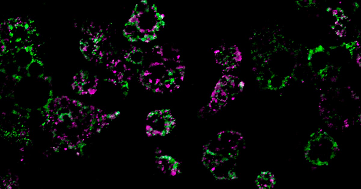
“Using a mouse model, we identified that this mutation led to an abnormal function in a group of immune cells known as regulatory T cells,” Dr. Cardinez, from the ANU John Curtin School of Medical Research (JCSMR), said.

“These cells are normally considered gatekeepers of the immune system. However, we found that this mutation alters the function of these cells, causing them to contribute to inflammation and promote the onset of disease.”

Rebecca Davey is one of at least 500,000 Australians that live with psoriasis. She also happens to have psoriatic arthritis and says the stiffness and pain she feels when she gets out of bed in the morning can be extreme.

“People don’t understand the debilitating effects these conditions can have on the individual and in fact a whole family when someone is in constant pain, has poor sleep from pain, and feels constantly fatigued,” Ms Davey said.

“My psoriatic arthritis drugs have largely reduced the larger outbreaks on my skin, but you do have to consider everything you put on your skin and the fabrics you wear. As a former nurse, even the constant hand washing that was required for work would cause my skin to flare up. It’s one of the reasons why I no longer work in the hospital system.”

Psoriasis and psoriatic arthritis are forms of autoimmune disease. These types of diseases occur when the immune system attacks healthy cells after wrongly perceiving them as a threat. According to Arthritis Australia, three out of every 10 Australians with psoriasis develop psoriatic arthritis.

Although there is no cure for psoriasis, there are treatments that can help manage the condition. In October 2023, the Pharmaceutical Benefits Scheme (PBS) listed a new, subsidized drug for Australians living with severe psoriasis.

Ms Davey, who is also CEO of Arthritis ACT, says it’s important to break down the stigma associated with these conditions. She says psoriasis is very misunderstood in the community.

“So many people are accused of having poor hygiene due to the plaques or even just minor skin lesions as they erupt. It’s not the individual’s fault that their skin is in the condition it’s in; psoriasis is a painful, debilitating condition,” she said.

“I had no idea what was causing my hands to flare up all the time. Our poor GPs often don’t recognize these conditions early.

“In regional and rural areas there is a drastic shortage of specialists both in dermatology and rheumatology to diagnose and treat these conditions, and people can wait over a year for an appointment if their symptoms are less dramatic.

“We must raise greater awareness of invisible disabilities such as those created by these conditions. A person might look ok from the outside, but in reality they are struggling on a daily basis.”

Dr. Cardinez said, “Studies have shown that delays in psoriatic arthritis diagnosis is linked to worse clinical outcomes for patients. Therefore, earlier detection and treatment of these immune diseases is key to improving health outcomes.

“By developing a better understanding of the IKBKB gene and the role it plays in promoting the onset of these diseases, it could bring us a step closer to one day finding a cure, which would offer new hope for hundreds of thousands of Australians.”

Deleting a Key Gene Shields Against Excess Weight Gain


A study from UC San Diego reveals that obesity causes mitochondrial dysfunction in fat cells, driven by a specific gene. Deactivating this gene in mice prevents obesity, suggesting new treatment approaches.

UC San Diego Study reveals key mechanism behind obesity-related metabolic dysfunction.

The number of people with obesity has nearly tripled since 1975, resulting in a worldwide epidemic. While lifestyle factors like diet and exercise play a role in the development and progression of obesity, scientists have come to understand that obesity is also associated with intrinsic metabolic abnormalities. Now, researchers from the University of California San Diego School of Medicine have shed new light on how obesity affects our mitochondria, the all-important energy-producing structures of our cells.

Researchers at the University of California San Diego School of Medicine have discovered that a high-fat diet causes mitochondrial fragmentation in fat cells, reducing their fat-burning capacity.

Mitochondrial Dysfunction in Obesity

In a study published today (January 29, 2024) in the journal Nature Metabolism, the researchers found that when mice were fed a high-fat diet, mitochondria within their fat cells broke apart into smaller mitochondria with reduced capacity for burning fat. Further, they discovered that this process is controlled by a single gene. By deleting this gene from the mice, they were able to protect them from excess weight gain, even when they ate the same high-fat diet as other mice.

“Caloric overload from overeating can lead to weight gain and also triggers a metabolic cascade that reduces energy burning, making obesity even worse,” said Alan Saltiel, PhD, professor in the Department of Medicine at UC San Diego School of Medicine. “The gene we identified is a critical part of that transition from healthy weight to obesity.”

These colored streaks are mitochondrial networks within fat cells. Researchers from UC San Diego discovered that a high-fat diet dismantles mitochondria, resulting in weight gain. Credit: UC San Diego Health Sciences

Adipose Tissue and Obesity

Obesity, which affects more than 40% of adults in the United States, occurs when the body accumulates too much fat, which is primarily stored in adipose tissue. Adipose tissue normally provides important mechanical benefits by cushioning vital organs and providing insulation. It also has important metabolic functions, such as releasing hormones and other cellular signaling molecules that instruct other tissues to burn or store energy.

In the case of caloric imbalances like obesity, the ability of fat cells to burn energy starts to fail, which is one reason why it can be difficult for people with obesity to lose weight. How these metabolic abnormalities start is among the biggest mysteries surrounding obesity.

The study was led by Alan Saltiel, PhD, professor in the Department of Medicine at UC San Diego School of Medicine. Credit: UC San Diego Health Sciences

Research Findings and Potential Therapies

To answer this question, the researchers fed mice a high-fat diet and measured the impact of this diet on their fat cells’ mitochondria, structures within cells that help burn fat. They discovered an unusual phenomenon. After consuming a high-fat diet, mitochondria in parts of the mice’s adipose tissue underwent fragmentation, splitting into many smaller, ineffective mitochondria that burned less fat.

In addition to discovering this metabolic effect, they also discovered that it is driven by the activity of single molecule, called RaIA. RaIA has many functions, including helping break down mitochondria when they malfunction. The new research suggests that when this molecule is overactive, it interferes with the normal functioning of mitochondria, triggering the metabolic issues associated with obesity.

“In essence, chronic activation of RaIA appears to play a critical role in suppressing energy expenditure in obese adipose tissue,” said Saltiel. “By understanding this mechanism, we’re one step closer to developing targeted therapies that could address weight gain and associated metabolic dysfunctions by increasing fat burning.”

By deleting the gene associated with RaIA, the researchers were able to protect the mice against diet-induced weight gain. Delving deeper into the biochemistry at play, the researchers found that some of the proteins affected by RaIA in mice are analogous to human proteins that are associated with obesity and insulin resistance, suggesting that similar mechanisms may be driving human obesity.

“The direct comparison between the fundamental biology we’ve discovered and real clinical outcomes underscores the relevance of the findings to humans and suggests we may be able to help treat or prevent obesity by targeting the RaIA pathway with new therapies,” said Saltiel “We’re only just beginning to understand the complex metabolism of this disease, but the future possibilities are exciting.”

Gene Linked to Mitochondrial Dysfunction in Obesity


Summary: Researchers made a breakthrough in understanding obesity’s impact on mitochondria, as detailed in a recent study.

They discovered that a high-fat diet causes fat cell mitochondria in mice to fragment into smaller, less efficient units, a process controlled by a single gene. By deleting this gene, the mice were protected from weight gain despite consuming the same high-fat diet.

This study offers new insights into the metabolic dysfunctions in obesity, paving the way for potential targeted therapies.

Key Facts:

  1. The study revealed that a high-fat diet leads to the fragmentation of mitochondria in fat cells, reducing their ability to burn fat.
  2. A single gene, associated with the molecule RaIA, was found to be responsible for this mitochondrial fragmentation and metabolic disruption in obesity.
  3. By removing this gene, researchers successfully protected mice from obesity induced by a high-fat diet, suggesting a new therapeutic target for obesity treatment in humans.

Source: UCSD

The number of people with obesity has nearly tripled since 1975, resulting in a worldwide epidemic. While lifestyle factors like diet and exercise play a role in the development and progression of obesity, scientists have come to understand that obesity is also associated with intrinsic metabolic abnormalities.

Now, researchers from the University of California San Diego School of Medicine have shed new light on how obesity affects our mitochondria, the all-important energy-producing structures of our cells.

This shows DNA.
How these metabolic abnormalities start is among the biggest mysteries surrounding obesity.

In a study published January 29, 2023 in Nature Metabolism, the researchers found that when mice were fed a high-fat diet, mitochondria within their fat cells broke apart into smaller mitochondria with reduced capacity for burning fat. Further, they discovered that this process is controlled by a single gene. By deleting this gene from the mice, they were able to protect them from excess weight gain, even when they ate the same high-fat diet as other mice.

“Caloric overload from overeating can lead to weight gain and also triggers a metabolic cascade that reduces energy burning, making obesity even worse,” said Alan Saltiel, PhD, professor in the Department of Medicine at UC San Diego School of Medicine. “The gene we identified is a critical part of that transition from healthy weight to obesity.”

Obesity, which affects more than 40% of adults in the United States, occurs when the body accumulates too much fat, which is primarily stored in adipose tissue. Adipose tissue normally provides important mechanical benefits by cushioning vital organs and providing insulation. It also has important metabolic functions, such as releasing hormones and other cellular signaling molecules that instruct other tissues to burn or store energy.

In the case of caloric imbalances like obesity, the ability of fat cells to burn energy starts to fail, which is one reason why it can be difficult for people with obesity to lose weight. How these metabolic abnormalities start is among the biggest mysteries surrounding obesity.

To answer this question, the researchers fed mice a high-fat diet and measured the impact of this diet on their fat cells’ mitochondria, structures within cells that help burn fat. They discovered an unusual phenomenon. After consuming a high-fat diet, mitochondria in parts of the mice’s adipose tissue underwent fragmentation, splitting into many smaller, ineffective mitochondria that burned less fat.

In addition to discovering this metabolic effect, they also discovered that it is driven by the activity of single molecule, called RaIA. RaIA has many functions, including helping break down mitochondria when they malfunction. The new research suggests that when this molecule is overactive, it interferes with the normal functioning of mitochondria, triggering the metabolic issues associated with obesity.

“In essence, chronic activation of RaIA appears to play a critical role in suppressing energy expenditure in obese adipose tissue,” said Saltiel. “By understanding this mechanism, we’re one step closer to developing targeted therapies that could address weight gain and associated metabolic dysfunctions by increasing fat burning.”

By deleting the gene associated with RaIA, the researchers were able to protect the mice against diet-induced weight gain. Delving deeper into the biochemistry at play, the researchers found that some of the proteins affected by RaIA in mice are analogous to human proteins that are associated with obesity and insulin resistance, suggesting that similar mechanisms may be driving human obesity.

“The direct comparison between the fundamental biology we’ve discovered and real clinical outcomes underscores the relevance of the findings to humans and suggests we may be able to help treat or prevent obesity by targeting the RaIA pathway with new therapies,” said Saltiel.

“We’re only just beginning to understand the complex metabolism of this disease, but the future possibilities are exciting.”

Co-authors of the study include: Wenmin Xia, Preethi Veeragandham, Yu Cao Yayun Xu, Torrey Rhyne, Jiaxin Qian, Ying Jones,Chao-Wei Hung, Zichen Wang, Hiroyuki Hakozaki and Johannes Schoneberg at UC San Diego, Peng Zhao at University of Texas Health Science Center, Hui Gao and Mikael Ryden at Karolinska Institute, Christopher Liddle, Ruth Yu, Michael Downes, Ronald Evans and Jianfeng Huang at Salk Institute for Biological Studies,Martin Wabitsch at Ulm University Medical Center and Shannon Reilly at Weill Medical College of Cornell University.

Funding: This study was funded, in part, by the National Institutes of Health (Grants P30DK063491, R01DK122804, R01DK124496, R01DK125820 and R01DK128796).


Abstract

Obesity causes mitochondrial fragmentation and dysfunction in white adipocytes due to RalA activation

Mitochondrial dysfunction is a characteristic trait of human and rodent obesity, insulin resistance and fatty liver disease. Here we show that high-fat diet (HFD) feeding causes mitochondrial fragmentation in inguinal white adipocytes from male mice, leading to reduced oxidative capacity by a process dependent on the small GTPase RalA.

RalA expression and activity are increased in white adipocytes after HFD. Targeted deletion of RalA in white adipocytes prevents fragmentation of mitochondria and diminishes HFD-induced weight gain by increasing fatty acid oxidation.

Mechanistically, RalA increases fission in adipocytes by reversing the inhibitory Ser637 phosphorylation of the fission protein Drp1, leading to more mitochondrial fragmentation. Adipose tissue expression of the human homolog of Drp1, DNM1L, is positively correlated with obesity and insulin resistance.

Thus, chronic activation of RalA plays a key role in repressing energy expenditure in obese adipose tissue by shifting the balance of mitochondrial dynamics toward excessive fission, contributing to weight gain and metabolic dysfunction.

Gene Identified That Plays a Role in People Who Recover from Dilated Cardiomyopathy


human heart

Researchers at the Mayo Clinic investigating factors related to recovery from dilated cardiomyopathy have identified a variant in a specific gene that plays a role in people who recover from the condition and those who don’t. Results of the genome-wide association study (GWAS), published in Circulation Research, is expected to spur future research to target the gene for future dilated cardiomyopathy treatments.

“We found genetic variation in the CDCP1 gene, a gene that no one has heard of in cardiology, and its link to improvement in heart function in these patients,” says lead author Naveen Pereira, MD, a Mayo Clinic cardiologist.

Cardiomyopathy is characterized by its effect on the heart’s left ventricle, making it more difficult to pump blood throughout the rest of the body.

According to the investigators, CDCP1 genetic variation leads to differences in the protein’s structure which may influence a person’s susceptibility to disease or response to specific drugs. Once the gene was associated with improving function in the left ventricle, the team examined why this occurs. A key finding was that the CDCP1 gene is often variably expressed in fibroblasts of people with dilated cardiomyopathy, and fibrosis—an excess of fibrous connective tissue in the heart—plays a central role in prognosis of patients with the condition. Further, Pereira noted that genetic variation in or near CDCP1 has a significant association with heart failure death.

Strengthening the case for CDCP1 as a drug target, the Mayo Clinic researchers observed that decreasing the expression of the gene in cardiac connective tissue decreased fibroblast proliferation in the heart. It also downregulated the IL1RL1 gene, which encodes a prominent heart failure biomarker aST2, which when found in high levels is associated with fibrosis and death. This suggests the importance of developing a better understanding of the relationship between CDCP1 and aST2, as researchers search for ways to treat heart failure.

This finding now comes as heart failure rates are expected to rise significantly over the rest of the decade. The American Heart Association forecasts that heart failure will affect eight million people in the U.S. by 2030, a 46% increase over current rates. Of those cases, between 30% and 40% are caused by dilated cardiomyopathy.

Building in this research, the Mayo Clinic team is continuing studies in animal models to better understand the role CDCP1 plays in heart failure, and they are developing molecules to assess their potential as treatments for dilated cardiomyopathy.

“By continuing with this research that started with a human population that we took to the molecular and now animal laboratory, we hope to find new avenues for treatments to take back to the human population we studied, to improve patients’ survival and quality of life ultimately,” says Pereira.

New Genes Tied to Alzheimer’s Disease


Senior men playing chess

Researchers identified 11 new genes that affect the risk for Alzheimer’s disease. The findings point to novel targets for preventing or delaying the disease.

Alzheimer’s disease is the most common cause of dementia in older adults. It affects more than 5 million Americans. A hallmark of the disease is the abnormal accumulation of amyloid protein in the brain. Until 2009, variants in only one gene, APOE, had been identified as a risk factor for late-onset Alzheimer’s disease, the most common form of the disorder. The list of known genetic risk factors has since grown to include several others.

Large-scale analyses are needed to gain the statistical power to identify additional genetic risk factors. Scientists in the International Genomic Alzheimer’s Project (IGAP) have been working together since 2011 on genome-wide association studies (GWAS), which involve thousands of DNA samples and shared datasets. GWAS are aimed at detecting subtle genetic differences that are statistically associated with disease.

IGAP’s latest effort involved scanning the DNA of more than 74,000 older volunteers from 15 countries. Participants included people with late-onset Alzheimer’s disease and those free of the disorder. It’s the largest genetic analysis yet conducted in Alzheimer’s research. The work was supported in part by NIH’s National Institute on Aging (NIA) and other NIH components. The findings were reported online in Nature Genetics on October 27, 2013.

The scientists confirmed many of the previously identified genes associated with the onset and progression of late-onset Alzheimer’s. In addition, they identified 11 new genes associated with the disease: HLA-DRB5/HLA-DRB1, SORL1, PTK2B, SLC24A4/RIN3, ZCWPW1, CELF1, NME8, FERMT2, CASS4, INPP5D, and MEF2C. The study also highlighted another 13 variants that merit further analysis.

The findings strengthen evidence about the involvement of certain pathways in Alzheimer’s disease, including the immune response, inflammation, cellular protein trafficking, and lipid transport. They also add to evidence for other pathways that may influence disease development, including synapse function, cytoskeletal function, and specialized cells in the brain called microglia.

“Interestingly, we found that several of these newly identified genes are implicated in a number of pathways,” says Dr. Gerard Schellenberg of the University of Pennsylvania School of Medicine, who directs one of the major IGAP consortia. “Alzheimer’s is a complex disorder, and more study is needed to determine the relative role each of these genetic factors may play.”

Scientists Have Pinpointed the Gene Responsible for Down Syndrome


IN BRIEF

A team of researchers from Singapore and the United Kingdom have discovered an enzyme that regulates sperm and egg cell production, which may be linked to Down Syndrome, Patau Syndrome, and other chromosomal aberrations.

UNDERSTANDING THE LINK

Humans have 23 pairs of chromosomes in every cell in their bodies, except for their reproductive cells or gamete cells (sperm or egg), which contain 23 chromosomes. The reason why chromosomes come in pairs is that one pair comes from the egg, and the other from the sperm. So when gametes fuse with each other, they end up as a single cell having two copies of each chromosome.

Gamete cells are produced by a process called meiosis — a type of cell division with two rounds of nuclear division, to make sure that the number of chromosomes in the parent cell is halved. Sometimes, though, errors occur during cell division, which may result to offsprings having abnormal number of chromosomes — a phenomenon called aneuploidy.

Aneuploidy causes Down Syndrome — the most common genetic condition, Patau Syndrome, and other genetic disorders. It is also the leading cause of miscarriage.

Image Credit: iStock/koya79

PLAYING A PIVOTAL ROLE

The research team, led by Dr. Prakash Arumugam from the National University of Singapore, noted how the process of meiosis can affect chromosomal irregularities: “Understanding how meiosis is regulated is of great importance to understanding the causes of aneuploidy and genetic disorders in human,” said Dr Gary Kerr and the team, writing in the journal Scientific Reports.

The researchers have discovered a particular enzyme which plays an essential role in chromosome segregation in meiosis. They identified this enzyme as PP2ACdc55, which is involved in various cellular processes. It was also shown from the research team’s previous findings that PP2ACdc55 plays a vital role in controlling the timing of meiosis, thus preventing the cells from prematurely exiting phases of cell division.

The scientists tracked the enzyme on yeast models using fluorescent tagging, and analyzed the resulting mutant yeast strains, characterized the mutations and determined the role of the Cdc55 gene. Their results suggest that the gene might have a role in meiotic chromosome segregation. This is, without a doubt, a step forward, but we still don’t know what causes the process to go wrong.

Scientists identify brain molecule that triggers schizophrenia-like behaviors, brain changes


Scientists at The Scripps Research Institute (TSRI) have identified a molecule in the brain that triggers schizophrenia-like behaviors, brain changes and global gene expression in an animal model. The research gives scientists new tools for someday preventing or treating psychiatric disorders such as schizophrenia, bipolar disorder and autism.

“This new model speaks to how schizophrenia could arise before birth and identifies possible novel drug targets,” said Jerold Chun, a professor and member of the Dorris Neuroscience Center at TSRI who was senior author of the new study.

The findings were published April 7, 2014, in the journal Translational Psychiatry.

What Causes Schizophrenia?

According to the World Health Organization, more than 21 million people worldwide suffer from schizophrenia, a severe psychiatric disorder that can cause delusions and hallucinations and lead to increased risk of suicide.

Although psychiatric disorders have a genetic component, it is known that environmental factors also contribute to disease risk. There is an especially strong link between psychiatric disorders and complications during gestation or birth, such as prenatal bleeding, low oxygen or malnutrition of the mother during pregnancy.

In the new study, the researchers studied one particular known risk factor: bleeding in the brain, called fetal cerebral hemorrhage, which can occur in utero and in premature babies and can be detected via ultrasound.

In particular, the researchers wanted to examine the role of a lipid called lysophosphatidic acid (LPA), which is produced during hemorrhaging. Previous studies had linked increased LPA signaling to alterations in architecture of the fetal brain and the initiation of hydrocephalus (an accumulation of brain fluid that distorts the brain). Both types of events can also increase the risk of psychiatric disorders.

“LPA may be the common factor,” said Beth Thomas, an associate professor at TSRI and co-author of the new study.

Mouse Models Show Symptoms

To test this theory, the research team designed an experiment to see if increased LPA signaling led to schizophrenia-like symptoms in animal models.

Hope Mirendil, an alumna of the TSRI graduate program and first author of the new study, spearheaded the effort to develop the first-ever animal model of fetal cerebral hemorrhage. In a clever experimental paradigm, fetal mice received an injection of a non-reactive saline solution, blood serum (which naturally contains LPA in addition to other molecules) or pure LPA.

The real litmus test to show if these symptoms were specific to psychiatric disorders, according to Mirendil, was “prepulse inhibition test,” which measures the “startle” response to loud noises. Most mice—and humans—startle when they hear a loud noise. However, if a softer noise (known as a prepulse) is played before the loud tone, mice and humans are “primed” and startle less at the second, louder noise. Yet mice and humans with symptoms of schizophrenia startle just as much at loud noises even with a prepulse, perhaps because they lack the ability to filter sensory information.

Indeed, the female mice injected with serum or LPA alone startled regardless of whether a prepulse was placed before the loud tone.

Next, the researchers analyzed brain changes, revealing schizophrenia-like changes in neurotransmitter-expressing cells. Global gene expression studies found that the LPA-treated mice shared many similar molecular markers as those found in humans with schizophrenia. To further test the role of LPA, the researchers used a molecule to block only LPA signaling in the brain.

This treatment prevented schizophrenia-like symptoms.

Implications for Human Health

This research provides new insights, but also new questions, into the developmental origins of psychiatric disorders.

For example, the researchers only saw symptoms in female mice. Could schizophrenia be triggered by different factors in men and women as well?

“Hopefully this animal model can be further explored to tease out potential differences in the pathological triggers that lead to disease symptoms in males versus females,” said Thomas.

In addition to Chun, Thomas and Mirendil, authors of the study, “LPA signaling initiates schizophrenia-like brain and behavioral changes in a mouse model of prenatal brain hemorrhage,” were Candy De Loera of TSRI; and Kinya Okada and Yuji Inomata of the Mitsubishi Tanabe Pharma Corporation.

Smoking Injurious to Genes Too


Here comes another shocker for those reluctant to kick the butt.

Smoking not only affects your health but also increases health risks of your children and grandchildren; today’s puffs of pleasure can permanently damage your genes, according to a new study.

Smoking can also affect the genes important for sperm quality or immune response.

The research findings from Uppsala University and Uppsala Clinical Research Center of Sweden showed that smoking alters several genes that can be associated with health problems for smokers, such as increased risk for cancer and diabetes.

The research, led by Asa Johansson, researcher at the Department of Immunology, Genetics and Pathology, said the genes of smokers as well tobacco users can change and expose them to more health risks.

However, according to the findings, tobacco itself may not be the cause of gene alterations, but the different elements that are formed when the tobacco is burnt.

“Our results therefore indicate that the increased disease risk associated with smoking is partly caused by epigenetic changes. A better understanding of the molecular mechanism behind diseases and reduced body function might lead to improved drugs and therapies in the future,” Johansson said.

The findings of the study have been published in the journal Human Molecular Genetics.

Designer Sperm Passes Selected Genes to Future Generations.


  • Frustrated by slow progress in gene therapy, a team of scientists opted for an unconventional approach. Instead of relying on the oocyte as a substrate for genetic modification, they took a closer look at male germ cells, including mature sperm. Sperm, owing to their accessibility, seemed to offer a convenient route to transgenesis.

    The scientists, based at the Royal Veterinary College in North Mimms, United Kingdom, used a viral vector to insert genetic material into mouse spermatozoa. Then the spermatozoa were used in an in vitro fertilization procedure. In the resulting embryos, the genetic material was found to be present and active—and inheritable. The genetic material that had been introduced to the spermatozoa was, the scientists confirmed, still functional after passing through at least three generations of mice.

    The scientists presented their results December 2 in The FASEB Journal, in an article entitled “Efficient generation of transgenic mice by lentivirus-mediated modification of spermatozoa.” In this article, the authors wrote, “When pseudotyped lentiviral vectors encoding green fluorescent protein (GFP) were incubated with mouse spermatozoa, these sperm were highly successful in producing transgenics.” Then, after embryo transfer, “≥42% of founders were found to be transgenic for GFP.”

    The authors also noted that they used inverse PCR for integration site analysis, which allowed them to show that at least one or two copies of GFP had been integrated in the transgenic animals, mapping to different chromosomes. GFP expression was detected in a wide range of murine tissues, including testis.

    This transgenic technology—if successful in humans—could lead to a new frontier in genetic medicine in which diseases and disorders are effectively cured, and new human attributes, such as organ regeneration, may be possible.

    “Transgenic technology is a most important tool for researching all kinds of disease in humans and animals, and for understanding crucial problems in biology,” said Anil Chandrashekran, Ph.D., a study author and research associate at the Royal Veterinary College.

    In detailing the more immediate applications of their work, the authors wrote, “This relatively simple, yet highly efficient, technique for generating transgenic animals by transducing spermatozoa with lentiviral vectors in vitro is a powerful tool for the study of fertilization/preimplantation development, vertical viral gene transmission, gene function and regulation, and epigenetic inheritance.

    Offering a more expansive view of the authors’ work, Gerald Weissmann, M.D., editor-in-chief of The FASEB Journal, noted that using modified sperm to insert genetic material has the potential to be a major breakthrough not only in future research, but also in human medicine.

    “It facilitates the development of transgenic animal models, and may lead to therapeutic benefits for people as well,” said Dr. Weissman. “For years we have chased effective gene therapies and have hit numerous speed bumps and dead ends. If we are able to able to alter sperm to improve the health of future generations, it would completely change our notions of ‘preventative medicine.'”


Gene responsible for hereditary cancer syndrome found to disrupt critical growth-regulating pathway.


 Whitehead Institute scientists report that the gene mutated in the rare hereditary disorder known as Birt-Hogg-Dubé cancer syndrome also prevents activation of mTORC1, a critical nutrient-sensing and growth-regulating cellular pathway.

This is an unexpected finding, as some cancers keep this pathway turned on to fuel their unchecked growth and expansion. In the case of Birt-Hogg-Dubé syndrome, the mutated gene prevents mTORC1 pathway activation early in the formation of tumors. Reconciling these opposing roles may give scientists a new perspective on how cancer cells can distort normal cellular functions to maintain their own harmful ways.

Cells use the mTORC1 (for “mechanistic target of rapamycin complex 1”) pathway to regulate growth in response to the availability of certain nutrients, including amino acids. Whitehead Member David Sabatini and other researchers have teased apart many components of this pathway, but the precise mechanism by which nutrient levels are actually sensed has remained elusive. Recently, Sabatini and his lab determined that a family of proteins known as Rag GTPases act as a switch for the pathway—when nutrients are present, the Rag proteins turn on the mTORC1 pathway.

Now, several members of the Sabatini lab, including graduate student Zhi-Yang Tsun, have determined that the FLCN protein acts as a trigger to activate the Rag protein switch. Their work is described in the November 7 issue of the journal Molecular Cell.

“Zhi has ascribed a molecular function to this protein, and that’s a major contribution,” says Sabatini, who is also a Howard Hughes Medical Institute investigator and a professor of biology at MIT. “For the first time, we have a biochemical function that’s associated with it. And in my view, that’s an important first step to understanding how it might be involved in cancer.”

Before Tsun’s work, very little was known about FLCN’s role in the cell. In the early 2000s, scientists determined that mutations in the gene coding for FLCN caused the rare cancer Birt-Hogg-Dubé syndrome, but the syndrome’s symptoms offered little insight into FLCN’s molecular function.

Birt-Hogg-Dubé syndrome causes unsightly but benign hair follicle tumors on the face, benign tumors in the lungs that can lead to collapsed lungs, and kidney cancer. The syndrome is an autosomal dominant disorder, which means that a child inheriting one mutated copy of the FLCN gene will eventually develop the syndrome. Currently, the disease is managed by treating symptoms, but no cure exists.

FLCN’s dual roles—as a cause of a rare cancer in its mutated form and as a trigger for a growth pathway that is often hijacked in cancer cells—has prompted Tsun and Sabatini to rethink how a mutation can push cells to become cancerous.

“Basically, the mTORC1 pathway is essential for life,” explains Tsun. “So when you lose this nutrient switch or if it can’t be turned on, then the cell seems to freak out and cause all other growth promoting pathways to be turned on to somehow overcompensate for this loss. And this is actually what we see in patient tumors.”

For Birt-Hogg-Dubé syndrome patients and their families, better understanding of FCLN’s function moves the field one step closer to developing a therapy.

“Usually diseases are first described, then the responsible gene or genes are identified, and then that gene’s molecular function is figured out,” says Tsun. “And you need to know the gene’s function before you can start working on drugs or therapy. We’ve done that third step, which is a very important discovery for these patients.”