Bio-inks are the key to amazing new medical applications for 3D printing


3D printing technology use cases have been somewhat lacking in the medical sector. Recently, a Ghent University spin-off project called Xpect-INX, started providing industry, clinicians, and researchers with unique materials (so-called ‘bio-inks’) to use for 3D printed cells, tissues, and possibly even entire organs.

The potential applications of  'bio-inks' are numerous. Ranging from converting 2D cell cultures in Petri dishes to more realistic 3D cultures, repairing or replacing damaged cells or tissues and growing 'mini organs-on-chip' to screen for new medic…
The potential applications of ‘bio-inks’ are numerous. Ranging from converting 2D cell cultures in Petri dishes to more realistic 3D cultures, repairing or replacing damaged cells or tissues and growing ‘mini organs-on-chip’ to screen for new medicines.

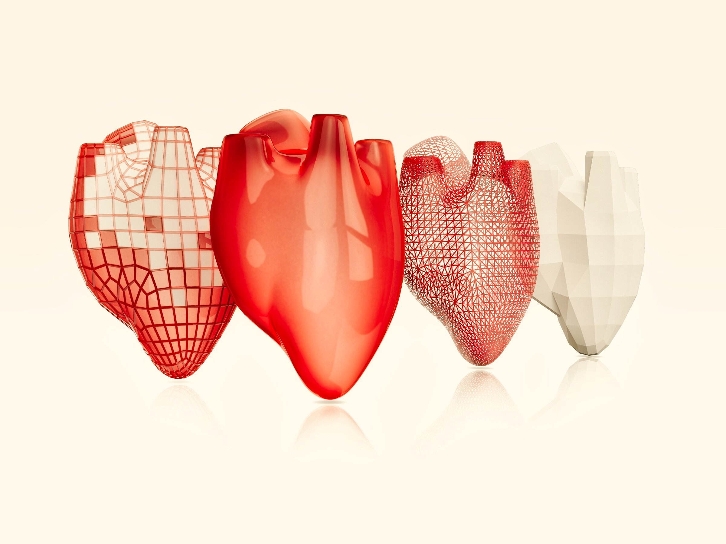
The Flanders region of Belgium has been a pioneer in the field of 3D printing technology for many years now, generating applications in space, fashion, and architecture. Sadly though, due to a lack of materials that can mimic the structure and function of biological tissues, the possibilities in the medical sector have been limited to 3D – printing of medical devices or anatomical models. However, with help from innovative materials developed at Ghent University, it is now possible to print human tissue.

In a Ghent University press release, Xpect-INX researcher and co-founder Jasper van Hoorick noted that 3D printing of cells and tissues is a very challenging process. 3D-printed cells must be able to grow and function like normal human cells in addition to being able to interact with existing cells until the body can fully integrate them. The key to this all is using appropriate and biocompatible materials, called “bio-inks.” Bio-inks are material mixtures that bear a strong resemblance to the natural cell environment. The bio-inks provide mechanical strength, a healthy growth environment for the cells, and they make it possible to process cells via various 3D printing techniques.

Image Credit: Wright Studio via Shutterstock / HDR tune by Universal-Sci

NEW OPPORTUNITIES

The potential applications of these ‘bio-inks’ are countless. They range from converting 2D cell cultures in Petri dishes to more realistic 3D cultures, repairing or replacing damaged cells or tissues due to illness or injury, and growing ‘mini organs-on-chip’ to screen for new medicines.

Currently, interventions such as breast reconstruction, replacement of a heart valve, or a meniscus often involve various complications. The combination of 3D printing with these bio-inks would offer a more sustainable and patient-specific solution. This way, damaged tissue can be replaced by new, body-specific tissue. The researchers expect that in the long term, even printing whole organs should be possible, which could overcome the shortage of donor organs.

Van Hoorick adds that this technology may provide opportunities to culture cells or mini-organs as a model for testing new drugs, substantially reducing the number of animal experiments.

SPECIFIC BIO-INKS FOR SPECIFIC APPLICATIONS

Every printer and application, from building an artificial cornea to the development of complex organs such as the liver, requires a specific, optimal ink. Currently, tissue biofabrication is still in its infancy, but it has come a long way. The Polymer Chemistry and Biomaterials research group, led by Peter Dubruel and Sandra van Vlierberghe, has been researching new polymers and bio-inks for these applications for more than ten years resulting in the Xpect-INX. 

As stated, the potential use cases for these bio-inks are innumerably providing us with hope for the future. Stay tuned as we keep an eye on this technology.

First functional human brain tissue produced through 3D printing


A team of researchers has created the first functional 3D-printed brain tissue to examine the brain’s function and study various neurological disorders. 

Representational image of signal transmitting neurons.

Representational image of signal transmitting neurons.

Get a daily digest of the latest news in tech, science, and technology, delivered right to your mailbox. Subscribe now.

The first functional 3D-printed brain tissue has been developed to examine the human brain’s function and study various neurological disorders. 

According to experts at the University of Wisconsin-Madison, printed tissue can “grow and function like typical brain tissue.”

This 3D-printed brain model might be useful in studying various neurological and neurodevelopmental problems, including Alzheimer’s and Parkinson’s disease.

“This could be a hugely powerful model to help us understand how brain cells and parts of the brain communicate in humans,” said Su-Chun Zhang, professor of neuroscience and neurology at UW–Madison’s Waisman Center.

“It could change the way we look at stem cell biology, neuroscience, and the pathogenesis of many neurological and psychiatric disorders,” added Zhang in the release. 

Use of horizontal 3D printing approach

Instead of using the conventional vertical layer stacking method, the researchers followed an innovative horizontal 3D printing approach in this development.

Neurons produced from induced pluripotent stem cells were carefully put in layers utilizing a softer bio-ink gel, creating a more favorable environment for growth.

“The tissue still has enough structure to hold together but it is soft enough to allow the neurons to grow into each other and start talking to each other,” Zhang said. 

First functional human brain tissue produced through 3D printing
Functional network model.

The developers mention that they deliberately maintained the tissue’s thinness to ensure optimal oxygen and nutrient intake for the neurons from the surrounding growth media.

The multilayer printing allowed the cells to form connections, resulting in networks similar to those observed in human brains.

Within these networks, neurons appeared to actively communicate by sending signals to one another. This communication occurs via neurotransmitters, chemical messengers that aid in passing signals between neurons.

“We printed the cerebral cortex and the striatum, and what we found was quite striking. Even when we printed different cells belonging to different parts of the brain, they could still talk to each other in a very special and specific way,” said Zhang in the press release.

The authors highlight that this method provides precision, allowing for control over cell types and arrangement. This feature is absent in brain organoids, which are miniature lab-grown organs created for brain research. 

This 3D print approach to emulate sophisticated communication and network development found in human brain tissue has great potential to provide insights into brain function and its disorders. 

The findings were published in the journal Cell Stem Cell.

Study abstract:

Probing how human neural networks operate is hindered by the lack of reliable human neural tissues amenable to the dynamic functional assessment of neural circuits. We developed a 3D bioprinting platform to assemble tissues with defined human neural cell types in a desired dimension using a commercial bioprinter. The printed neuronal progenitors differentiate into neurons and form functional neural circuits within and between tissue layers with specificity within weeks, evidenced by the cortical-to-striatal projection, spontaneous synaptic currents, and synaptic response to neuronal excitation. Printed astrocyte progenitors develop into mature astrocytes with elaborated processes and form functional neuron-astrocyte networks, indicated by calcium flux and glutamate uptake in response to neuronal excitation under physiological and pathological conditions. These designed human neural tissues will likely be useful for understanding the wiring of human neural networks, modeling pathological processes, and serving as platforms for drug testing.

Closer to 3D Printing Life-Like Organs


Summary: Researchers have developed a new technique that transforms medical images from MRI scans into detailed 3D computerized models. This new advance is an important step toward creating realistic 3D models of human organs for research and medical training.

Source: University of Colorado

A team of University of Colorado researchers has developed a new strategy for transforming medical images, such as CT or MRI scans, into incredibly detailed 3D models on the computer.

The advance marks an important step toward printing lifelike representations of human anatomy that medical professionals can squish, poke and prod in the real world.

The researchers describe their results in a paper published in December in the journal 3D Printing and Additive Manufacturing.

The discovery stems from a collaboration between scientists at CU Boulder and CU Anschutz Medical Campus designed to address a major need in the medical world: Surgeons have long used imaging tools to plan out their procedures before stepping into the operating room. But you can’t touch an MRI scan, said Robert MacCurdy, assistant professor of mechanical engineering and senior author of the new study. 

His team wants to fix that, giving doctors a new way to print realistic, and graspable, models of their patients’ various body parts, down to the detail of their tiny blood vessels—in other words, a model of your very own kidney entirely fabricated from soft and pliable polymers.

“Our method addresses the critical need to provide surgeons and patients with a greater understanding of patient-specific anatomy before the surgery ever takes place,” said Robert MacCurdy, senior author of the new paper and an assistant professor of mechanical engineering at CU Boulder.

The latest study gets the team closer to achieving that goal. In it, MacCurdy and his colleagues lay out a method for using scan data to develop maps of organs made up of billions of volumetric pixels, or “voxels”—like the pixels that make up a digital photograph, only three-dimensional.  

The researchers are currently experimenting with how they can use 3D printers to turn those maps into physical models that are more accurate than those available through existing tools.

The project, which is led by MacCurdy and CU Anschutz’ Nicholas Jacobson, is funded by AB Nexus, a grant program that seeks to spur new collaborations between the two Colorado campuses.

“In my lab we look for alternative ways of representation that will feed, rather than interrupt, the thinking process of surgeons,” said Jacobson, a clinical design researcher at the Inworks Innovation Initiative. “These representations become sources of ideas that help us and our surgical collaborators see and react to more of what is in the available data.”

Slicing the orange

Human organs are complicated—made up of networks of tissue, blood vessels, nerves and more, all with their own texture and colors. 

Currently, medical professionals try to capture these structures using “boundary surface” mapping, which, essentially, represents an object as a series of surfaces.

This shows a cross section of a kidney
Voxel map of a cross section of a human kidney.

“Think of existing methods as representing an entire orange by only considering the exterior orange peel,” MacCurdy said. “When viewed that way, the entire orange is peel.”

His team’s method, in contrast, is all juicy insides.

The approach begins with a Digital Imaging and Communications in Medicine (DICOM) file, the standard 3D data that CT and MRI scans produce. Using custom software, MacCurdy and his colleagues convert that information into voxels, essentially slicing an organ into tiny cubes with a volume much smaller than a typical tear drop. 

And, MacCurdy said, the group can do all that without losing any information about the organs in the process—something that’s impossible with existing mapping methods.

To test these tools, the team took real scan data of a human heart, kidney and brain, then created a map for each of those structures. The resulting maps were detailed enough that they could, for example, distinguish between the kidney’s fleshy interior, or medulla, and its outer layer or, cortex—both of which look pink to the human eye. 

“Surgeons are constantly touching and interacting with tissues,” MacCurdy said, “So we want to give them models that are both visual and tactile and as representative of what they’re going to face as they can be.”

Abstract

Defining Soft Tissue: Bitmap Printing of Soft Tissue for Surgical Planning

Nearly all applications of 3D printing for surgical planning have been limited to bony structures and simple morphological descriptions of complex organs due to the fundamental limitations in accuracy, quality, and efficiency of the current modeling paradigms and technologies.

Current approaches have largely ignored the constitution of soft tissue critical to most surgical specialties where multiple high-resolution variations transition gradually across the interior of the volume. Differences in the scales of organization related to unique organs require special attention to capture fine features critical to surgical procedures.

We present a six-material bitmap printing technique for creating 3D models directly from medical images, which are superior in spatial and contrast resolution to current 3D modeling methods, and contain previously unachievable spatial fidelity for soft tissue differentiation.

3D printing: potential clinical applications for personalised solid dose medications


Three‐dimensional printing or additive manufacturing has the potential to transform personalised medicine

Personalised medicine aims to move gold‐standard care away from empiric prescribing for a typical patient towards tailored treatment for the patient as an individual.1 It is well known that the effect of a medicine on an individual can vary based on factors including sex, genetics and even hormones. Currently, the personalisation of medicines to adjust for factors such as these is limited by the doses and combinations that are commercially available. This inflexibility makes it difficult for clinicians to tailor the medication for individual needs. One technology that could revolutionise personalised medicine is a process called additive manufacturing. In this process, a three‐dimensional (3D) object is produced by fusing thin layers of materials on top of each other until the complete object is formed. This 3D printing method could be applied to medicines to include several drugs in a single tablet at entirely customisable doses set by the clinician, such as the proof of concept five‐in‐one polypill developed in 2015.2

What are 3D‐printed medicines?

The field of 3D‐printed medicines is rapidly emerging. Spritam (Aprecia Pharmaceuticals) is an orodispersible levetiracetam tablet that completely dissolves in the mouth within 10 seconds; and in 2015, it became the first 3D‐printed medicine to be approved by the Food and Drug Administration.3 Since then, there have been several studies that have confirmed the safety, efficacy and feasibility of 3D‐printed medicines against their traditionally manufactured counterparts.4 With the right techniques, 3D‐printed medicines can be made to mimic the immediate or sustained release profiles of conventional tablets or even accommodate several medicines with different release profiles in the same tablet.2,5 These dosage forms are of a high quality and accuracy, and they can mimic all the characteristics of conventionally made dosage forms but with the ability to adjust and fine‐tune the dose of each medicine in the tablet.5

3D‐printed tablets can be produced using common fused deposition modelling 3D printers, which are portable, easy to use, and cost‐effective. Fused deposition modelling printers melt a filament through a heated nozzle to draw a two‐dimensional cross‐section on the build plate, then the third dimension is constructed by depositing layers of material on top of one another consecutively until a complete object is formed (Box 1).3,5 The materials used in fused deposition modelling 3D printers are commonly thermoplastic polymers — materials that soften upon heating but return to their previous solid state when cooled. For pharmaceutical 3D printing, a variety of biodegradable and biocompatible polymers such as hypromellose, povidone and polyvinyl alcohol can be used, all of which are already used in traditionally manufactured medicines.6,7

Box 1

Schematic representation of a fused deposition modelling three‐dimensional (3D) printer consecutively layering several active ingredients into a single tablet

What will this look like in practice?

3D‐printed dosage forms have the potential to address many of the problems encountered with conventional dosage forms, including customisable dose titration, reducing pill burden, removing barriers to medicine adherence, modifiable excipients, and improving accessibility in times of disaster (Box 2).

Box 2

Three‐dimensional (3D)‐printed tablets as a solution to help overcome challenges faced with traditionally manufactured products

The ability to titrate doses of medicines slowly is a particular benefit of 3D‐printed dosage forms. This is especially applicable to medicines prone to adverse events on initiation and dose increases, including serotonergic effects on initiation of antidepressants such as serotonin and noradrenaline reuptake inhibitors or dizziness associated with antihypertensive drugs such as angiotensin receptor blockers.8,9 Initiation doses could be started lower than currently marketed strengths and could be increased at increments smaller than is possible with existing dosage forms.

Perhaps the most exciting opportunity is the potential to reduce the tablet burden of our ageing population, with an example being the five‐in‐one polypill.2 In practice, a combination of several drugs is often used to achieve optimal patient outcomes for many conditions, such as in heart failure and secondary prevention of cardiovascular disease. In these complicated treatments, an advanced 3D‐printed medication regime could simultaneously help the patient take the right medicines at the right doses at the right time, while also reducing the number of pills the patient must swallow. Certain patient populations, especially older people, can be increasingly non‐adherent to medication regimens due to reasons such as forgetfulness, difficulty managing medicines, and the cost of medicines.10 If multiple medicines in individualised doses could be combined into one single tablet or capsule, it could ease this difficulty in managing medicines and even reduce the cost of medicines.

3D printing would also allow for advancements in the aesthetics of tablet design. Braille or visual descriptors, such as a heart symbol for cardiovascular medicines, could be used to assist the visually impaired. Flavoured or coloured shells could be implemented with the intention to improve adherence in children. It has been shown that pharmaceutical 3D printing processes are possible without excipients,11 not only reducing the cost of consumables but also allowing smaller tablets to be produced. Patients and their carers often resort to splitting or crushing tablets and opening capsules in order to reduce the size and allow incorporation with food and drinks to facilitate swallowing.12 Combining smaller tablet sizes with an individual’s preferred shape and flavour characteristics could improve swallowability and, consequently, adherence.

The ability to 3D‐print tablets with fewer components11 also potentially provides a solution for medicine supply shortages in times of disaster. During the coronavirus disease 2019 (COVID‐19) pandemic, many patients have been unable to obtain adequate supply of several commonly used medicines. In Australia, this may be worsened by long supply chains, where medications are made and processed overseas before being freighted to Australia.13 For example, the blood thinner combination dipyridamole–aspirin was unavailable in any brand for almost an entire year, forcing doctors and pharmacists to find alternate solutions.14 A compounding pharmacy or hospital with access to extruders, 3D printers and bulk quantities of raw medicine and polymer base could manufacture replacements on site to ensure adequate supply of regular medications during unexpected shortages.

Challenges

The goal of 3D‐printed personalised medicine is an admirable target, but the logistics of this type of manufacturing must be considered. For example, the current state of the technology has limited throughput, requiring significant time to produce a single dose. This raises issues of scalability, requiring multiple 3D printers to quickly produce enough doses for a patient or to provide personalised medicines for multiple patients. In addition, although the materials used for 3D printing are relatively inexpensive, the printers themselves range from a few hundred dollars to tens of thousands of dollars, contributing significantly to the cost of this type of operation. As the technology continues to develop, however, it is likely that production speed will improve while costs will fall. When 3D‐printed pharmaceuticals are integrated into practice, health professionals administering the service will require training. Initially, this training may be as specific additional qualifications, but as it enters mainstream use, we may see incorporation into university degree programs for relevant health professions.

Although the active ingredients themselves remain the same as in our current medications, the formulation, including excipients; the number of active ingredients; and their doses will be new. There are many factors to consider when designing each new dose form, not least the potential for interactions between medications and interactions between a medication and an excipient. Compatibility testing could be supported through the development of software and machine‐learning tools to develop formulae for consistent and compatible filaments that relate to different combinations of drugs and release profiles. These tools could be used to optimise the material and process variables, which in this application could be the quantity of filament, nozzle temperature, and printing speed calculated from the intrinsic properties of the filament.15 A concern about this rapid development is that regulatory requirements lag behind. Quality control may become a topic of debate, as each batch of products made via mass manufacturing can be tested, but this is unlikely to be possible for custom 3D‐printed tablets produced in small quantities for an individual patient. This will require the development of regulations and standards of practice by the Therapeutic Goods Administration with input from prescribers, producers and users of 3D‐printed dosage forms.

Conclusion

3D printing has the potential to be a disruptive technology by revolutionising the status quo of oral dosage form design, but many barriers exist to its broader acceptance in practice.16 The Food and Drug Administration approval of Spritam is a promising sign for 3D‐printed medicines, but there is a still a long way to go. Spritam is not available in truly personalised doses, but in fixed strengths just like the traditionally manufactured counterpart. This highlights the fact that pharmaceutical companies stand to gain little from the integration of 3D‐printed medicines into the current health care environment. Rather, the true potential for 3D‐printed medicines is for prescribers and their patients through fully customisable doses, unique medication combinations, and with design considerations to improve patient adherence. Although the patient and practitioner benefits are apparent, without regulatory approval or further interest of pharmaceutical companies in 3D printing, seeing the technology in practice may not be a reality over the next 5–10 years. Hopefully, personalised medicine will become available, all in a dosage form 3D‐printed at a local hospital or pharmacy at the click of a button.

Scientists turn spinach leaf into working heart tissue


Worcester Polytechnic Institute Grows Heart Tissue on Spinach Leaves
Spinach is good for your heart 

Researchers have managed to turn a spinach leaf into working heart tissue and are on the way to solving the problem of recreating the tiny, branching networks of blood vessels in human tissue.

Until now, scientists have unsuccessfully tried to use 3D printing to recreate these intricate networks.

Now, with this breakthrough, it seems turning plants with their delicate veins into human tissue could be the key to delivering blood via a vascular system into the new tissue.

 Scientists have managed in the past to create small-scale artificial samples of human tissue, but they have struggled to create it on a large scale, which is what would be needed to treat injury.

Researchers have suggested that eventually this technique could be used to grow layers of healthy heart muscle to treat patients who have suffered a heart attack.

Watch the video. URL:

Plants and animals of course have very different ways of transporting chemicals around the body.

However, the networks by which they do so are quite similar.

The authors of the study are publishing their findings in research journal Biomaterials in May

The scientists, from the Worcester Polytechnic Institute wrote: “The development of decellularized plants for scaffolding opens up the potential for a new branch of science that investigates the mimicry between plant and animal.”

In order to create the artificial heart, the scientists stripped the plant cells from the spinach leaves, sending fluids and microbeads similar to human blood cells through the spinach vessels and then “seeded” the human cells which are used to line blood vessels into it.

 Glenn Gaudette, professor of biomedical engineering at Worcester Polytechnic Institute, said:  “We have a lot more work to do, but so far this is very promising.

“Adapting abundant plants that farmers have been cultivating for thousands of years for use in tissue engineering could solve a host of problems limiting the field.”

Source:http://www.telegraph.co.uk/

As 3D Printing Takes Off, Who Are The Leaders in the Software Market?


3D printing is here to stay. It’s all around us in manufacturing, and there are a few companies that seem to know what they’re doing when it comes to 3D printing software and have a good hold on the market. The following will take you through a quick guide of some of the top names to look out for (i.e. the big players) and what makes them so hot.

Autodesk:  This company is mainly known for its 3D modeling software, but when it acquired Netfabb in 2015 that gave Autodesk the power to tackle the 3D printing industry too.  Netfabb is a great 3D printing software that offers both a free version that can repair damaged files or holes in models and a pro version that allows the optimization of 3D printed structures too.  There’s also free software called Meshmixer that’s available from Autodesk that’s been designed to work best with low-end 3D printers.

netfabb

Materialise: This is a Belgian company that works in close cooperation with HP, as well as many others in the 3D printing world. With over 25 years experience in developing 3D printing software and providing these services, the team at Materialise certainly know what they’re doing. They make their own Build Processor available to a range of manufacturers and have also developed software that effectively manages 3D printer networks and prepares medical data for printing among a whole heap of other functions.

materialise-mimics 490 × 300

3DSIM:  Private startup, 3DSIM is working on algorithmic software that has the ability to perform physics simulations during the printing process. This allows operators to effectively predict problems such as how the laser will affect printing depending on what material is used.


Cloud-based Software:  3DPrinterOS is a startup that focuses on managing 3D printer networks from the cloud while also utilizing 3D printing apps and is probably the leader in this area. Another company that is also popular is Authentise, who again offer 3D printer management, but unlike 3DPrinterOS, they charge a hefty fee for their services.

grabcad

Desktop Print Management Software:  One of the leading companies in this area is Ultimaker with their open source program, Cura. It’s for use in desktop 3D printing and is used by many. Another that is very similar is Simplify3D that allows great control over printing and the ability to anticipate problems before the process begins, but is a paid alternative.

simplify3d 533 × 265

The World of Prosthetics Gets Even More Incredible With Help from 3D Printing


A new project is underway, courtesy of American designer William Root, which looks to redefine and remodel the whole concept of the prosthesis. His creation is the Exo Prosthetic leg and could be used to replace traditional robot prosthetics that are used today. It’s with thanks to 3D printing that making these prosthetics has become so simple, fast, and cost-effective.

With over 2 million amputees in the United States alone, this is fantastic news and could help a great number of people. Unlike most traditional prosthetics, Root’s Exo-Prosthetic leg uses a combination of 3D printing, 3D scanning, and 3D modeling software to create a product that is affordable, customizable, and beautiful on its right.

exo-prosthetic-leg-6 760 × 553

exo-prosthetic-leg-8 760 × 527

exo-prosthetic-leg-3

exo-prosthetic-leg-9

exo-prosthetic-leg-4

First, 3D scanning is used to create a virtual model of the limb. Then 3D software is used to create a raw model of the prosthetic and then customize it to suit the patient’s taste and aesthetics. Finally, 3D printing is used to print out the leg using titanium dust particles, and the finishing touches are made by hand at the end.

The Exo Prosthetic leg is a great alternative to the traditional prosthetics that are currently on offer. It looks better than most other examples around, is far more eco-friendly to produce than most, it can be customized to your own specification and taste, and will cost far less too. So, what is there to say no about?

3D printing may safe coral reefs.


Coral reefs are enduring the longest and most pervasive bleaching event on record. Warmer waters are cooking coral, sapping reefs of their color and life.

Ecologists are scrambling to save coral reefs before it’s too late. The best solution could come from 3D printing.

What’s going on with coral?

Coral colonies are comprised of tiny, squishy polyps that attach to rocks on the sea floor. Polyps secrete calcium carbonate at their base. Those secretions turn into hard coral, providing the structure of coral reefs. Algae live inside polyps, supplying nutrients and lending corals their vivid color.


When stressed, coral polyps eject their algae and turn white. That’s coral bleaching. If the bleaching isn’t reversed, the coral could die. Unusually warm waters are causing mass bleaching around the world. Making matters worse, carbon pollution is turning oceans more acidic, making it harder for polyps to absorb the calcium needed to produce hard coral.

How does a 3D printed reef work?

For years, humans have created artificial reefs by sinking ships or dropping concrete blocks into shallow waters, providing a rock-like surfaces where coral — along with algae, barnacles, anemones and other species — could make a home. 3D printing improves this process, producing reefs that better imitate hard coral.

Marine biologist Kristen Marhaver explained in a TED Talk that baby coral polyps are drawn to “white and pink, the colors of a healthy reef,” and they “they prefer crevices and grooves and holes, where they will be safe from being trampled or eaten by a predator.” 3D printers are working to recreate this environment.

Teams in Bahrain and Monaco have manufactured pastel-colored sandstone reefs with the same shape and texture of coral. Sandstone’s neutral pH makes the artificial reefs an attractive destination for baby coral polyps. A forthcoming model from Reef Design Lab will feature a porcelain coating that more closely resembles the chemical makeup of coral.

Some designers have taken a different tack, using artificial coral as a salve for ocean acidification. In her last year at the Royal College of Art in London, artist Nell Bennett 3D printed artificial coral made of calcium carbonate. When placed in a reef, the coral would slowly dissolve, turning the surrounding waters less acidic.

Why does this matter?

Coral reefs occupy less than 1 percent of the sea floor, but they underpin roughly a quarter of all ocean life — not just algae and zooplankton, but countless species along the food chain, from crabs to sea turtles to human beings. Reefs support fishing and tourism and guard against coastal erosion. Half a billion people worldwide depend on coral reefs for food or income.

Now, rising temperatures are devastating reefs. Even drastic cuts to carbon pollution will do little to slow their demise. Warming of just 1.5º C would put 90 percent of coral reefs at risk.

Facing a crisis of that magnitude, the only option is to innovate. 3D printing could protect the countless animals that depend on coral.

Constructing the Future: Homes Can Now Be Made In A Day With 3D Printing


IN BRIEF

Companies around the globe are using 3D printing to create structures that are both safe and cost effective. It’s revolutionizing how we live, and it could allow us to provide housing for our poorest citizens.

While 3D printing is excelling in many different fields, it has been rather low key in the one field in which you may assume it would be rather prominent and well known: Construction. While developments in that area are not scarce, 3D printing is not as famous for building houses as it is building organs and human cells.

That looks like its going to change, as companies around the world are diving into building 3D printed housing projects, and the buildings are cropping up seemingly everywhere. Take this new project for example: a company has 3D printed a Suzhou-style Chinese courtyard in less than two months.

Built in Binzhou City, in East China’s Shandong Province, 3D printing construction company WinSun built the house fully from a 3D printer, and it comes with all of the amenities, including air conditioning.

The house was built with a massive printer 150 meters (492) in length, 20 meters (65 ft) in width, and 6 meters (16 ft) in height. The technology wastes little of the concrete ink it uses, increasing efficiency and environmental friendliness.

This was built layer-upon-layer, each between 0.6cm and 3cm in thickness. The prior modeling has allowed the printer to make the walls of the house hollow, giving planners the freedom to put insulation or load bearing material into the walls.

undefined

On another part of the planet, DUS Architects 3D-printed a tiny guesthouse in Amsterdam, which is meant to provide housing to short-term visitors. And best of all, it is right on the canal. Ultimately, the company notes that they hope to one day utilize these methods to help provide affordable and safe homes to fast-growing urban cities.

The current production is just 8 sq m (86 sq ft), and as our population continues to grow, space will obviously be key.

DUS

Then there is Dubai’s 3D-printed building, which is a 2,700-square-foot, single-story structure printed out of a special mix of concrete, fiber reinforced plastic, and glass fiber reinforced gypsum. And it looks astounding.

It will serve to promote innovation and enhance communication, specifically as a meeting place for international delegates. It will also house the Dubai Future Foundation, a government initiative dedicated to solving modern society’s biggest challenges.

UAE

Ultimately, 3D printing allows for a more personalized architecture, letting homeowners and business planners have the freedom to dictate how they want their houses to turn out. This frees them from the confines of traditional construction.

And the price isn’t too heavy—indeed, it is one of the primary benefits. For the Chinese home, it cost just 5,000 yuan (or $750) per square meter. Moreover, that is expected to lower as the technology develops.

3D Printing May Have Helped Solve A 3-Million-Year-Old Death.


By studying 3D-prints and X-Rays of human ancestor Lucy’s bones, scientists determined her likely cause off death.

Animal sculptor Emmanuel Janssens Casteels works on a Lucy replica

It’s one of the oldest, coldest cases in forensic science. Lucy, an early hominid who lived 3.2 million years ago, mysteriously died in her prime and remained buried in a shallow Ethiopian stream until researchers discovered her well-preserved remains in the year 1974. Was she attacked by wild beasts? Did she succumb to an ancient disease? Was she murdered by a fellow Australopithecus?

Now, a new study in the journal Nature suggests that Lucy took a 50-foot plunge to her death, likely after slipping off a tree branch while climbing or sleeping. The findings are based on a forensic and medical analysis of detailed X-Ray scans and 3D-printed replicas of Lucy’s broken bones, now available online for researchers around the world to study from the comfort of their labs.

“It’s rarely the case that the skeleton actually preserves evidence of how an individual died,” coauthor John W. Kappelman of the University of Texas at Austin told CNN. “What we’re proposing here is the first hypothesis that’s out there, and we’ve had her for 42 years now, about how she died.”

“I am not aware that anyone else has ever [done that].”

Maybe that’s how she fell to her death. For this new study, researchers scanned Lucy’s skeleton in a High-Resolution X-ray Computed Tomography Facility and printed the results using a 3D printer, so that they could study the fossils without having to travel to Lucy’s permanent home in Ethiopia. “We scanned nonstop, 24/7, for 10 days,” Kappelman told The Washington Post. “We were exhausted. I was happy to see her come, but I was happy to see her go.”

Scanning and 3D-printing is gaining traction worldwide as a tool for paleontologists and archaeologists who can use the tech to study, manipulate, and even damage faux samples without worrying about destroying priceless artifacts. “Sometimes the originals are in another museum [and] display of the specimens can make further scientific study difficult,” Kenneth Lacovara of Drexel University told The Verge in 2012, when the practice was first gaining traction. “Science has always been open source…[3D-printing is a] “platform for global collaboration among paleontologists.”

After analyzing the X-Rays and faux fossils, Kappelman noticed that Lucy’s clean fractures and slivers of damaged bone fragments looked a lot like the sort of injuries orthopedic surgeons see after patients have fallen from great heights. Of particular interest are Lucy’s arm fractures, which look very much like what happens when falling humans instinctively put out their arms to break a nasty fall. To check his work, Kappelman consulted with nine orthopedic surgeons who agreed with his analysis. Kappelman suspects that Lucy’s upright posture may have worked against her.

“The point we argue is that it may well be the evolution of these traits for bipedalism [walking upright] that compromised her ability to climb as safely and efficiently in the trees,” Kappelman told CNN. “That may have meant that her species was more subject to a higher frequency of falls.”

Kappelman’s ancient coroner’s report tells a sobering story about Lucy’s final moments. She was likely conscious when she fell 50 feet — as evidenced by the fact that she tried to break her fall — and upon impact with what may have been the rocky bed of a shallow stream, her neck twisted to the side as her ankles, knees, hip, and shoulder shattered. Fortunately, Kappelman suspects that Lucy died relatively quickly. “Lucy probably bled out pretty fast after falling,” he told Science News.

News of Lucy’s tragic demise, however, has been met with some skepticism from the anthropology community. Paleoanthropologist Tim White of the University of California, Berkeley told Science News that the Kappelman’s study is, “a classic example of paleoanthropological storytelling being used as clickbait for a commercial journal eager for media coverage” — a fairly strong indictment. In his rebuttal, White refers to reams of prior research that suggest Lucy’s bone fractures occurred after her death. In fact, White points out, broken bones much like Lucy’s can be found in ancient samples of gazelles, hippos, and rhinos — none of which typically climb (or fall from) trees.

The good news is that the Ethiopian government has released the 3D files of Lucy’s bone scans so that scientists around the world can print their own Lucy replicas to help solve the mystery behind her untimely death once and for all. As for Kappelman, the results speak for themselves. “It’s like putting yourself there at someone’s death and being able to picture that, almost as if understanding that drops us into a time machine and we fly back through 3 million years so we’re there observing how this little individual died,” he told CNN.

“It was in understanding her death that she became alive for me.”