How the gut microbiome may impact the immune response


New research dives into the relationship between myalgic encephalomyelitis/chronic fatigue syndrome and the gut microbiome. Image credit: Lea Suzuki/The San Francisco Chronicle via Getty Images.

  • Myalgic encephalomyelitis/chronic fatigue syndrome (ME/CFS) is a chronic illness affecting up to 24 million people globally, for which there is currently no specific treatment or cure.
  • Two new studies show a link between the gut microbiome and ME/CFS.
  • Researchers believe these study results may aid in new treatments and diagnostic tools for the disease.

In recent years, scientists have come to understand the large role the body’s microbiome plays in our overall health.

Previous research shows that a person’s microbiota directly impacts a variety of diseasesTrusted Source, including celiac disease, inflammatory bowel disease, asthma, diabetes, heart disease, and cancer.

Now two recently published studies in the journal Cell Host & Microbe — one on the gut microbiome-host interactionsTrusted Source, and another on butyrate production in the gutTrusted Source — show a link between the gut microbiome and myalgic encephalomyelitis/chronic fatigue syndrome (ME/CFS).

What is ME/CFS?

ME/CFS is a chronic illness affecting up to 24 million people around the world.

The main symptom of the disease is a feeling of intense fatigue that does not improve with rest. This feeling of tiredness may worsen after both physical and mental activities.

Other symptoms of ME/CFS include:

There is currently no specific treatment or cure for ME/CFS. Different treatments and lifestyle changes are aimed at helping to alleviate and manage symptoms, and these include:

Lack of butyrate

In one of the studiesTrusted Source, researchers compared microbiome samples from 74 people with ME/CFS that had been diagnosed in the previous four years to samples from 75 people who had received their ME/CFS diagnosis at least 10 years prior. Scientists also included samples from 79 healthy participants who acted as the study’s control.

Upon analysis, the research team found patients with shorter-term disease had changes in their microbiome with regard to diversity, including a lower number of microbes known to produce butyrateTrusted Source.

Butyrate plays an important roleTrusted Source in the health of the gut microbiome, and previous research shows it is a source of sleep-promoting signalsTrusted Source in the body.

“This points to potential mechanisms by which the microbiome can impact the immune system, as immune irregularities have been observed in ME/CFS,” Dr. Julia Oh, an associate professor at the Jackson Laboratory and senior author of this study, explained to Medical News Today.

“Butyrate, tryptophanTrusted Source, and other microbial metabolites have been linked to mucosal immune regulation, and our team previously showed a striking immune dysbiosis in different blood immune markers,” she told us.

“This is an exciting association because each of these cell types has been linked to bacterial or fungal infections, responds to microbial metabolites, and has been linked to the pathogenesis ofautoimmuneTrusted Sourceorchronic inflammatory diseasesTrusted Source. Thus, it is possible that the microbiome primes or sustains an aberrant immune response following disease onset.”

– Dr. Julia Oh

Fatigue and butyrate

In the second studyTrusted Source, researchers examined the microbiomes of 106 people with ME/CFS and 91 participants without the disease matched by age, sex, geography, and socioeconomic status. Additionally, scientists examined the microbial species levels in stool samples from all participants.

Scientists found a correlation between the severity of a participant’s fatigue symptoms and the levels of specific species of gut bacteria, mainly Faecalibacterium prausnitziiTrusted Source.

Past studiesTrusted Source have shown that F. prausnitzii is one of the body’s main producers of butyrate, and it has anti-inflammatory properties.

F. prausnitzii is an important, abundant bacteria in the microbiome, with health-promoting properties associated with its role in butyrate production and controlling inflammation,” Dr. Brent L. Williams, an assistant professor of epidemiology, pathology, and cell biology in the Center for Infection and Immunity at Columbia University’s Mailman School of Public Health and senior author of this study explained to MNT.

“Deficiency of F. prausnitzii has been associated with a range of other human diseases and may be considered as a biosensor of human health,” he pointed out.

“It has also been previously associated with fatigue experienced by some individuals with inflammatory bowel disease,” Dr. Williams continued. “Thus, it perhaps should not be a complete surprise that F. prausnitzii is also correlated with fatigue symptoms experienced by individuals with ME/CFS.”

“Of course,” he cautioned, “this is just a correlation and doesn’t show causation. Further research will be needed to establish any causal link.”

Suggested research next steps

MNT also spoke with Dr. Hector Bonilla, medical director of the Stanford ME/CFS Clinic, about these two studies.

He commented these studies help explain when people with ME/CFS change their diet, their symptoms improve.

“What people eat affects the gut flora — the microbiome,” he detailed. “The human gut microbiota is a powerful modulator of host immune responses and metabolism.”

“There is increasing evidence that chronic low-grade inflammation plays a significant role in mortality and the pathogenesis of chronic inflammatory-related diseases such as ischemic heart diseaseTrusted Source, diabetes, stroke, cancer, chronic kidney diseaseTrusted Source, non-alcoholic fatty liver diseaseTrusted Source, and autoimmune and neurodegenerative diseasesTrusted Source,” Dr. Bonilla noted.

“In ME/CFS, analysis of the composition of the gut microbiomes had shown a decreased bacterial diversity (Firmicutes phylumTrusted Source), and changes such as increased AlistipesTrusted Source and reduced Faecalibacterium, can be considered a top biomarker of ME/CFS, more specifically the abundance of F. prausnitzii was inversely associated with fatigue severity,” he continued.

“Furthermore, those changes in the microbiome population are associated with an increase in pro-inflammatory species and a reduction in anti-inflammatory species.”

For the next steps in this research, Dr. Bonilla said he would like researchers to validate these studies in different populations and “see what symptoms are more associated with changes in the microbiome.”

“And besides bacteria, see what kind of proteins have been linked with these panel changes, because knowledge can help us start to develop some targets for therapeutics,” he added.

Brain health: Can cinnamon improve learning and memory?


A study suggests that cinnamon may positively affect learning and memory, but further research is needed. Jeff Wasserman/Stocksy

  • Researchers analyzed 40 studies investigating how cinnamon affects cognitive function.
  • They found that consuming cinnamon may improve learning and memory.
  • Further studies are needed before conclusions can be made.

Cinnamon has a long history in culinary use, as an aroma, and in herbal medicine.

Studies show that cinnamon confers cognitive benefitsTrusted Source and anti-inflammatory, anticancer, and immunomodulatory properties.

Some researchTrusted Source shows that cinnamon potentially has neuroprotective effects, including against Alzheimer’s disease.

A compound in cinnamon known as cinnamaldehyde, for example, has been shown to inhibitTrusted Source the buildup of amyloid-beta plaques in the brain- a key sign of Alzheimer’s.

Further study of cinnamon’s potential cognitive benefits could aid the development of preventive strategies for cognitive decline.

Recently, researchers conducted a meta-analysis of 40 studies investigating the effects of cinnamon on cognitive function.

They found that cinnamon significantly improves cognitive function, described as learning and memory.

The study appears in Nutritional Neuroscience.

Cinnamon and cognitive function

For the study, the researchers analyzed 40 studies detailing the relationship between cinnamon and cognitive function.

For the analysis, they included two clinical studies, five in vitro studies, and 33 in vivo studies, including 17 involving rats, 15 in mice, and one in the common fruit fly.

To begin, the researchers analyzed studies involving cinnamon extract or powder.

In one clinical study, the researchers found that chewing cinnamon gum for 40 days positively affected memory in adolescents. The other clinical studyTrusted Source, however, reported no significant changes in memory when taken orally.

Most in vivo studies found cinnamon positively affected learning and memory. One study, however, found that cinnamon decreased learning and short memory.

Meanwhile, an in-vivo studyTrusted Source found that methanol extract from cinnamon bark can inhibit amyloid-beta production.

Next, the researchers investigated cinnamon components such as eugenol, cinnamic acid, and cinnamaldehyde.

They found that eugenol has cognitive protective effects due to its antioxidant properties and ability to inhibit amyloid plaques.

They further found that compounds known as cinnamaldehyde and trans-cinnamaldehyde have anti-cell death and anti-inflammatory effects that protect against cognitive impairmentTrusted Source in animal models.

The researchers noted that their results were not dose-dependent, meaning that both lowTrusted Source and high doses of cinnamon conferred positive effects.

Limited clinical data on cinnamon use

When asked about limitations to the findings, Molly Rapozo, RDN, Registered Dietician Nutritionist & Senior Nutrition and Health Educator at Pacific Neuroscience Institute in Santa Monica, CA, not involved in the study, told Medical News Today:

“Only 2 clinical studies were part of this review, and one of the two did not show a positive effect. Most of the literature included were rodent models. Therefore more clinical studies are needed. Furthermore, there were many variations among the studies for duration, dosage, and cinnamon components used.”

When reflecting on why one of the two clinical trials included reported positive effects, she said: “Perhaps cinnamon didn’t show a positive effect in one of the clinical studies because the dosage, duration, or cinnamon used wasn’t as impactful as the combination used in the positive study.”

Dr. Karen D. Sullivan, board certified neuropsychologist and creator of I CARE FOR YOUR BRAIN, not involved in the study, also told MNT:

“The main limitations are the low quality of many of the included studies which even the authors describe as “imprecise.” Numerous variables were [poorly-defined, including] the cognitive abilities tested, the use of different components of cinnamon and varying exposures to the compounds.”

“The data on cinnamon inhibiting the pathophysiological mechanisms seen in Alzheimer’s disease is very weak and limited to very small sample sizes in laboratory samples,” she added.

Dr. Jonathan J. Rasouli, Director of Complex and Adult Spinal Deformity Surgery at Staten Island University Hospital, also not involved in the study, told MNT that the human studies did not have adequate control groups, meaning it is hard to draw a firm conclusion from the research.

“In order to definitively say there is a benefit, we will need a prospective, randomized controlled trial, and that is still pending,” Dr. Rasouli noted.

Including cinnamon in diet 

Cinnamon comes in two types: Ceylon and cassia. Both can be healthy additions to one’s diet, however, cassia cinnamon contains a toxin that can be harmful if you eat too much of it.

“Eating high amounts of cassia cinnamon can hurt liver function, increase the risk of lung, liver and kidney cancer, medication-interactions and due to high amounts of coumarin. Also, eating too much ground cinnamon of either kind at once cause coughing and difficulty breathing because the very fine texture of the spice can get caught in the vacuoles of the lungs. This is especially concerning for people with asthma,” Dr. Sullivan cautioned.

Rapozo, however, noted that cinnamon is considered to be a safe herbal medicine and has a long history in various cultures. This means it could easily be included as a part of an accessible whole foods diet for a broad range of people.

“I recommend culinary herbs and spices as part of a brain-healthy anti-inflammatory diet. Cinnamon makes a wonderful hot or cold tea, tastes great with whole grains and fruit, as well as being essential to savory spice blends throughout the world,” she concluded.

Patent Urachus with Posterior Urethral Valves


A 1-month-old boy was transferred to a tertiary care hospital for the management of obstructive uropathy. He had initially been brought to a local hospital for progressive abdominal distention. At the local hospital, laboratory studies had shown kidney injury, and ultrasonography of the abdomen showed a thickened bladder wall, ascites, hydronephrosis of both kidneys, and a perinephric collection around the left kidney. He was subsequently transferred for further care. On arrival at the tertiary care hospital, the physical examination was notable for a distended abdomen, reducible umbilical hernia, and rash on the lower abdomen (Panel A). When the abdomen was palpated, urine flowed from the umbilicus (Panel A and video). A voiding cystourethrogram (Panel B, lateral view) showed a dilated posterior urethra (arrow), bladder diverticula (arrowhead), and vesicoureteral reflux (asterisk) on both sides. A diagnosis of posterior urethral valves was made. Posterior urethral valves — congenital obstructing membranes in the lumen of the urethra — are a common cause of urinary tract obstruction in male infants. In severe cases, retrograde accumulation of urine can result in vesicoureteral reflux, bladder diverticula, urinomas, urinary ascites, and a patent urachus — all of which were seen in this case. Urachus ligation, urinoma drainage, urethral valve ablation, and a vesicostomy with later closure were performed. During 2 years of subsequent follow-up, the patient’s development and kidney function remained normal.

Case 5-2023: A 67-Year-Old Man with Interstitial Lung Disease, Fever, and Myalgias


Presentation of Case

Dr. Peter W. Croughan (Medicine): A 67-year-old man was admitted to this hospital because of fever, myalgias, and one previous episode of vomiting.

Nine months before this admission, the patient was admitted to another hospital with cough and shortness of breath, and coronavirus disease 2019 (Covid-19) pneumonia was diagnosed. He received remdesivir, dexamethasone, and supplemental oxygen, which was delivered through a high-flow nasal cannula. His clinical condition gradually improved, and he was discharged home with supplemental oxygen, which was to be administered through a nasal cannula at a rate of 4 liters per minute.

While the patient was recovering at home, cough persisted and shortness of breath worsened, despite treatment with prednisone. Two weeks after discharge from the hospital, organizing pneumonia was diagnosed. Pulmonary-function testing revealed a severe restrictive ventilatory defect with impaired gas exchange, and findings on computed tomography (CT) of the chest were consistent with organizing pneumonia superimposed on fibrotic interstitial lung disease. Treatment with pirfenidone and mycophenolate mofetil was initiated, and the dose of prednisone was increased. Despite the use of these treatments, there was no decrease in dyspnea; consequently, treatment with pirfenidone was stopped, and the dose of prednisone was tapered over the course of the 2 months before the current admission. Because of persistent severe dyspnea on exertion, the patient was listed for lung transplantation 2 weeks before the current admission.

Two days before the current admission, myalgias and nausea developed, and an episode of vomiting occurred. The tympanic temperature, as measured by the patient at home, was 40.0°C. There was also a slight increase in shortness of breath, and the patient began to administer supplemental oxygen at an increased rate of 5 liters per minute. One day before the current admission, a home test for severe acute respiratory syndrome coronavirus 2 (SARS-CoV-2) antigen was negative. The patient presented to the emergency department of this hospital for further evaluation.

In the emergency department, the patient reported a chronic dry cough that was unchanged. Other medical history included gastroesophageal reflux disease, obstructive sleep apnea, and prostate cancer that had been treated surgically 4 years before this admission. Medications included albuterol, calcium carbonate, cholecalciferol, ipratropium–albuterol delivered by nebulizer, mycophenolate mofetil, pantoprazole, prednisone, and trimethoprim–sulfamethoxazole. He had no known drug allergies. He drank three or four alcoholic drinks per week and was a lifelong nonsmoker. He lived in a rural area of New England on five acres of land with woods. He used well water at home, but he boiled drinking water before consumption. He owned a dog, and he spent time outdoors, including mowing the lawn 1 week before this admission. He had had mosquito bites but no known tick bites. He was retired from previous work as a welder.Table 1. Laboratory Data.

On examination, the temporal temperature was 36.6°C, the blood pressure 126/67 mm Hg, the pulse 91 beats per minute, the respiratory rate 28 breaths per minute, and the oxygen saturation 97% while the patient was receiving supplemental oxygen through a nasal cannula at a rate of 5 liters per minute. The body-mass index (the weight in kilograms divided by the square of the height in meters) was 24.8. Respirations were slightly labored, and the patient spoke in short sentences. He was alert and oriented and answered questions appropriately. No oral thrush was present. There were crackles at the bases of both lung fields. There was no hepatosplenomegaly. The remainder of the examination was normal. The blood levels of electrolytes and glucose were normal, as were the results of kidney-function tests. Urinalysis results were normal. The blood level of 1,3-β-d-glucan was less than 31 pg per milliliter (reference value, <60), and the blood galactomannan index was 0.13 (reference range, 0.00 to 0.49). Tests for Lyme disease and human immunodeficiency virus (HIV) were negative; other laboratory test results are shown in Table 1. A respiratory viral panel that included testing for SARS-CoV-2 nucleic acids was negative. Imaging studies were obtained.Figure 1. CT of the Chest on Admission.

Dr. Amita Sharma: Chest radiography revealed bilateral peripheral reticular opacities that were similar in appearance to those observed in studies obtained 7 and 9 months earlier and were consistent with the patient’s reported history of interstitial lung disease, with no new focal consolidation. CT of the chest (Figure 1), performed without the administration of intravenous contrast material, showed resolution of multifocal air-space opacities and new peripheral curvilinear opacity. There was peripheral reticulation that was unchanged from that seen in the studies obtained 7 and 9 months earlier, basilar-predominant architectural distortion, and traction bronchiectasis with evidence of honeycombing in the lung bases. There was a slight decrease in mediastinal lymphadenopathy as compared with the earlier studies and a single new paraesophageal lymph node on the right side.

Dr. Croughan: Blood and urine cultures were obtained, and treatment with intravenous cefepime was initiated. Twelve hours after the patient presented to the emergency department, the temporal temperature was 38.8°C, the blood pressure 82/53 mm Hg, the pulse 78 beats per minute, the respiratory rate 20 breaths per minute, and the oxygen saturation 99% while the patient was receiving supplemental oxygen through a nasal cannula at a rate of 5 liters per minute. One liter of intravenous fluids was administered, and the blood pressure increased to 108/52 mm Hg. Owing to the patient’s acute illness, the dose of prednisone was increased, and he was admitted to the hospital.

On hospital day 1, fever persisted; the maximum temporal temperature was 39.9°C. The blood pressure remained stable; treatment with intravenous vancomycin was added.

On hospital day 2, pancytopenia developed, and the results of liver-function tests had worsened; laboratory test results are shown in Table 1. Blood and urine cultures showed no growth. A diagnostic test was performed.

Differential Diagnosis

Dr. Hemal N. Sampat: This immunosuppressed 67-year-old man with interstitial lung disease presented with high fever, worsening hypoxemia and dyspnea, myalgias, nausea, and vomiting. Subsequently, pancytopenia developed, along with elevation of aminotransferase levels. Each of these presenting signs and symptoms has a potentially broad differential diagnosis, but the combination of fever, pancytopenia, and elevation of aminotransferase levels suggests a state of immune activation, so I will begin by constructing a differential diagnosis that focuses on immune activation and fever.

Fever

Causes of fever generally fall into three broad categories: infection, cancer, and inflammation. Initial investigations for infection in this patient revealed negative blood and urine cultures, a negative respiratory viral panel, a normal blood level of 1,3-β-d-glucan, a normal blood galactomannan index, and negative tests for HIV and Lyme disease. In consideration of cancer as a cause of fever in this patient, there was no smoking history, no weight loss or night sweats, and no palpable lymphadenopathy or hepatosplenomegaly on examination. Other than an increase in lymphadenopathy, findings on chest radiography and chest CT were unchanged from previous studies. Without evidence of an apparent infection or cancer in this patient, I will first focus on inflammatory causes of fever.

Hemophagocytic Lymphohistiocytosis and Recency Bias

Before I initially reviewed this patient’s clinical presentation, I had recently attended a case conference in which a colleague discussed a patient who had had a similar presentation of fever, pancytopenia, and elevated aminotransferase levels and had received a diagnosis of hemophagocytic lymphohistiocytosis (HLH). HLH is a hyperinflammatory syndrome in which the presence of various triggers results in the failure of the immune system to downregulate activated macrophages and cytotoxic T cells.1 It primarily affects children, but it has been described in adults up to 70 years of age. HLH is characterized by fever, both anemia and thrombocytopenia (from immune destruction of red cells and platelets), an elevated blood level of ferritin, and hepatosplenomegaly. Recency bias probably contributed to my early consideration of HLH. However, this patient had relatively mild anemia and clinically significant leukopenia, which make HLH an unlikely diagnosis.

Complications of Covid-19

Could this patient’s history of Covid-19 be contributing to his current illness? Shortly after the Covid-19 pandemic began, a new hyperinflammatory syndrome similar to Kawasaki’s disease was described in children with Covid-19: multisystem inflammatory syndrome in children (MIS-C).2 Soon thereafter, a similar Covid-19–related complication was described in adults: multisystem inflammatory syndrome in adults (MIS-A). In addition to fever, the diagnosis of MIS-A requires the presence of either severe cardiac illness or both rash and nonpurulent conjunctivitis, plus several secondary criteria.3 The diagnosis also requires a recent positive SARS-CoV-2 test and elevation of at least two inflammatory markers. Although this patient’s C-reactive protein level was markedly elevated, MIS-A is an unlikely cause of his fever, given that Covid-19 was diagnosed 9 months before the current admission, and there is no cardiac illness, rash, or nonpurulent conjunctivitis.

Treatment with Mycophenolate Mofetil

There are case reports of an acute inflammatory syndrome that is associated with the use of mycophenolate mofetil. It is characterized by fever, arthralgia, arthritis, myalgia, and elevated inflammatory markers in the absence of infection.4-8 However, this syndrome typically occurs within 1 week after the initiation of mycophenolate mofetil therapy or after a change in the dose. This patient had been receiving a stable dose of mycophenolate mofetil for 6 months. Although an inflammatory syndrome related to mycophenolate mofetil is possible in this patient, this syndrome is rare.

Infection

Given that this patient had been treated for interstitial lung disease with mycophenolate mofetil and prednisone, he was at risk for various infections that are associated with immunosuppressive therapy. Leukopenia could be caused by an acute illness, or it could be an effect of mycophenolate mofetil therapy. Although the prednisone dose had been tapered over the course of the 2 months preceding this admission, I consider this patient to be moderately immunocompromised.

What other clues can help determine whether this patient had an infectious disease? He had chronic hypoxemia and dyspnea due to interstitial lung disease, but these symptoms had worsened abruptly. A nonpulmonary source of infection could cause worsening dyspnea and hypoxemia by further stressing his already chronically ill lungs. However, the worsening dyspnea and hypoxemia could also be clues to a pulmonary source of infection.

Pulmonary Infection

Chest imaging showed no evidence of bacterial, viral, or atypical pneumonia or of pulmonary tuberculosis. The patient had been receiving trimethoprim–sulfamethoxazole prophylactically for Pneumocystis jiroveci pneumonia, which makes this diagnosis unlikely. The blood level of 1,3-β-d-glucan was not elevated, which further argues against a diagnosis of P. jiroveci pneumonia. In addition, the blood galactomannan index was normal. How do these negative fungal markers help refine the differential diagnosis, when considering pathogens such as aspergillus?

The polysaccharide 1,3-β-d-glucan is a constituent of the cell wall of many fungi. The 1,3-β-d-glucan assay to diagnose invasive fungal infection has a sensitivity of 50 to 77% and a specificity of 85 to 99%.9,10 Galactomannan is a more specific polysaccharide constituent of the cell wall of aspergillus species and several other fungi. The galactomannan assay to identify aspergillus infection or colonization has a sensitivity of 81% and a specificity of 82%.11 Thus, a normal blood level of 1,3-β-d-glucan and a normal blood galactomannan index do not rule out invasive fungal infection in this patient. However, invasive fungal infections are seen primarily in patients who are severely immunocompromised, such as those with leukemia or those who have undergone bone marrow transplantation. In a moderately immunocompromised patient such as this one, 1,3-β-d-glucan or galactomannan testing is typically not indicated, given the low likelihood of an invasive fungal disease.12

Systemic Infection

Could this patient have had a nonpulmonary source of infection? Indeed, there was an important finding on the chest CT that is suggestive of a systemic infection. The degree of mediastinal lymphadenopathy was mildly decreased as compared with that on previous studies, but the finding of a new paraesophageal lymph node was notable. Paraesophageal lymph nodes drain the esophagus and areas below the diaphragm — not the lungs.13 The presence of a new paraesophageal lymph node suggests either an abdominal source of infection or a systemic source. Although the patient had nausea, vomiting, and elevated aminotransferase levels, there are no other elements of the history or physical examination that would suggest an abdominal process.

Potential systemic sources of infection in this patient with concurrent leukopenia, thrombocytopenia, and elevated aminotransferase levels include viral infections such as Epstein–Barr virus (EBV) infection, cytomegalovirus (CMV) infection, and viral hepatitis. However, I often find it helpful to ask the question, “Why now?” Are there recent events that can be correlated with the onset of his illness?Table 2. Clinical Features of Tickborne and Mosquito-borne Illnesses as Compared with This Patient’s Illness.

It is interesting to note that this patient spent substantial time outdoors and that he had mowed his lawn the week before this admission. In addition, he lived in rural New England, had a dog, and reported recent mosquito bites. Although he had had no known tick bites, he was certainly at risk for them. Although it would be prudent to test for EBV infection, CMV infection, and viral hepatitis in this patient, a mosquito-borne or tickborne illness is more likely to explain his clinical syndrome, given the history of outdoor activity (Table 2).

Infection with a mosquito-borne virus such as West Nile virus or eastern equine encephalitis virus is unlikely in this patient because he did not have encephalopathy. Rocky Mountain spotted fever is also unlikely because he did not have headache or diffuse rash that includes the palms and soles. Babesiosis is unlikely, given the absence of hemolytic anemia.

Ehrlichiosis and anaplasmosis are tickborne illnesses that are characterized by fever, leukopenia, thrombocytopenia, and elevated aminotransferase levels. Persons with anaplasmosis often have pronounced lymphopenia,14 as was seen in this patient. Ehrlichiosis is associated with rash in 33% of affected patients and with encephalopathy in approximately 20% of patients; rash and encephalopathy are rare in patients with anaplasmosis.15,17,23 The vector for ehrlichia species is the lone star tick (Amblyomma americanum), and ehrlichiosis is most common in the southern and central United States.24 Anaplasma phagocytophilum, the bacterium that causes anaplasmosis, is transmitted by the deer tick (Ixodes scapularis), which is also the vector for Lyme disease and babesiosis, in the northeastern and upper midwestern United States.25 In accordance with the increased activity of their vectors, both ehrlichiosis and anaplasmosis have the highest prevalence in the late spring and early summer months. Although the season during which this patient presented is not specified, we can assume that it was the spring or summer when he would be outside mowing his lawn.

Both illnesses are associated with a high incidence of hospitalization — 57% for ehrlichiosis and 36% for anaplasmosis — and can progress to septic shock or a toxic shock–like illness if left untreated.26,27 Ehrlichiosis is fatal in 1.0 to 2.7% of affected patients, and anaplasmosis is fatal in 0.3% of patients.28 Risk factors for severe illness include immunocompromise, an older age, and delayed initiation of treatment.

Clinically, ehrlichiosis and anaplasmosis are often indistinguishable, and they are treated the same way. Given the much higher prevalence of anaplasmosis in New England, as well as the presence of lymphopenia in this patient, I suspect that this patient had anaplasmosis. To confirm this diagnosis, I would obtain a specimen of blood for nucleic acid testing for anaplasma, and I would initiate treatment with doxycycline immediately while awaiting the results of that diagnostic test.

Dr. Hemal N. Sampat’s Diagnosis

Anaplasmosis.

Microbiologic Discussion

Dr. E. Zachary Nussbaum: Nucleic acid amplification testing (NAAT) for ehrlichia species and CMV was negative, as was an examination of thick and thin blood smears for babesia and plasmodium species. Blood tests for hepatitis A, B, and C viruses were also negative. Results of serologic testing for EBV were consistent with previous infection. Ultimately, the diagnosis in this case was confirmed by the presence of A. phagocytophilum nucleic acids in a specimen of whole blood.

The diagnosis of anaplasmosis can be achieved by means of direct and indirect methods. Direct diagnostic methods include NAAT and microscopy. The sensitivity of NAAT ranges from 70 to 90% and is highest when the test is performed early in the disease course. The specificity approaches 100%. Given these testing characteristics, NAAT is considered to be the test of choice, particularly during the early stages of disease.29,30 Microscopic examination of a peripheral-blood or buffy-coat smear (stained with Wright–Giemsa stain) can detect intracytoplasmic inclusions (morulae) in granulocytes, which can support the diagnosis. However, microscopy is time-consuming and highly operator-dependent. The overall sensitivity of microscopic analysis ranges from 25 to 75%.29

Indirect diagnostic tests include serologic testing as well as evaluation of the peripheral-blood count and tests of liver function. Serologic testing requires paired serum samples that are obtained during the acute and convalescent phases of infection; test sensitivity is particularly limited in early infection. Supportive, although less specific, laboratory abnormalities noted during this patient’s presentation included elevated aspartate aminotransferase and alanine aminotransferase levels as well as leukopenia, thrombocytopenia, and an elevated C-reactive protein level. Increased aminotransferase levels are observed in approximately 80% of affected patients. Thrombocytopenia and leukopenia occur in 75% and 55% of patients, respectively, and C-reactive protein elevations are seen in more than 90% of patients.31-33 Thrombocytosis and leukocytosis are highly unlikely to occur in patients with anaplasmosis, unless such abnormalities are caused by an alternative process.34 Anemia occurs in less than one third of patients,16 and therefore this finding can help to distinguish anaplasmosis from babesiosis, a parasitic infection transmitted by the same ixodes tick species in a similar geographic distribution. Anemia is expected in cases of babesiosis, and the relatively normal hemoglobin level in this patient was a clue that babesiosis was a less likely diagnosis.

Microbiologic Diagnosis

Anaplasma phagocytophilum infection.

Discussion of Management

Dr. Nussbaum: Antimicrobial therapy is warranted in all patients with a confirmed diagnosis of anaplasmosis. This patient received doxycycline for a total of 10 days. Doxycycline is the preferred treatment for adults with anaplasmosis, and there is no documented resistance to this agent.30 This observation is based primarily on retrospective data, since data from randomized, controlled trials are lacking. Tetracyclines are bactericidal against anaplasma species and are associated with relatively few toxic effects. In addition, coinfection with Borrelia burgdorferi, the causative agent of Lyme disease, which is transmitted by the same ixodes tick species that transmits A. phagocytophilum, can occur in up to 12% of patients.35 The risk of coinfection offers additional rationale for the use of doxycycline, which is active against both bacterial species. Rifamycins have shown in vitro activity against anaplasma species and can be considered if an alternative to doxycycline is needed.

Doxycycline is typically administered for 7 to 10 days. Persistent infection after treatment is extremely uncommon. Counseling about preventive measures to avoid future tick bites, such as the use of repellents and protective clothing, should be provided. No licensed vaccines are currently available, and previous infection does not provide protective immunity against reinfection.

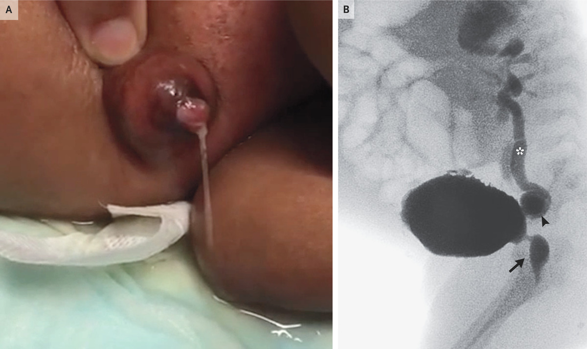
This patient’s fever resolved within 24 hours after the initiation of doxycycline therapy. The abnormalities in hematologic function and liver function resolved within several days. One month after the resolution of anaplasmosis, the patient underwent successful bilateral lung transplantation for his underlying interstitial lung disease.

Final Diagnosis

Human granulocytic anaplasmosis.

Omission of Radiotherapy in Early Breast Cancer


BCR ABL Genetic Test


What is a BCR-ABL genetic test?

A BCR-ABL genetic test looks for a genetic mutation (change) on a specific chromosome.

Chromosomes are the parts of your cells that contain your genes. Genes are parts of DNA passed down from your mother and father. They carry information that determines your unique traits, such as height and eye color.

People normally have 46 chromosomes, divided into 23 pairs, in each cell. One of each pair of chromosomes comes from your mother, and the other pair comes from your father.

BCR-ABL is a mutation that is formed by the combination of two genes, known as BCR and ABL. It’s sometimes called a fusion gene.

  • The BCR gene is normally on chromosome number 22.
  • The ABL gene is normally on chromosome number 9.
  • The BCR-ABL mutation happens when pieces of BCR and ABL genes break off and switch places.
  • The piece of chromosome 9 that breaks off includes part of the ABL gene. When this piece moves over to chromosome 22, part of the ABL gene attaches to the BCR gene. The merged gene is called the BCR-ABL fusion gene. 
  • The changed chromosome 22, which contains the BCR-ABL gene, is called the Philadelphia chromosome because that’s the city where researchers first discovered it.
  • The BCR-ABL gene is not the type of mutation that is inherited from your parents. It is a type of somatic mutation, which means you are not born with it. You get it later in life.

The BCR-ABL gene shows up in patients with certain types of leukemia, a cancer of the bone marrow and white blood cells. BCR-ABL is found in almost all patients with a type of leukemia called chronic myeloid leukemia (CML). Another name for CML is chronic myelogenous leukemia. Both names refer to the same disease.

The BCR-ABL gene is also found in some patients with a form of acute lymphoblastic leukemia (ALL) and rarely in patients with acute myelogenous leukemia (AML).

Certain cancer medicines are especially effective in treating leukemia patients with the BCR-ABL gene mutation. These medicines also have fewer side effects than other cancer treatments. The same medicines are not effective in treating different types of leukemia or other cancers.

Other names: BCR-ABL1, BCR-ABL1 fusion, Philadelphia chromosome

What is it used for?

A BCR-ABL test is most often used to diagnose or rule out chronic myeloid leukemia (CML) or a specific form of acute lymphoblastic leukemia (ALL) called Ph-positive ALL. Ph-positive means a Philadelphia chromosome was found. The test is not used to diagnose other types of leukemia.

The test may also be used to:

  • See if cancer treatment is effective.
  • See if a patient has developed a resistance to certain treatment. That means a treatment that used to be effective is no longer working.

Why do I need a BCR-ABL genetic test?

You may need a BCR-ABL test if you have symptoms of chronic myeloid leukemia (CML) or Ph-positive acute lymphoblastic leukemia (ALL). These include:

  • Fatigue
  • Fever
  • Weight loss
  • Night sweats (excessive sweating while sleeping)
  • Joint or bone pain

Some people with CML or Ph-positive ALL have no symptoms, or very mild symptoms, especially in the early stages of the disease. So your health care provider may order this test if a complete blood count or other blood test showed results that were not normal. You should also let your provider know if you have any symptoms that concern you. CML and Ph-positive ALL are easier to treat when found early.

You may also need this test if you are currently being treated for CML or Ph-positive ALL. The test can help your provider see if your treatment is working.

What happens during a BCR-ABL genetic test?

A BCR-ABL test is usually a blood test or a procedure called a bone marrow aspiration and biopsy.

If you are getting a blood test, a health care professional will take a blood sample from a vein in your arm, using a small needle. After the needle is inserted, a small amount of blood will be collected into a test tube or vial. You may feel a little sting when the needle goes in or out. This usually takes less than five minutes.

If you are getting a bone marrow aspiration and biopsy, your procedure may include the following steps:

  • You’ll lie down on your side or your stomach, depending on which bone will be used for testing. Most bone marrow tests are taken from the hip bone.
  • Your body will be covered with cloth, so that only the area around the testing site is showing.
  • The site will be cleaned with an antiseptic.
  • You will get an injection of a numbing solution. It may sting.
  • Once the area is numb, the health care provider will take the sample. You will need to lie very still during the tests.
    • For a bone marrow aspiration, which is usually performed first, the health care provider will insert a needle through the bone and pull out bone marrow fluid and cells. You may feel a sharp but brief pain when the needle is inserted.
    • For a bone marrow biopsy, the health care provider will use a special tool that twists into the bone to take out a sample of bone marrow tissue. You may feel some pressure on the site while the sample is being taken.
  • It takes about 10 minutes to perform both tests.
  • After the test, the health care provider will cover the site with a bandage.
  • Plan to have someone drive you home, since you may be given a sedative before the tests, which may make you drowsy.

Will I need to do anything to prepare for the test?

You usually don’t need any special preparations for a blood or bone marrow test.

Are there any risks to the test?

There is very little risk to having a blood test. You may have slight pain or bruising at the spot where the needle was put in, but most symptoms go away quickly.

After a bone marrow test, you may feel stiff or sore at the injection site. This usually goes away in a few days. Your health care provider may recommend or prescribe a pain reliever to help.

What do the results mean?

If your results show you have the BCR-ABL gene, as well as an abnormal amount of white blood cells, you will probably be diagnosed with chronic myeloid leukemia (CML) or Ph-positive, acute lymphoblastic leukemia (ALL).

If you are currently being treated for CML or Ph-positive ALL, your results may show:

  • The amount of BCR-ABL in your blood or bone marrow is increasing. This may mean your treatment is not working and/or you’ve become resistant to a certain treatment.
  • The amount of BCR-ABL in your blood or bone marrow is decreasing. This may mean your treatment is working.
  • The amount of BCR-ABL in your blood or bone marrow has not increased or decreased. This may mean your disease is stable.

If you have questions about your results, talk to your health care provider.

Learn more about laboratory tests, reference ranges, and understanding results.

Is there anything else I need to know about a BCR-ABL genetic test?

Treatments for chronic myeloid leukemia (CML) and Ph-positive, acute lymphoblastic leukemia (ALL) have been successful in patients with these forms of leukemia. It’s important to see your health care provider regularly to make sure your treatments continue to work. If you become resistant to treatment, your provider may recommend other types of cancer therapy.

EGFR and Lung Cancer


When tumor tissue is looked at under a microscope, physicians can see what type of cancer it is. But physicians can also look for changes in the DNA of the tumor that might be causing the tumor to grow. Sometimes these changes are called biomarkers or molecular markers.

Watch the brief video. URL https://youtu.be/CgcjlB1P7zI

One way to think about it is that our DNA is like an instruction manual. If there is a typo in the instruction manual, the cell receives wrong instructions and can grow into cancer. Biomarker testing looks for those typos, so physicians know if you are a candidate to receive a targeted therapy that directly addresses those typos. 

A mutation in the EGFR gene is one biomarker that physicians look for in non-small cell lung cancer. If you have non-small cell lung cancer, it is important to talk to your doctor about comprehensive biomarker testing to see if you have an EGFR mutation or another biomarker. The results of this testing influence your treatment options. To learn more about biomarker testing, visit Lung.org/biomarker-testing

What is EGFR-positive cancer?

EGFR (epidermal growth factor receptor) is a protein on cells that helps them grow. A mutation in the gene for EGFR can make it grow too much, which can cause cancer.  

There are different types of EGFR mutations. If you think of a mutation as a typo in the DNA, you can have missing or added words in the DNA, sometimes called deletions or insertions. You can also have places where the DNA is misspelled, which is called a point mutation. 

On your test results you might see that you have an EGFR 19 deletion or an EGFR L858R point mutation (the most common type of EGFR mutations that we know of). That information helps tell physicians exactly where the typo is in the DNA. EGFR 19 deletions and EGFR L858R point mutations are typically treated the same way. 

Amongst the EGFR mutations that are tested for in lung cancer, there are a few rare types that are treated differently than the more common EGFR mutations. The major example of this in lung cancer is EGFR exon 20 insertions. This is a type of EGFR mutation that doesn’t respond to the typical treatment for EGFR-positive lung cancer, which are called tyrosine kinase inhibitors, or TKIs. 

How do you know if you have EGFR-positive lung cancer?

In general, there are two ways to detect EGFR mutations. The best way is through comprehensive next-generation sequencing (NGS). This type of testing places tissue from a patient’s tumor (gathered from a biopsy) in a machine that looks for a large number of possible biomarkers at one time. There may be some situations where a patient can’t undergo the biopsy needed to perform NGS, and so liquid biopsy is recommended. A liquid biopsy can look for certain biomarkers in a patient’s blood. Talk to your doctor to make sure one of these tests was performed.  

Learn more about the different types of biomarker tests here.

Who is likely to have EGFR-positive lung cancer?

EGFR-positive lung cancer represents about 10-15% of lung cancer in the United States and generally appears in  adenocarcinoma subtype of non-small cell lung cancer. Patients with lung cancers with EGFR mutations tend to have minimal to no smoking history. But EGFR mutations can appear in lung cancer patients with different subtypes and smoking histories.  

What are the treatment options for someone with EGFR-positive cancer?

Knowing if you have EGFR-positive lung cancer has the most treatment implications for stage four patients. Most patients with stage four EGFR-positive lung cancer will likely be prescribed a pill called an EGFR targeted tyrosine kinase inhibitor (TKI) or EGFR-inhibitor to begin with. In addition to patients with stage 4 lung cancer, patients with stage IB-III lung cancer who have had their lung cancer removed through surgery are also eligible to receive an EGFR inhibitor after surgery.

The choice of a particular EGFR inhibitor depends on your oncologist’s preference, your specific type of lung cancer, and your treatment goals. Talk through the pros and cons of each option with your doctor. In general, based on studies showing that osimertinib is better tolerated and more effective than other EGFR inhibitors (erlotinib, gefitinib), most patients will receive osimertinib as their first EGFR inhibitor.

EGFR inhibitors can often control the cancer for several months or even multiple years depending on the patient and the drug, but it will not cure the cancer. Ultimately the cancer learns away around these treatments, which is called “acquired resistance”. When this happens, it is recommended that your doctor repeat biomarker testing, either through a tissue or liquid biopsy to see if you have a new biomarker or mutation that will help your doctor know which treatment to suggest next. 

Below are the treatments available for EGFR-positive lung cancer patients. 

Tyrosine Kinase Inhibitors (TKIs)

  • Afatinib (approved for stage 4 lung cancer)
  • Dacomitinib (approved for stage 4 lung cancer)
  • Erlotinib (approved for stage 4 lung cancer)
  • Gefitinib (approved for stage 4 lung cancer)
  • Osimertinib (approved for stage 1-4 lung cancer)

Others treatment options for EGFR-positive lung cancer include:

  • Ramucirumab with Erlotinib. 

Ramucirumab is a medication given through an IV that works with the EGFR-inhibitor to control the cancer. Ramucirumab blocks proteins needed by cancer cells to form blood vessels to help support their growth and spread.

Exon 20 Insertions

  • Patients with EGFR exon 20 insertions may receive chemotherapy, immunotherapy or the targeted therapy drug Amivantamab-vmjw.

EGFR Inhibitors after Surgery

  • Some EGFR-positive patients whose lung cancer is caught early may be eligible for surgery to remove the tumor. 
  • To keep the cancer from coming back, some patients may be given chemotherapy followed-by osimertinib (an EGFR inhibitor pill). 

Work with your doctor to discuss your goals and options each time you have to make a treatment decision. The three big questions to ask are:

  1. What is the goal of this treatment?
  2. What are the potential side effects?
  3. What other options do I have?

Research is happening at a rapid pace and your doctor should be up to date on the recommendations for your specific type of lung cancer. If you don’t feel comfortable with the answers you are receiving, do not hesitate to seek out a second opinion.  

9 Common Questions About Cancer Biomarkers


Cancer biomarkers, as the name suggests, indicate the characteristics of cancer. (Gorodenkoff/Shutterstock)

Cancer biomarkers, as the name suggests, indicate the characteristics of cancer.

Gene-targeted therapy and immunotherapy can be regarded as the most cutting-edge methods in current cancer treatment, saving the lives of many cancer patients. An essential indicator in these new types of cancer treatment is cancer biomarker testing.

Here are answers to nine frequently asked questions about cancer markers.

1. What Is a Cancer Biomarker?

The full name of biomarker is biological marker; a cancer biomarker or cancer marker, as the name suggests, indicates the characteristics of cancer.

Modern medicine is moving toward precision medicine, which is also known as personalized medicine. Medical experts are not only concerned about the general tissue classification of cancer, but have also begun to pay attention to how the same type of cancer manifests in different patients. According to the National Cancer Institute, these characteristics reflect different changes in genes, proteins, etc., and these differences can affect the efficacy of cancer treatment. Therefore, doctors can develop targeted treatments based on these differences.

These genes, proteins, and other molecules that represent the characteristics of cancer in different patients are called cancer markers.

There are other names for cancer biomarker testing, such as tumor genetic testing, tumor subtyping, molecular profiling, etc.

2. What Is the Purpose of Cancer Biomarker Testing?

An important purpose of cancer biomarker testing is to seek targeted cancer treatments.

For example, there are mutated EGFR genes in the cancer cells of many lung cancer patients, and there are BCR-ABL genes in cancer cells of patients with a type of chronic leukemia; these two mutated genes are cancer markers for corresponding cancer patients, and they provide important survival signals for the cancer cells. Hence, drugs targeting these genes can be an effective treatment.

3. How Is Cancer Biomarker Testing Done?

In cancer biomarker testing, doctors first need to take a small sample of cancer tissue from the patient. For solid tumors, they can take a sample during surgery; if the patient does not require surgery, a sample is obtained through biopsy.

For example, for lung cancer biopsy, doctors generally use minimally invasive methods such as bronchoscopy and thoracoscopy. Experienced surgeons may use video-assisted thoracoscopic surgery (VATS), which involves making a small incision between the patient’s ribs, and inserting a tiny camera and surgical tools into the chest cavity to take tissue samples under video guidance.

However, if necessary, the doctor may also recommend a thoracotomy to obtain tissue samples.

For leukemia patients, a sample of cancer cells can be obtained through a blood draw (liquid biopsy).

Typically, cancer biomarker testing also requires healthy cells in the patient’s blood, saliva, or skin to compare against abnormal changes found in cancer cells.

After a patient’s tissue sample is taken and sent for testing, it can be screened for dozens or even hundreds of cancer markers. However, doctors may also choose to check for only a few common cancer markers. One example the National Cancer Institute gives is the case of Oncotype DX, which is a breast cancer marker test that measures the activity of 21 different genes to predict whether chemotherapy is likely to work for the patient.

Generally, the results of a tumor marker test are available within 72 hours, although some may take longer.

In addition to testing directly with cancer tissue samples, the U.S. Food and Drug Administration has also approved the use of two noninvasive liquid biopsy tests that look for cancer markers from cancer cells in blood or other fluids, but they have a higher rate of false negatives.

4. What Are Some Common Cancer Markers?

There are different types of cancer markers for different types of cancer. For example, the main cancer markers for lung cancer include EGFR, ALK, and KRAS. The main cancer markers for breast cancer include ER, PR, and HER2, and the main markers for ovarian cancer include CA125, CEA, LDH, β-hCG, etc.

5. Is Cancer Biomarker Testing the Same as Genetic Testing?

Most cancer biomarker tests require genetic testing, while some look for proteins or other substances. Most cancer biomarker tests check for one or more genes, and there are two tests that use whole-genome sequencing to look at the entire DNA sequence in cancer cells.

However, according to the National Cancer Institute, cancer biomarker testing is different from general genetic testing for cancer risk. Cancer susceptibility genes are present in healthy cells of the patient and can be passed on to offspring. Cancer biomarkers, on the other hand, denote mutated genes in cancer cells, and these mutated genes will not be passed on to offspring.

6. When Should Cancer Biomarker Testing Be Done?

Although cancer biomarkers reflect the characteristics of cancer, a biopsy is usually required before a diagnosis of cancer can be made. Some clinicians suggest that patients should consider cancer biomarker testing at this stage; if a cancer diagnosis is confirmed, a subsequent biopsy may not necessarily yield enough tissue samples, so it is better to make good use of the first biopsy or surgery.

7. Is One Biopsy Enough for Cancer Marker Testing?

For various reasons, multiple biopsies may be required during cancer biomarker testing. For example, an insufficient tissue sample from the first biopsy requires a second biopsy. There may also be new dominant genetic mutations in cancer recurrence, which also require repeat biopsies to check for new cancer markers.

8. What Does a Positive Cancer Biomarker Test Result Indicate?

If a cancer patient tests positive for a cancer marker, it may be considered lucky in a way, because it means that the patient can receive the corresponding targeted drug therapy. Before the test, patients can discuss with their doctors which cancer markers are worth checking. The scope of testing may not be limited to cancer markers targeted by existing approved drugs; new drugs and corresponding markers in clinical trials may also be considered.

9. What Does a Negative Cancer Biomarker Test Result Indicate?

A negative result of a cancer biomarker test only means that the corresponding targeted therapy is not suitable for use; the patient still has many other treatment options, such as immunotherapy.

9 Reasons to Use Apple Cider Vinegar Everyday


(Shutterstock*)

Apple cider vinegar has a multitude of benefits, many of which more and more people are beginning to realize. It’s great for your hair, cleaning, digestion, acid reflux and more. See why apple cider vinegar is the latest craze. Don’t miss the powerful benefits of this substance…

1. Sooth a Sore Throat

You can mix about 1/3 of a cup of apple cider vinegar with warm water and use it to gurgle with. Of the numerous natural sore throat remedies many resort to this one. You can also mix it with honey and hot water for more of a tea like drink.

2. As a Natural Home Cleaner & Detoxifier

Many store bought cleaners contain harmful chemicals and toxins. You can ditch them and use apple cider vinegar instead. This vinegar is a great alternative to conventional cleaning products.

3. Give You Shiny Hair

To see how apple cider vinegar can be used to improve your hair shine simply use it after you wash your hair with shampoo.

4. Improve Skin Health

Apple cider vinegar can be used to treat skin issues from eczema to dry skin and psoriasis. Simply rub some on the affected areas with a cotton ball.

5. Apple Cider Vinegar Can Help With Weight Loss

Studies have shown that adding apple cider vinegar to your diet can lower body fat, weight and waist circumference. You can simply add 2 teaspoons to 16 ounces of water each day.

6. Treatment for Acid Reflux & Heartburn

Acid reflux is not caused by too much acid. In many cases it’s not enough acid that is causing the problem. Apple cider vinegar can help balance out the missing acid in your system. Use a teaspoon followed by a glass of water.

7. Apple Cider Vinegar Freshens Your Mouth and Removes Stains From Teeth.

To use for bad breath, mix with water and swish. For teeth stains apply apple cider vinegar directly to the teeth and rub. Rinse with water.

8. Relieves Itching

All it takes is a small bit of apple cider vinegar on a cotton ball to relieve the itch of a mosquito bite. Just dab a little bit on the bite and voila!

9. Excellent for Your Lymphatic System

One of the powerful benefits of apple cider vinegar is its ability to reduce mucous and sinus congestion. Thus, it can help fight off sinus infections and cleanse the lymph system.

Mastering the Art of Letting Go


Putting down our ideas of how things should be—including ourselves—can free of us a certain kind of suffering

Mastering the Art of Letting Go

One of the keys to living a life of calm and purpose is letting go.

If you’d like a more peaceful life, it’s powerful to look at what disturbs that peace and practice letting go of whatever you’re holding onto that’s causing you anxiety and frustration.

If you’d like a life of purposeful focus, it’s powerful to examine what’s standing in the way of that … and let go of whatever is blocking you.

Letting go can seem quite simple, but it isn’t necessarily easy. We have attachments that we cling to tightly, and we don’t want to let them go.

In this article, I’ll share the deeper part of the practice of letting go. Then, I’ll talk about how you might practice.

The Heart of Letting Go

Why do we cling to something that creates resistance in us to our purposeful action or disturbs our calm?

It’s usually because of some kind of idea, notion, or narrative we have in our minds. Let’s look at some examples:

  • We often think it’s something outside of us—that person over there did something that upsets, frustrates, or annoys me. But the other person isn’t the real cause—they’re just doing something. The real cause is that we have the idea that they shouldn’t be the way they are.
  • Sometimes we think we’re the problem—we shouldn’t be so lazy, or undisciplined, or something like that. We blame ourselves, feel bad about ourselves, and then try not to think about it. But what if the cause of our feeling bad is that we don’t accept ourselves as we are?
  • We might think that the problem is with the task or activity—we’re resisting because we don’t like that task. But what if the cause of resistance is that we think the activity should feel some other way than it does?

You can see in these examples that I’m pointing to an idea that things should be different than they are. People will resist this because they want things to be different than they are. They want change. And that’s understandable, we want to change what we don’t like. But what if we accepted what things are and then created change from a different place—from wanting to create, play, or explore?

How to Let Go of Notions

All of this stems from having an idea of how things should be that’s different than how they are. There may be nothing wrong with this idea—but it’s just an idea. And to the extent that it’s causing difficulties, we can see how it would be helpful to let it go.

Imagine that you’re frustrated with yourself, someone else, or a situation you’re facing.  Imagine that this frustration or feeling stems from an idea that things should be different than they are.

Now imagine letting go of that idea. You’re just left with the experience of this moment, just as it is.

Notice how freeing that can be. It’s not about letting someone “off the hook” or letting go of accountability or commitment to change. It’s about freeing ourselves from the attachment to an idea that’s causing some kind of suffering (frustration, resistance, feeling bad).

We’re freeing ourselves by letting go of the idea we’re holding onto.

The key realization is that the idea is just an idea. It isn’t that it’s wrong or bad, but it’s a mental conception, rather than reality. We can use mental conceptions when they’re helpful, but let go of them if they aren’t.

Our idea of other people, of ourselves, of any situation is simply a notion. What if we freed ourselves by realizing that we’ve created this notion and we don’t need it right now?

It can simply evaporate if we let it. Our conception of how things should be can become cloud-like, with little influence on our movement through life.

Try it right now: Whatever you think you should be, whatever you think someone else is, is just a notion you’ve created. Can you let it go in this moment and see what you’re left with?

How to Practice

First, notice when there’s difficulty and feelings of frustration, resistance, self-blame, annoyance, or anxiety. Noticing these feelings is key to being able to play with them.

Second, without needing to judge how you’re feeling, could you simply be with it? For example, if you’re feeling frustration, could you just let yourself feel the frustration as a physical experience in your body (as opposed to getting caught up in the narrative of frustration)? Give yourself compassion if you can. But there’s nothing wrong with feeling what you’re feeling. Often it’s useful to simply let ourselves feel the emotion, rather than trying to fix it.

Third, if you’d like to free yourself, you can let go of the notion that’s causing the difficulty. It’s usually an idea of how you think things should be. What if you could just let it evaporate, and let yourself be free? Play around with it.

Fourth, you might just experience the moment free of the notion. Just pure experience. Is there something in this moment you can be curious about? Be grateful for? Can you feel the wonder of this moment?

Fifth, once you’re free, you can take action if any is needed. For example, you can take on the task that you’re resisting, once you’re free of the idea that the task should feel different. Or you can have a conversation with someone, once you’ve let go of your frustration with them. Being free doesn’t mean we don’t take action—we just do so from a different place.

Would you like to take on this freeing practice?