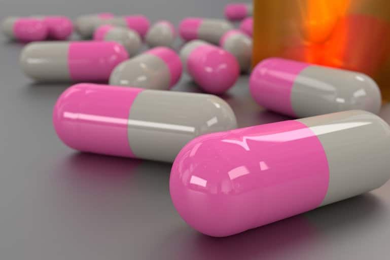
Black seed contains an abundance of nutrients that can help improve your health by balancing blood pressure, controlling blood sugar, and targeting obesity.
Short-term use of black seed oil may reduce chronic inflammation and is being tested in the treatment of COVID-19.
STORY AT-A-GLANCE
- Black seed oil may be an alternative herbal treatment for combating COVID-19
- The bioactive compounds have an antihistamine effect and anti-inflammatory properties that downregulate interferon regulatory factor 3 activation and promote autophagy
- Researchers hypothesize that several of the terpenes in the oil are similar in structure to chloroquine and may act as a zinc ionophore; evidence also suggests one compound may block ACE2 receptors, which the virus uses to release viral RNA into the cell
- Black seed oil has many traditional medicinal uses, including as an antidiabetic, antimicrobial, bronchodilator and antioxidant
- The Front Line doctors list black seed oil as an alternative therapeutic agent in the treatment of COVID-19. However, the oil is high in linoleic acid and so should not be used preventively against COVID
Black seed oil has been used for its therapeutic benefits for thousands of years. Since the pandemic began, researchers have been evaluating the effect it may have on COVID-19.[1] The seeds come from the Nigella sativa (N. sativa) plant that grows in Southern Europe, Southwest Asia and the Middle East.[2] Traditional medicine healers have used the seeds in different forms.
Black seed is coal-black with a dull surface and shaped like tiny Brazil nuts. The bioactive components include thymoquinone, α-hederin, alkaloids and omega-6 fatty acids.
Black seed oil has also been used for dermatological applications. For example, a review of the literature in the Journal of Dermatology & Dermatologic Surgery[3] found black seed oil could promote wound healing in farm animals and reduce the impact of vitiligo in lizards.
In one clinical study[4] in humans, lotion infused with 10% of N. sativa oil reduced acne vulgaris after two months with 67% of the patients fully satisfied and 28% partially satisfied with the results.
In the current research,[5] scientists believe the oil may have promising benefits in the treatment and prevention of COVID-19. However, while short-term use for treatment may be advantageous, it is wise to avoid long-term use for prevention.
Black Seed Oil May Be a Promising Option for COVID-19
Historically, black seed oil has been used to help balance the inflammatory response in the body as well as reduce oxidative stress, inflammation and ischemia. Using recent computational findings, one research group found the active ingredients in N. sativa were “strongly suggestive of combating emerged COVID-19 pandemic.”[6]
The paper suggests that the active ingredients, especially thymoquinone, α-hederin, and nigellidine, may be an alternative herbal treatment in the combat against COVID-19. The paper reviews several pathways that the active ingredients in black seed oil may use to protect health.
For example, the researchers reviewed past studies that demonstrated the actions the active ingredients in black seed oil have on the immune system, which may help reduce the severity of COVID-19. The active compounds have a significant antihistamine effect in animal studies and thymoquinone downregulates interferon regulatory factor 3 activation, which plays a critical role in innate immune responses.[7]
Autophagy is a mechanism that clears out damaged cells in the body. Data has shown that COVID-19 suppresses autophagy and that pharmacological agents used to induce the process may therefore have an antiviral effect. One study[8] published in 2018 indicated thymoquinone promotes autophagy in the heart muscle.
The researchers suggest that further study should be undertaken to determine if thymoquinone has a similar effect with COVID-19. Severe COVID-19 is characterized by cytokine storms that may require intensive care.[9] Animal studies have demonstrated that the bioactive compound α-hederin has anti-inflammatory activity and can decrease histamine levels.
The researchers reported one study had found thymoquinone helped inhibit two enzymes that produce inflammatory leukotriene and prostaglandins. Researchers suggest that these actions may potentially make the active compounds in N. sativa useful in the treatment and prevention of SARS-CoV-2 viral infections.
Finally, in their review of comorbid conditions associated with COVID-19, the researchers found that N. sativa may have a positive effect against diabetes, high blood pressure, heart disease, autoimmune and auto inflammatory diseases and bacterial infections associated with COVID-19.
After a review of the evidence, the researchers suggest that further experiments with the active compounds found in black seed oil are required to determine if they have preventive potential or may provide a new treatment modality.[10]
Zinc Combinations Help Treat COVID-19
Research into medications that may influence COVID-19 have included antivirals. One such antiviral being studied is favipiravir. Data show the drug was approved and has been used for flu infections in Japan.[11] In late 2020, a study published in PNAS[12] revealed that when given in high doses to hamsters favipiravir had promising antiviral activity against SARS-CoV-2.
However, a computer-model molecular docking study published in Biological and Medicinal Chemistry[13] revealed that the bioactive compounds nigellidine and α-hederin found in black seed oil were able to inhibit SARS-CoV-2 with a greater potential than favipiravir, chloroquine and hydroxychloroquine.
Evidence from a second paper that reviewed the biological effects of the active compounds in black seed oil suggested that thymoquinone may block ACE2 receptors,[14] which is where the SARS-CoV-2 binds to the cell and releases the viral RNA into the cytoplasm.
Contrary to what the PNAS hamster study found, these researchers hypothesized that chloroquine, and potentially the derivative hydroxychloroquine, may also interfere with the virus’s ability to bind with the ACE2 receptors.
This is one pathway the researchers suggest that black seed oil may use in the treatment of COVID-19. Another pathway is as a zinc ionophore. The body uses zinc in several pathways to support the immune system, including:[15]
- Proliferation and activation of natural killer cells, macrophages, neutrophils, and T and B cells
- Mediating protection against reactive oxygen species produced during an inflammatory response
- Stopping recombinant RNA dependent RNA polymerase activity needed to replicate SARS-CoV-2
- Inhibiting replicase processing
Therefore, moving zinc into the cytoplasm is crucial to help prevent the replication of the SARS-CoV-2 virus and thus effectively stop infected cells from infecting other cells.
Oral supplementation of zinc alone is not sufficiently effective since zinc cannot move easily across the cell wall. It needs another compound to provide transportation. The second compound is called a zinc ionophore. The application of zinc with an ionophore has demonstrated improved outcomes in patients hospitalized with COVID.[16]
Research has identified several zinc ionophores, including chloroquine,[17] hydroxychloroquine,[18] quercetin and EGCG.[19] Scientists have suggested that several of the terpenes in black seed oil, such as nigellimine, are similar in structure to chloroquine. They hypothesize that this may mean they can function in a similar manner as a zinc ionophore.[20]
Thus, recent data have shown that the active ingredients found in black seed oil may have greater potential in the treatment of COVID-19 than antiviral drugs, may act as a zinc ionophore and may help block the ACE2 receptors the virus uses to infect cells.
Black Seed Oil Has Traditional Medicinal Uses
N. sativa is a popular and traditional medicinal and has a long history of use in several diseases and ailments.[21] Traditional medicine has used black seed oil as a diuretic, antidiarrheal, appetite stimulant, analgesic, lower blood pressure, liver tonic and skin disorder treatment.[22] However, research has also explored the pharmacological activity of black seed oil and found it has many properties, including:[23]
- Antidiabetic
- Anticancer
- Immunomodulator
- Analgesic
- Antimicrobial
- Anti-inflammatory
- Spasmolytic
- Bronchodilator
- Hepato-protective
- Renal protective
- Gastro-protective
- Antioxidant
The immune-modulating activities of black seed oil help to regulate the immune system and maybe another pathway in the treatment for COVID-19. In addition to the studies using black seed oil, researchers have also separated the bioactive ingredients and evaluated their potential benefit against SARS-CoV-2.
A data review[24] on thymoquinone was published in February 2021, in which the researchers reviewed the bioactivity of the compound found in past research. They wrote that thymoquinone increased the activity and number of natural killer cells, macrophages, lymphocytes and cytokines suppressors.
Additionally, they cite past research that showed the active ingredient had antiviral potential against other viruses, including human immunodeficiency virus, other coronaviruses, Epstein-Barr virus, cytomegalovirus and hepatitis C. In addition, they reviewed an Egyptian study in which thymoquinone demonstrated antiviral activity in a strain of SARS-CoV-2 isolated in patients and the inhibitory effect it has on the viral protease, which may reduce viral replication.
One study[25] from Saudi Arabia evaluated the effectiveness of black seed oil as a supplement in patients with mild COVID-19 who were between 18 and 65 years. The intervention group received 500 mg of soft gel capsules twice daily for 10 days in addition to their standard treatment.
Initial results were published on Clinical Trials.[26] The primary outcome measurement was the percentage of participants who showed clinical recovery within 14 days after treatment began. The team reported 62.1% of those receiving the black seed oil demonstrated recovery from mild COVID-19 while 36% of the control group recovered within 14 days.
Consider Oil for Short-Term Use to Reduce Health Risk
The Front Line COVID-19 Critical Care Alliance (FLCCC)[27] lists black seed oil as an alternative for prevention and treatment of COVID-19. They stress there is no “magic bullet” for COVID-19, yet:[28]
“… a number of therapeutic agents have shown great promise for both the prevention and treatment of this disease including Ivermectin, Vitamin D, quercetin, melatonin, fluvoxamine, corticosteroids, curcumin (turmeric), Nigella sativa and antiandrogen therapy.”
The team recommends taking N. sativa with honey as they both have antimicrobial, antiviral, immunomodulatory and anti-inflammatory effects with proven safety profiles. They list N. sativa and honey in the prevention protocol for children and adolescents, and as an alternative for first-line treatment in the early treatment protocol at home.
While short-term use of black seed oil may be efficacious in the treatment of COVID-19, long-term use for prevention may have other unwanted effects. One study[29] of the chemical composition of black seed oil shows that the majority of fatty acids are from linoleic acid, an omega-6 polyunsaturated fat or (PUFA).
As I have discussed in an interview with Tucker Goodrich,[30] linoleic acid (LA) is likely the leading contributing cause of virtually all chronic diseases we have encountered in the last century.
When LA is consumed in excessive amounts, it acts as a metabolic poison. Chances are you’re getting an excess amount of this dangerous fat from foods that you may even consider healthy. For example, olive oil and chicken, which are fed LA-rich grains, increase your levels of omega-6 fatty acids.
Many are aware that the omega-3 to omega-6 ratio is important and should be close to equal. However, it is not as simple as raising omega-3 fatty acids. You need to minimize omega-6. fats A compelling report in the journal Gastroenterology[31] has offered a logically sound explanation as to why some patients with COVID-19 develop life-threatening organ failure.
The data indicate that the mortality rates appear to be heavily influenced by the amount of unsaturated fats you eat, such as LA. To date, there have been no clinical studies on the treatment they believe may help reduce the rate of organ failure and ICU admissions, namely the administration of calcium and egg albumen. It appears these compounds can bind the unsaturated fats and reduce injury to vital organs.
Take Care to Lower Your Omega-6 Intake
Fortunately, you can analyze your food for LA from the convenience of your home. All you need to do is accurately enter your food intake into Cronometer — a free online nutrition tracker — and it will provide you with your total LA intake. The key is to carefully weigh your food with a digital kitchen scale so you can enter the weight of your food to the nearest gram.
Cronometer.com is free to use as a desktop version. If you feel the need to use your cellphone (which is not recommended), then you will need to purchase a subscription. Personally, I have used the desktop version exclusively for the last five years as it has greater functionality and allows me to avoid electromagnetic fields from my phone.
Ideally, it is best to enter your food before you eat it. The reason for this is quite simple: It’s impossible to delete the food once you have already eaten it, but you can easily delete it from your menu if you find something that pushes you over the ideal limit.
Once you’ve entered the food for the day, go to the “Lipid” section on the lower left side. To find out how much LA is in your diet for that day, you merely need to see how many grams of omega-6 are present since roughly 90% of the omega-6 you eat is LA.
Thus, you can see that chronic use of black seed oil for prevention against COVID-19 will likely increase your risk of other chronic diseases that increase your risk of severe disease. If you are interested in using black seed oil, consider the extracts of the bioactive ingredients and not the whole oil.
If you are seeking an alternative for prevention and treatment, consider a combination of quercetin and zinc. Quercetin also has antiviral properties[32] and is a zinc ionophore.[33] While safe to take for about two weeks when you’re ill, it is important you are careful with zinc supplements as you may offset your zinc/copper balance and negatively impact your immune system.