Summary: Deep learning technology can accurately reflect a person’s risk of cognitive decline and Alzheimer’s disease based on brain age.
The human brain holds many clues about a person’s long-term health — in fact, research shows that a person’s brain age is a more useful and accurate predictor of health risks and future disease than their birthdate.
Now, a new artificial intelligence (AI) model that analyzes magnetic resonance imaging (MRI) brain scans developed by USC researchers could be used to accurately capture cognitive decline linked to neurodegenerative diseases like Alzheimer’s much earlier than previous methods.
Brain aging is considered a reliable biomarker for neurodegenerative disease risk. Such risk increases when a person’s brain exhibits features that appear “older” than expected for someone of that person’s age. By tapping into the deep learning capability of the team’s novel AI model to analyze the scans, the researchers can detect subtle brain anatomy markers that are otherwise very difficult to detect and that correlate with cognitive decline.
Their findings, published on Tuesday, January 2, in the journal Proceedings of the National Academy of Sciences, offer an unprecedented glimpse into human cognition.
“Our study harnesses the power of deep learning to identify areas of the brain that are aging in ways that reflect a cognitive decline that may lead to Alzheimer’s,” said Andrei Irimia, assistant professor of gerontology, biomedical engineering, quantitative & computational biology and neuroscience at the USC Leonard Davis School of Gerontology and corresponding author of the study.
“People age at different rates, and so do tissue types in the body. We know this colloquially when we say, ‘So-and-so is forty, but looks thirty. The same idea applies to the brain. The brain of a forty-year-old may look as ‘young’ as the brain of a thirty-year-old, or it may look as ‘old’ as that of a sixty-year-old.”
A more accurate alternative to existing methods
Irimia and his team collated the brain MRIs of 4,681 cognitively normal participants, some of whom went on to develop cognitive decline or Alzheimer’s disease later in life.
Using these data, they created an AI model called a neural network to predict participants’ ages from their brain MRIs. First, the researchers trained the network to produce detailed anatomic brain maps that reveal subject-specific patterns of aging. They then compared the perceived (biological) brain ages with the actual (chronological) ages of study participants. The greater the difference between the two, the worse the participants’ cognitive scores, which reflect Alzheimer’s risk
The results show that the team’s model can predict the true (chronological) ages of cognitively normal participants with an average absolute error of 2.3 years, which is about one year more accurate than an existing, award-winning model for brain age estimation that used a different neural network architecture.
“Interpretable AI can become a powerful tool for assessing the risk for Alzheimer’s and other neurocognitive diseases,” said Irimia, who also holds faculty positions with the USC Viterbi School of Engineering and USC Dornsife College of Letters, Arts and Sciences.
“The earlier we can identify people at high risk for Alzheimer’s disease, the earlier clinicians can intervene with treatment options, monitoring, and disease management. What makes AI especially powerful is its ability to pick up on subtle and complex features of aging that other methods cannot and that are key in identifying a person’s risk many years before they develop the condition.”
Brains age differently according to sex
The new model also reveals sex-specific differences in how aging varies across brain regions. Certain parts of the brain age faster in males than in females, and vice versa.
Males, who are at higher risk of motor impairment due to Parkinson’s disease, experience faster aging in the brain’s motor cortex, an area responsible for motor function. Findings also show that, among females, typical aging may be relatively slower in the right hemisphere of the brain.
An emerging field of study shows promise for personalized medicine
Applications of this work extend far beyond disease risk assessment. Irimia envisions a world in which the novel deep learning methods developed as part of the study are used to help people understand how fast they are aging in general.
“One of the most important applications of our work is its potential to pave the way for tailored interventions that address the unique aging patterns of every individual,” Irimia said.
Brain aging is considered a reliable biomarker for neurodegenerative disease risk.
“Many people would be interested in knowing their true rate of aging. The information could give us hints about different lifestyle changes or interventions that a person could adopt to improve their overall health and well-being. Our methods could be used to design patient-centered treatment plans and personalized maps of brain aging that may be of interest to people with different health needs and goals.”
About the study
Authors on the study include Phoebe Imms, Anar Amgalan, Nahian F. Chowdhury, Roy J. Massett, and Nikhil N. Chaudhari of the USC Leonard Davis School of Gerontology; and Chenzhong Yin, Mingxi Cheng, Xinghe Chen, Paul M. Thompson, and Paul Bogdan of the USC Viterbi School of Engineering; and colleagues from the Alzheimer’s Disease Neuroimaging Initiative.
A.I. is grateful to Kenneth H. Buetow, Caleb E. Finch, Margaret Gatz, and Mara Mather for useful discussions.
The Canadian Institutes of Health Research is providing funds to support ADNI clinical sites in Canada. Private sector contributions are facilitated by the Foundation for the NIH (http://www.fnih.org).
The ADNI grantee organization is the Northern California Institute for Research and Education, and the study is coordinated by the Alzheimer’s Therapeutic Research Institute at the University of Southern California. ADNI data are disseminated by the Laboratory for Neuro Imaging at the University of Southern California. Data for this study were provided, in part, by the Human Connectome Project, WUMinn Consortium (Principal Investigators: David Van Essen and Kamil Ugurbil; 1 U54 MH 091657) funded by the 16 NIH Institutes and Centers that support the NIH Blueprint for Neuroscience Research and by the McDonnell Center for Systems Neuroscience at Washington University. Research reported in this publication was also supported by the National Institute on Aging of the NIH under award U01 AG 052564.
CamCAN funding was provided by the UK Biotechnology and Biological Sciences Research Council (grant number BB/H008217/1), together with support from the UK Medical Research Council and the University of Cambridge, UK. This research has been conducted using the UK Biobank Resource, under application numbers 11559 and 47656.
The views, opinions, and/or findings contained in this article are those of the authors and should not be interpreted as representing the official views or policies, either expressed or implied by the NIH, DARPA, DoD, NSF, or any other entity acknowledged here.
The funding sources had no role in the study design; in the collection, analysis, and interpretation of data; in the writing of the report; and in the decision to submit the article for publication.
Abstract
Anatomically interpretable deep learning of brain age captures domain-specific cognitive impairment
The gap between chronological age (CA) and biological brain age, as estimated from magnetic resonance images (MRIs), reflects how individual patterns of neuroanatomic aging deviate from their typical trajectories. MRI-derived brain age (BA) estimates are often obtained using deep learning models that may perform relatively poorly on new data or that lack neuroanatomic interpretability.
This study introduces a convolutional neural network (CNN) to estimate BA after training on the MRIs of 4,681 cognitively normal (CN) participants and testing on 1,170 CN participants from an independent sample. BA estimation errors are notably lower than those of previous studies.
At both individual and cohort levels, the CNN provides detailed anatomic maps of brain aging patterns that reveal sex dimorphisms and neurocognitive trajectories in adults with mild cognitive impairment (MCI, N = 351) and Alzheimer’s disease (AD, N = 359). In individuals with MCI (54% of whom were diagnosed with dementia within 10.9 y from MRI acquisition), BA is significantly better than CA in capturing dementia symptom severity, functional disability, and executive function. Profiles of sex dimorphism and lateralization in brain aging also map onto patterns of neuroanatomic change that reflect cognitive decline.
Significant associations between BA and neurocognitive measures suggest that the proposed framework can map, systematically, the relationship between aging-related neuroanatomy changes in CN individuals and in participants with MCI or AD. Early identification of such neuroanatomy changes can help to screen individuals according to their AD risk.
Summary: A new study reveals community gardening helps lower stress and anxiety, and reduces cancer risks. Researchers found those who gardened had elevated fiber intake and increased physical activity.
Get more exercise. Eat right. Make new friends.
As we compile our lists of resolutions aimed at improving physical and mental health in 2023, new CU Boulder research suggests one addition could have a powerful impact: Gardening.
Funded by the American Cancer Society, the first-ever, randomized, controlled trial of community gardening found that those who started gardening ate more fiber and got more physical activity—two known ways to reduce risk of cancer and chronic diseases. They also saw their levels of stress and anxiety significantly decrease.
The findings were published Jan. 4 in the journal Lancet Planetary Health.
“These findings provide concrete evidence that community gardening could play an important role in preventing cancer, chronic diseases and mental health disorders,” said senior author Jill Litt, a professor in the Department of Environmental Studies at CU Boulder.
Filling the research gap
Litt has spent much of her career seeking to identify affordable, scalable and sustainable ways to reduce disease risk, especially among low-income communities.
Gardening seemed an ideal place to start.
“No matter where you go, people say there’s just something about gardening that makes them feel better,” said Litt, who is also a researcher with the Barcelona Institute for Global Health.
But solid science on its benefits is hard to come by. Without evidence, it’s hard to get support for new programs, she said.
Some small observational studies have found that people who garden tend to eat more fruits and vegetables and have a healthier weight. But it has been unclear whether healthier people just tend to garden, or gardening influences health.
Only three studies have applied the gold standard of scientific research, the randomized controlled trial, to the pastime. None have looked specifically at community gardening.
To fill the gap, Litt recruited 291 non-gardening adults, average age of 41, from the Denver area. More than a third were Hispanic and more than half came from low-income households.
After the last spring frost, half were assigned to the community gardening group and half to a control group that was asked to wait one year to start gardening.
The gardening group received a free community garden plot, some seeds and seedlings, and an introductory gardening course through the nonprofit Denver Urban Gardens program and a study partner.
Both groups took periodic surveys about their nutritional intake and mental health, underwent body measurements and wore activity monitors.
A fiber boost
By fall, those in the gardening group were eating, on average, 1.4 grams more fiber per day than the control group—an increase of about 7%.
The authors note that fiber exerts a profound effect on inflammatory and immune responses, influencing everything from how we metabolize food to how healthy our gut microbiome is to how susceptible we are to diabetes and certain cancers.
While doctors recommend about 25 to 38 grams of fiber per day, the average adult consumes less than 16 grams.
“An increase of one gram of fiber can have large, positive effects on health,” said co-author James Hebert, director of University of South Carolina’s cancer prevention and control program.
The gardening group also increased their physical activity levels by about 42 minutes per week. Public health agencies recommend at least 150 minutes of physical activity per week, a recommendation only a quarter of the U.S. population meets. With just two to three visits to the community garden weekly, participants met 28% of that requirement.
Study participants also saw their stress and anxiety levels decrease, with those who came into the study most stressed and anxious seeing the greatest reduction in mental health issues.
Only three studies have applied the gold standard of scientific research, the randomized controlled trial, to the pastime. None have looked specifically at community gardening.
The study also confirmed that even novice gardeners can reap measurable health benefits of the pastime in their first season. As they have more experience and enjoy greater yields, Litt suspects such benefits will increase.
Blooming relationships
The study results don’t surprise Linda Appel Lipsius, executive director of Denver Urban Gardens (DUG), a 43-year-old nonprofit that helps about 18,000 people each year grow their own food in community garden plots.
“It’s transformational, even life-saving, for so many people,” Lipsius said.
Many DUG participants live in areas where access to affordable fresh fruits and vegetables is otherwise extremely limited. Some are low-income immigrants now living in apartments—having a garden plot allows them to grow food from their home country and pass on traditional recipes to their family and neighbors.
The social connection is also huge.
“Even if you come to the garden looking to grow your food on your own in a quiet place, you start to look at your neighbor’s plot and share techniques and recipes, and over time relationships bloom,” said Litt, noting that while gardening alone is good for you, gardening in community may have additional benefits. “It’s not just about the fruits and vegetables. It’s also about being in a natural space outdoors together with others.”
Litt said she hopes the findings will encourage health professionals, policymakers and land planners to look to community gardens, and other spaces that encourage people to come together in nature, as a vital part of the public health system. The evidence is clear, she said.
Gardening works.
Researchers from the Colorado School of Public Health, Colorado State University and Michigan State University also contributed to this study.
Abstract
Effects of a community gardening intervention on diet, physical activity, and anthropometry outcomes in the USA (CAPS): an observer-blind, randomised controlled trial
Background
Unhealthy diet, physical inactivity, and social disconnection are important modifiable risk factors for non-communicable and other chronic diseases, which might be alleviated through nature-based community interventions. We tested whether a community gardening intervention could reduce these common health risks in an adult population that is diverse in terms of age, ethnicity, and socioeconomic status.
Methods
In this observer-blind, randomised, controlled trial, we recruited individuals who were on Denver Urban Garden waiting lists for community gardens in Denver and Aurora (CO, USA), aged 18 years or older, and had not gardened in the past 2 years. Participants were randomly assigned (1:1), using a randomised block design in block sizes of two, four, or six, to receive a community garden plot (intervention group) or remain on a waiting list and not garden (control group). Researchers were masked to group allocation. Primary outcomes were diet, physical activity, and anthropometry; secondary outcomes were perceived stress and anxiety. During spring (April to early June, before randomisation; timepoint 1 [T1]), autumn (late August to October; timepoint 2 [T2]), and winter (January to March, after the intervention; timepoint 3 [T3]), participants completed three diet recalls, 7-day accelerometry, surveys, and anthropometry. Analyses were done using the intention-to-treat principle (ie, including all participants randomly assigned to groups, and assessed as randomised). We used mixed models to test time-by-intervention hypotheses at an α level of 0·04, with T2 and T3 intervention effects at an α level of 0·005 (99·5% CI). Due to potential effects of the COVID-19 pandemic on outcomes, we excluded all participant data collected after Feb 1, 2020. This study is registered with ClinicalTrials.gov, NCT03089177, and data collection is now complete.
Findings
Between Jan 1, 2017, and June 15, 2019, 493 adults were screened and 291 completed baseline measures and were randomly assigned to the intervention (n=145) or control (n=146) groups. Mean age was 41·5 years (SD 13·5), 238 (82%) of 291 participants were female, 52 (18%) were male, 99 (34%) identified as Hispanic, and 191 (66%) identified as non-Hispanic. 237 (81%) completed measurements before the beginning of the COVID-19 pandemic. One (<1%) participant in the intervention group had an adverse allergic event in the garden. Significant time-by-intervention effects were observed for fibre intake (p=0·034), with mean between-group difference (intervention minus control) at T2 of 1·41 g per day (99·5% CI –2·09 to 4·92), and for moderate-to-vigorous physical activity (p=0·012), with mean between-group difference of 5·80 min per day (99·5% CI –4·44 to 16·05). We found no significant time-by-intervention interactions for combined fruit and vegetable intake, Healthy Eating Index (measured using Healthy Eating Index-2010), sedentary time, BMI, and waist circumference (all p>0·04). Difference score models showed greater reductions between T1 and T2 in perceived stress and anxiety among participants in the intervention group than among those in the control group.
Interpretation
Community gardening can provide a nature-based solution, accessible to a diverse population including new gardeners, to improve wellbeing and important behavioural risk factors for non-communicable and chronic diseases.
Summary: Study reveals almost one-third of chronic pain patients use cannabis to help manage their symptoms.
As more U.S. states legalize cannabis (also known as marijuana) for medical and recreational use, increasing numbers of people are experimenting with it for pain relief.
According to a new study published in JAMA Network Open, almost a third of patients with chronic pain reported using cannabis to manage it.
More than half of the 1,724 adults surveyed reported that using cannabis led them to decrease the use of pain medications, including prescription opioids and over-the-counter analgesics.
Cannabis also effected the use of other non-drug related pain relief methods to various degrees: some people indicated that cannabis led them to turn less often to techniques that many clinical guidelines recommend as first-line therapies such as physical therapy and cognitive behavioral therapy, while others with chronic pain increased their use of such treatments.
More than half of the 1,724 adults surveyed reported that using cannabis led them to decrease the use of pain medications, including prescription opioids and over-the-counter analgesics.
“The fact that patients report substituting cannabis for pain medications so much underscores the need for research on the benefits and risk of using cannabis for chronic pain,” said Mark Bicket, M.D., Ph.D., Assistant Professor in the Department of Anesthesiology and Co-Director of the Michigan Opioid Prescribing Engagement Network.
Abstract
Use of cannabis and other pain treatments among adults with chronic pain in US states with medical cannabis programs
Introduction
Most states have enacted laws allowing individuals to treat chronic pain with cannabis. Evidence is mixed about whether medical cannabis serves as a substitute for prescription opioids or other pain treatments. Accurate estimates of cannabis use or its substitution in place of pain treatments among adults with chronic noncancer pain are, to our knowledge, not available.
Methods
In this cross-sectional study, we surveyed a representative sample of adults aged 18 years or older with chronic pain who lived in the 36 states (and Washington, DC) with active medical cannabis programs in March to April 2022. We fielded the survey using the National Opinion Research Center (NORC) AmeriSpeak panel. This probability-based panel includes about 54 000 members and is sourced from a sample covering 96% of US households with a recruitment rate of 34%. We defined chronic noncancer pain using the National Health Interview Survey (NHIS) criterion of pain unrelated to cancer on most days or every day in the past 6 months. The survey was conducted from March 3, 2022, to April 11, 2022. A screener survey consented and identified people with chronic pain (response rate: 75.4%), who were invited to complete the full survey.
We assessed self-reported use (ever, past 12 months, past 30 days) of medical cannabis, pharmacologic treatments (prescription opioids, prescription nonopioid analgesics, and over-the-counter analgesics), common nonpharmacologic treatments (physical therapy, meditation, cognitive behavioral therapy), and substitution of cannabis in place of these treatments for chronic pain. All analyses incorporated survey sampling weights to generate estimates representative of the 36 included states and Washington, D.C. The investigation was approved by the Johns Hopkins Bloomberg School of Public Health institutional review board and followed the STROBE reporting guideline for cross-sectional studies. Statistical analysis was performed using Stata statistical software version 15 (StataCorp).
Results
Of the 1724 individuals identified as having chronic pain, 1661 (96.3%) completed the full survey (948 [57.1%] female; mean [SD] age, 52.3 [16.9] years); 31.0% (95% CI, 28.2%-34.1%) of adults with chronic pain reported having ever used cannabis to manage their pain; 25.9% (95% CI, 23.2%-28.8%) reported using cannabis to manage their chronic pain in the past 12 months, and 23.2% (95% CI, 20.6%-26.0%) reported using cannabis in the past 30 days. Most people who reported using cannabis to manage chronic pain also reported having used either at least 1 other pharmacologic (94.7%; 95% CI, 91.3%-96.8%) or nonpharmacologic pain treatment (70.6%; 95% CI, 64.8%-75.7%).
More than half of adults who used cannabis to manage their chronic pain reported that use of cannabis led them to decrease use of prescription opioid, prescription nonopioid, and over-the-counter pain medications, and less than 1% reported that use of cannabis increased their use of these medications. Fewer than half of respondents reported that cannabis use changed their use of nonpharmacologic pain treatments. Among adults with chronic pain in this study, 38.7% reported that their used of cannabis led to decreased use of physical therapy (5.9% reported it led to increased use), 19.1% reported it led to decreased use of meditation (23.7% reported it led to increased use), and 26.0% reported it led to decreased used of cognitive behavioral therapy (17.1% reported it led to increased use).
Discussion
Among adults with chronic pain in states with medical cannabis laws, 3 in 10 persons reported using cannabis to manage their pain. Most persons who used cannabis as a treatment for chronic pain reported substituting cannabis in place of other pain medications including prescription opioids. The high degree of substitution of cannabis with both opioid and nonopioid treatment emphasizes the importance of research to clarify the effectiveness and potential adverse consequences of cannabis for chronic pain. Our results suggest that state cannabis laws have enabled access to cannabis as an analgesic treatment despite knowledge gaps in use as a medical treatment for pain. Limitations include the possibility of sampling and self-reporting biases, although NORC AmeriSpeak uses best-practice probability-based recruitment, and changes in pain treatment from other factors (eg, forced opioid tapering).
Adjuvant oxaliplatin-containing chemotherapy improves survival in patients with colorectal cancer (CRC).1 Chemotherapy-induced peripheral neuropathy (CIPN) from oxaliplatin can be debilitating and is a reason for early discontinuation or dose reduction of adjuvant treatment. Acute neuropathy symptoms occur in 89% of patients, who experience sensory disturbances or pain in the extremities.2 Chronic CIPN is cumulative to oxaliplatin dose and has long-term functional implications, including impaired mobility and risk of falls.3 Symptoms can worsen even after discontinuation of treatment, a phenomenon known as coasting.2
There are no effective preventive agents and few evidence-based treatment options for CIPN, and therefore the mainstay of management is to limit exposure to oxaliplatin at the risk of attenuating treatment efficacy and affecting survival outcomes.4 Multiple risk factors for CIPN have been proposed but are inconsistently defined and reported. Research into the clinical and genetic factors predisposing to CIPN has been performed to try to identify patients at risk to allow individualization of treatment and balance toxicity and efficacy.5–7 Comorbidities such as the metabolic syndrome and diabetes can also cause peripheral neuropathy and are thought to contribute to an individual’s risk.8
CIPN is an important functional concern for patients, with those affected experiencing deficits in mobility and balance with associated risk of falls and disability.9,10 Effect of CIPN on quality of life (QoL) and distress has been reported in multiple CRC populations worldwide, with long-term worsening of patient-reported QoL and function in those with ongoing sensory and motor symptoms.11,12
Recent evidence from a multicenter randomized trial suggests that 3 versus 6 months of oxaliplatin-containing adjuvant chemotherapy provides clinically equivalent benefit, with reduction in CIPN and other toxicities.13 Our recent systematic review and meta-analysis (9,853 patients from 27 studies) estimated that approximately one-half of patients treated with adjuvant oxaliplatin for CRC have CIPN at 1 year, and one-quarter of patients have CIPN at 3 years following chemotherapy. Most patients included in the meta-analysis were planned to receive 6 months of oxaliplatin.14 Analysis of patients who received 3 months of adjuvant oxaliplatin identified considerable and pervasive CIPN.15
The aim of this study was to assess patient-reported CIPN and its associations in real-world CRC survivors, and to review the impact of neurotoxicity on dose delivery of oxaliplatin at a major tertiary cancer center, allowing for comparison with clinical trial populations.
Discussion
This study describes CIPN symptoms in a real-world cohort of CRC survivors and examines the precipitating factors and outcomes of CIPN. Prevalence of CIPN symptoms was 57% in our population of CRC survivors. CIPN was strongly associated with the use of adjuvant oxaliplatin, with 74% of patients reporting neuropathy following adjuvant therapy containing oxaliplatin. A higher proportion of patients required dose modifications than reported in clinical trials, with neuropathy as the most common recorded reason in 61%. Survivors with any numbness or tingling had lower QoL scores in the physical, emotional, and functional domains. Numbness was associated with cardiovascular comorbidities, and severe numbness was associated with the presence of pain.
Delivery of oxaliplatin in this real-world cohort was substantially less than reported in clinical trials, with a mean of 4 cycles of oxaliplatin, and 80% of patients needing dose reduction or early cessation of therapy. A treatment completion rate exceeding 80% was reported in the phase III MOSAIC trial that showed a survival benefit for oxaliplatin containing adjuvant chemotherapy.24 The ACHIEVE trial reported treatment completion of 72%.25 The SCOT trial reported 83% treatment completion in patients assigned to 3 months of chemotherapy and 59% in patients assigned to receive 6 months. Of the patients who discontinued treatment early, neuropathy was the cited reason for 27%.26 In comparison, early cessation occurred in 54% of our cohort, with neuropathy recorded as the reason in 61% of patients. This discrepancy may be explained by the difference in populations, with real-world patients typically a less selected sample with more comorbidities than clinical trial participants.
Our results highlight the persistence of CIPN symptoms long after completion of therapy, with 40% of our real-world respondents reporting symptoms >36 months after completing chemotherapy. This was higher than the long-term follow-up of the initial MOSAIC trial cohort, which identified a CIPN rate of 18% with any CIPN at 3 years and 15% at 4 years.1 Our recent systematic review of clinical trials and cohort studies showed the rate of any grade CIPN after adjuvant oxaliplatin was 58% at 6 months after completion of chemotherapy, decreasing by 26% per year.14 In comparison, our real-world data cohort indicates a higher burden of CIPN in the oxaliplatin-treated population, with 72.6% of patients reporting neuropathy symptoms at a mean follow-up of 6 months.
Our findings showed that development of neuropathy was relatively agnostic to age, gender, and other comorbidities. Lifestyle factors and medical comorbidities have been examined as potential risk factors for CIPN. In a cross-sectional online survey of 986 cancer survivors with predominantly breast cancer (58.9%), severe CIPN was associated with older age, higher BMI, and comorbidities, in contrast to our findings in the CRC population.27 Lifestyle factors have been examined as a risk factor for peripheral neuropathy in a systematic review of cross-sectional and cohort studies, which supported an increased severity or incidence of oxaliplatin-induced neuropathy in patients with markers of obesity, although some studies used BMI and others used body surface area.8
Consistent with our results, other studies have found poorer health-related QoL in CRC survivors with higher CIPN scores compared with those with minimal symptoms.12,28
The strengths of this study are the high completion rates of PRO measures as well as clinical information, allowing a comprehensive dataset encompassing a large number of CRC survivors in routine (nonclinical trial) practice. The longitudinal subset of patients provides additional data on the persistence of symptoms over time. The use of PROs to evaluate neuropathy symptoms is another strength of our study. Differences in clinician-assessed and patient-reported CIPN have been examined in several cohort studies. The findings have suggested that patient-reported measures may be more sensitive at intermediate levels of neuropathy and at identifying functional impairment.19,29 The score used in our cohort was a single question asking about “numbness and tingling.” It is intended as a screening tool to prompt clinicians to focus on relevant patient concerns. The authors appreciate that there are several validated patient-reported questionnaires that comprehensively assess neuropathy, including impact on driving, walking, and erectile function,30,31 but these were not feasible to use during an initial visit to SCSC where patients are already completing several PRO measures. Although consensus is lacking on the best way to measure CIPN, research into an optimal screening method suggested that a brief PRO measure without the use of an objective test provided adequate screening.32
A limitation of this analysis is that prechemotherapy symptom data were not available, and therefore neuropathy symptoms were not necessarily attributable to chemotherapy. Neuropathy reported by those who did not receive oxaliplatin could have been related to concurrent medical conditions, such as diabetic neuropathy or degenerative spinal and joint conditions, which are more prevalent in older age groups.33,34 The single-item question has not been validated against other measures of neuropathy. Although hand-foot syndrome (HFS) from fluoropyrimidines may manifest as sensory changes, the natural history of HFS is rapid resolution following cessation of treatment.35 Given that our assessment occurred at a median of 6 months following chemotherapy, it is unlikely that HFS is implicated in the “numbness and tingling” reported. We observed that patients receiving fluoropyrimidines alone were older and had more documented comorbidities, which may discourage clinicians from using oxaliplatin-containing chemotherapy in this group and may explain this observation, given that preexisting neuropathy is a relative contraindication to oxaliplatin.
Another limitation is that longitudinal data were only available for a subset of patients (45% of the total cohort), which broadens the precision of the estimates. Selection bias is another potential limitation in our dataset, because individuals with more symptoms, better health literacy, or greater motivation to improve their health may be more likely to attend the SCSC and comply with symptom reporting, omitting those who are either too unwell or lack the language proficiency to complete the questionnaires. However, we estimate that approximately 90% of eligible patients with CRC treated at our center attend the SCSC.
The clinical implications of this study are that the true burden of real-world CIPN is greater than reported in clinical trials. The persistence of symptoms and the impact on QoL domains is also demonstrated, highlighting the need for more effective prevention and treatment strategies. This study raises the potential utility of screening for patient-reported neuropathy symptoms using a single question. Pending further validation, this simple measure may be adequate to identify patients who may benefit from interventions for CIPN or prompt a more comprehensive assessment. A clinical pathway for management of such patients has been described.36 SCSC patients with CIPN can access multidisciplinary care, including allied health, pharmacologic therapy, or specialist referral as needed; outcomes have been reported separately.37
The results indicate the challenges in delivering treatment proven to be beneficial in trials into the everyday clinical context. Combined with recent phase III results indicating that 3 months of adjuvant chemotherapy provides comparable benefit to 6 months, with substantially less neurotoxicity,13 and data indicating that early discontinuation of oxaliplatin does not affect disease-free or overall survival,38 clinicians and patients can be reassured that survival outcomes are not compromised if oxaliplatin needs to be discontinued due to toxicity.
Conclusions
CIPN is a common and often persistent problem for survivors of CRC, strongly associated with use of oxaliplatin. In our cohort of CRC survivors, patient-reported neuropathy was associated with the presence of pain and reduction in QoL. As the true impact of CIPN is underreported in clinical trials, data from this and other real-world cohorts have an essential role in quantifying the prevalence and impact of CIPN, to allow more comprehensive research into its evaluation, prevention, and treatment.
Long-distance plane travel is infamous for being inconvenient and uncomfortable. Due to the logistics of check-in, the stress of security lines, and hours being stuck in a confined space, many people find extended plane trips to be seriously taxing.
Jet lag frequently contributes to the physical burden of long flights. Jet lag refers to the misalignment of your body’s internal clock with the local time at your destination. This phenomenon often occurs when flying across three or more time zones.
Jet lag can throw off your sleep and cause other bothersome symptoms that persist for days or even weeks after a flight. Whether you’re traveling for business or pleasure, jet lag can negatively impact your trip.
For travelers, knowing about jet lag — including its symptoms, causes, and ways of reducing them — can make long-distance trips more pleasant and less disruptive to sleep and overall health.
Under normal circumstances, a person’s circadian rhythm aligns with daylight, promoting alertness during the day and sleep at night. This internal clock synchronizes with the 24-hour day to promote quality sleep as well as physical and mental health. A person’s geographic location affects their circadian rhythm since sunrise and sunset occur at different times in different locations.
Jet leg generally happens when a person travels east or west across three or more time zones. For example, if you fly from Los Angeles to New York and arrive at 8 p.m., your body might still operate as if it’s in L.A. at 5 p.m. This jet lag can cause you to stay up later than you’d like, sleep at odd hours, or feel more tired than usual, among other symptoms.
Symptoms of Jet Lag
The most common symptomsTrusted SourceMedline Plus MedlinePlus is an online health information resource for patients and their families and friends. medlineplus.gov of jet lag include:
Sleeping problems: It may be hard to fall asleep when you want to, or you may wake up earlier than planned. Jet lag can also cause sleep to be fragmented.
Daytime sleepiness: Jet lag frequently causes you to feel drowsy or tired during the day.
Impaired thinking: You may experience problems with attention or memory or simply feel like your thinking is slowed.
Hampered physical function: Your body may feel tired, and peak physical performance may be affected, which is especially notable for traveling athletes.
Emotional difficulties: Some people with jet lag feel irritable, and evidence indicates that jet lag can exacerbate mental health problemsTrusted SourceNational Library of Medicine, Biotech Information The National Center for Biotechnology Information advances science and health by providing access to biomedical and genomic information. pubmed.ncbi.nlm.nih.gov , such as mood disorders.
General malaise: Jet lag may make you feel malaiseTrusted SourceMSD Manuals First published in 1899 as a small reference book for physicians and pharmacists, the Manual grew in size and scope to become one of the most widely used comprehensive medical resources for professionals and consumers. msdmanuals.com , which is a general feeling of discomfort, illness, or uneasiness
Stomach problems: Jet lag can induce gastrointestinal problems like reduced appetite, nausea, or even constipation and irritable bowel syndromeTrusted SourceNational Library of Medicine, Biotech Information The National Center for Biotechnology Information advances science and health by providing access to biomedical and genomic information. pubmed.ncbi.nlm.nih.gov .
Sleep paralysis and seizures: In rare circumstances, jet lag may impact sleep architecture which may increase the risk of sleep paralysis and nighttime seizuresTrusted SourceNational Library of Medicine, Biotech Information The National Center for Biotechnology Information advances science and health by providing access to biomedical and genomic information. pubmed.ncbi.nlm.nih.gov .
These symptoms arise after long flights to different time zones because the disruption to your circadian rhythm impacts how and when your body produces hormonesTrusted SourceMSD Manuals First published in 1899 as a small reference book for physicians and pharmacists, the Manual grew in size and scope to become one of the most widely used comprehensive medical resources for professionals and consumers. msdmanuals.com that affect sleep and other bodily processes.
People with jet lag experience one or more of the symptoms listed above. Symptoms can begin immediately or set in a few days after arrival. Many people sleep well the first night after a flight only to encounter sleep problems in the following days.
Jet lag lasts anywhere from a few days to a few weeksTrusted SourceAmerican Academy of Sleep Medicine (AASM) AASM sets standards and promotes excellence in sleep medicine health care, education, and research. aasm.org . In general, symptoms persist for 1-1.5 days per time zone crossed, but the duration of symptoms varies depending on the person and their trip details.
Can Jet Lag Have Long-Term Consequences?
Jet lag is usually a short-term problem that goes away once the body’s circadian rhythm has adjusted to the local time. For people who frequently take long-distance flights, such as pilots, flight attendants, and business travelers, jet lag can become a chronic problemTrusted SourceNational Library of Medicine, Biotech Information The National Center for Biotechnology Information advances science and health by providing access to biomedical and genomic information. pubmed.ncbi.nlm.nih.gov .
A chronically out-of-sync circadian rhythm can create persistent sleep problems that may give rise to insomnia. A healthy internal clock is important for the overall health of the bodyTrusted SourceNational Institute of General Medical Sciences (NIGMS) Blog The NIGMS supports basic research that increases our understanding of biological processes and lays the foundation for advances in disease diagnosis, treatment, and prevention. biobeat.nigms.nih.gov , therefore chronic circadian rhythm disruption may raise the risk of disorders like diabetes and depressionTrusted SourceNational Library of Medicine, Biotech Information The National Center for Biotechnology Information advances science and health by providing access to biomedical and genomic information. pubmed.ncbi.nlm.nih.gov as well as some types of cancerTrusted SourceNational Library of Medicine, Biotech Information The National Center for Biotechnology Information advances science and health by providing access to biomedical and genomic information. pubmed.ncbi.nlm.nih.gov .
Causes of Jet Lag
Plane travel that crosses three or more time zones causes jet lag. Symptoms may be more pronounced as more time zones are crossed.
Most people find that jet lag is worse when traveling east than it is when traveling westTrusted SourceNational Library of Medicine, Biotech Information The National Center for Biotechnology Information advances science and health by providing access to biomedical and genomic information. pubmed.ncbi.nlm.nih.gov . Jet lag differs based on the direction of travel because it’s generally easier to delay your internal clock than advance it. Jet lag does not occur on north-south flights that do not cross multiple time zones.
Not everyone who takes a long-distance flight gets jet lag. Multiple factors influence the likelihood and severity of jet lag:
Trip details: The total distance, amount of layovers, time zones crossed, direction of travel, local daylight hours, length of time at the destination, and other specifics of a trip can affect jet lag.
Arrival time: When you arrive at your destination may affect your circadian rhythm. For eastward travel, some evidence indicates that jet lag is reduced with afternoon arrivalsTrusted SourceNational Library of Medicine, Biotech Information The National Center for Biotechnology Information advances science and health by providing access to biomedical and genomic information. pubmed.ncbi.nlm.nih.gov compared to those in the early morning.
Age: A person’s age may play a role in jet lag, although studies have found mixed results. People over 60 experience circadian changes that can make it harder for them to recover from jet lagTrusted SourceNational Library of Medicine, Biotech Information The National Center for Biotechnology Information advances science and health by providing access to biomedical and genomic information. pubmed.ncbi.nlm.nih.gov , but some research in pilots found jet lag to be worse in younger peopleTrusted SourceNational Library of Medicine, Biotech Information The National Center for Biotechnology Information advances science and health by providing access to biomedical and genomic information. pubmed.ncbi.nlm.nih.gov .
Sleep before travel: Poor sleep in the days leading up to a flight can increase a person’s propensity for jet lag after traveling.
Stress: Being stressed-out can keep the mind and body on-edge in ways that interfere with sleep and make it harder to cope with jet lag.
Use of alcohol and caffeine: Many people drink alcohol and coffee during flights, and these substances affect the brain in ways that can disrupt sleep.
Past history of jet lag: People who have previously had jet lag are prone to have it againTrusted SourceNational Library of Medicine, Biotech Information The National Center for Biotechnology Information advances science and health by providing access to biomedical and genomic information. pubmed.ncbi.nlm.nih.gov .
Individual variation: For reasons that aren’t fully understood, some people are more likely to experience circadian rhythm disruption with long-distance flights than others.
Because there are many factors involved, it is hard to know exactly who will develop jet lag, how severe it will be, and how long it will last. However, it is common for at least mild jet lag to occur when more than three time zones are crossed during flight.
How is Jet Lag Different From Travel Fatigue?
It’s normal to feel wiped out after you’ve had a long travel day. While this can be confused with jet lag, it’s often a result of travel fatigueTrusted SourceNational Library of Medicine, Biotech Information The National Center for Biotechnology Information advances science and health by providing access to biomedical and genomic information. pubmed.ncbi.nlm.nih.gov . Travel fatigue includes symptoms like tiredness and headaches that can arise because of the physical toils of travel.
Airplane cabins, which have cool, dry, low-pressure air, can cause dehydrationTrusted SourceNational Library of Medicine, Biotech Information The National Center for Biotechnology Information advances science and health by providing access to biomedical and genomic information. pubmed.ncbi.nlm.nih.gov and susceptibility to respiratory problemsTrusted SourceNational Library of Medicine, Biotech Information The National Center for Biotechnology Information advances science and health by providing access to biomedical and genomic information. pubmed.ncbi.nlm.nih.gov . Air pressure changes can lead to bloating, and long-term sitting can cause leg swelling. It’s often difficult to sleep upright in an airplane seat, especially with in-flight interruptions, so getting quality rest in transit can be challenging.
All of these factors contribute to feeling exhausted after a long flight; however, this is distinct from jet lag.
Unlike jet lag, travel fatigue does not involve circadian rhythm disruption. For that reason, while travel fatigue usually goes away after a good night’s sleep, jet lag can persist for days or weeks until a person’s internal clock becomes realigned.
It is possible to have both travel fatigue and jet lag after a long-haul flight, but jet lag is far more likely to cause lasting and extensive symptoms.
Jet lag can have ruinous effects on a vacation, business trip, or athletic competition. As a result, travelers of all kinds strive to minimize the effects of jet lag.
The key to preventing and reducing jet lag is quickly realigning your circadian rhythm to synchronize with the time zone of your destination. Until this is achieved, steps can be taken to manage symptoms.
For very short trips, you may be able to avoid jet lag by scheduled activities, including sleep, to keep your circadian rhythm aligned with your home time zoneTrusted SourceNational Library of Medicine, Biotech Information The National Center for Biotechnology Information advances science and health by providing access to biomedical and genomic information. pubmed.ncbi.nlm.nih.gov . In this way, you avoid circadian disruption both during the trip and after you’ve returned home.
For travel lasting more than a few days, minimizing jet lag requires acclimating to the day-night cycle at your destination. The following sections address methods of reorienting your circadian rhythm and practical tips for reducing jet lag.
Light Exposure
Light is the most powerful influence on circadian rhythm, and strategic light exposure may help adjust your internal clock to avoid or reduce jet lag.
The effect on circadian rhythm depends on the level and timing of light exposure. Sunlight has the highest level of illumination and the strongest circadian effects. Different types of artificial light can also influence circadian timing to a lesser degree.
Indiscriminate light exposure doesn’t resolve jet lag because the timing is critical. At certain times, light exposure can either advance or delay your internal clock.
Properly timed periods of both daylight and darkness can help sync your circadian rhythm with local time. When access to natural light is limited, light therapy lamps, also known as lightboxes, can deliver bright light exposure with greater circadian influence.
Melatonin and Sleep Aids
Melatonin is a hormone that the body produces that helps to both make you feel sleepy and govern your circadian rhythm. Melatonin is normally produced in the evening, a few hours before bedtime, but this schedule can get thrown off by jet lag.
There are both prescription medicationsTrusted SourceNational Library of Medicine, Biotech Information The National Center for Biotechnology Information advances science and health by providing access to biomedical and genomic information. pubmed.ncbi.nlm.nih.gov and dietary supplements that boost the body’s levels of melatonin, and some research suggests melatonin can reduce jet lag.
Other types of sleeping pills, including prescription and over-the-counter drugs and natural sleep aids, may help you fall asleep or stay asleep, but they do not work to change your circadian rhythm. In some cases, they may even mask an ongoing case of jet lag.
Sleep aids can have side effects, including a heightened risk of falls and accidents if they increase drowsiness. Before taking melatonin or any other sleep medication, it’s best to consult with a doctor, ideally prior to your trip, to discuss the benefits and risks pertaining to your specific situation.
Pre-Adjusting Your Internal Clock
Some methods of preventing jet lag are based on modifying your sleep schedule in the days leading up to your trip so that when you arrive at your destination there is less of a discrepancy between your circadian rhythm and the local time.
In addition to changing your bedtime, this approach often involves carefully timed melatonin and light exposure to proactively alter your circadian rhythm.
While this approach may be beneficial in some cases, it may not be practical depending on your daily schedule, and professional, family, and social obligations.
Creating a Plan for Overcoming Jet Lag
The optimal plan to avoid jet lag depends on many factors including the direction of your flight, the number of time zones crossed, how long you will remain at your destination, and your schedule and obligations during your trip.
Taking these factors into account, you can create a personalized plan to reduce jet lag. Light and melatonin together can help you realign your circadian rhythm, but without proper timing, they can exacerbate rather than reduce jet lag.
A doctor, travel nurse, or sleep specialist may be available to help you prepare a plan for managing jet lag. Several online resources and apps can help you generate tailored schedules to help reduce jet lag based on your trip detaails.
Practical Tips for Reducing Jet Lag
A number of practical tipsTrusted SourceCenters for Disease Control and Prevention (CDC) As the nation’s health protection agency, CDC saves lives and protects people from health threats. cdc.gov for before, during, and after your flight can help reduce sleep disruptions and travel fatigueTrusted SourceMedline Plus MedlinePlus is an online health information resource for patients and their families and friends. medlineplus.gov so that you make the most of your trip.
Before Traveling
Schedule the first days of your trip: Make sure to give yourself time to sleep and follow your plan for light exposure. Build buffers into your schedule just in case you feel sluggish, and if possible, try to arrive days in advance of an important meeting or event so that you have time to acclimate.
Minimize travel stress: Don’t wait until the last minute to pack or leave for the airport. Being in a rush can heighten stress and make your travels more difficult.
Get quality sleep: Focus on getting quality rest for at least a few nights before your trip so that you’re not already sleep-deprived at the beginning of the trip.
During Flight
Stay hydrated: Drink water to replenish fluids and counteract the dehydration that can occur in-flight.
Limit alcohol and caffeine: Reduce alcohol and caffeine intake on-board or skip them entirely.
Eat smart: Reduce the risk of digestive problems by eating healthy and light. Opt for fruits and vegetables over heavy, calorie-rich, fatty snacks.
Stand up and move: Blood clots and stiffness can occur if you are seated for too long. Walking, standing, and gently stretching a few times during the flight may reduce these risks.
After Arrival
Exercise: Find time for a walk or other light physical activity. Exercising outside to receive appropriately timed daylight exposure will help recalibrate your circadian rhythm.
Limit alcohol, caffeine, and heavy meals: Avoid excessive caffeine, alcohol, or heavy and calorie-rich meals.
Nap with caution: Avoid the temptation to take an extra long nap. Try to keep naps less than 30 minutes and only nap eight or more hours before your planned bedtime.
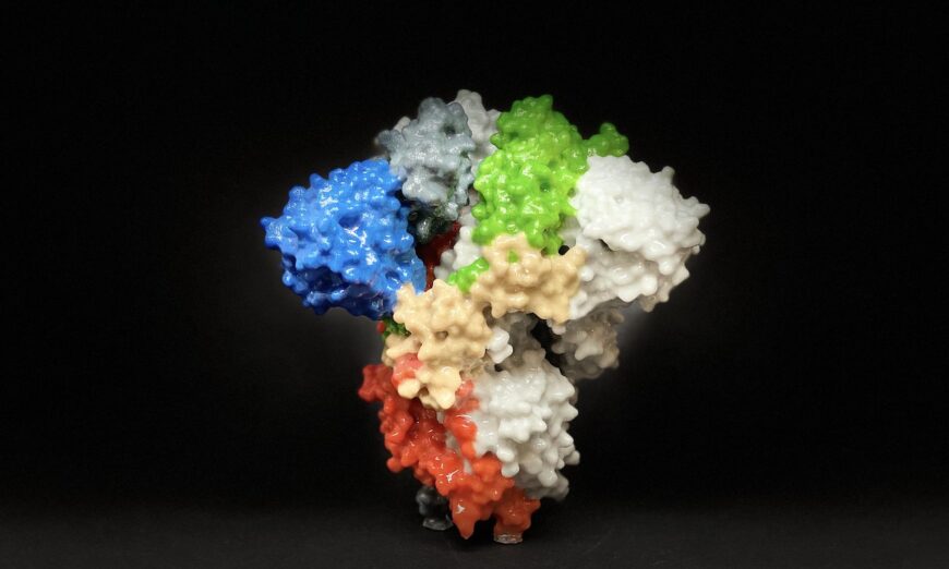
3D print of a spike protein on the surface of SARS-CoV-2. Spike proteins cover the surface of SARS-CoV-2 and enable the virus to enter and infect human cells. (NIH)
0:004:28
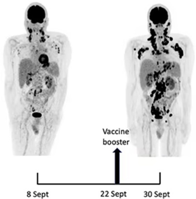
People who suffered from myocarditis after receiving an mRNA-based COVID-19 vaccine were found to have persistently higher levels of circulating spike protein compared to those who also received an mRNA-based COVID-19 vaccine but did not develop myocarditis, according to findings from a new study.
The study, published Jan. 4 in the Journal of the American Heart Association, sought to understand better the immune profiles—also referred to as immunoprofiles—of people who suffered from myocarditis after receiving an mRNA-based vaccine.
From January 2021 through February 2022, researchers prospectively obtained blood samples from 61 adolescents and young adults, all of whom had been vaccinated with either the Pfizer or Moderna COVID-19 mRNA vaccine.
Of the group, 16 had been hospitalized with myocarditis after COVID-19 vaccination, and had presented to Massachusetts General for Children or Boston Children’s Hospital with chest pain. The other 45 people were used as “healthy, asymptomatic, age-matched” control subjects in the study; they did not have symptoms of myocarditis after having been vaccinated. The researchers did not include any unvaccinated people as control subjects in the study.
“We performed extensive antibody profiling, including tests for SARS-CoV-2–specific humoral responses and assessment for autoantibodies or antibodies against the human-relevant virome, SARS-CoV-2–specific T-cell analysis, and cytokine and SARS-CoV-2 antigen profiling,” the authors stated.
Findings
The researchers flagged what they called a “notable finding,” which was that “markedly elevated levels of full-length spike protein (33.9±22.4 pg/mL), unbound by antibodies, were detected in the plasma of individuals with postvaccine myocarditis, whereas no free spike was detected in asymptomatic vaccinated control subjects (unpaired t test; P<0.0001).”
A p-value of less than 0.05 would be considered statistically significant.
Otherwise, they found that in the myocarditis group, “adaptive immunity and T-cell responses were essentially indistinguishable from those of asymptomatic age-matched vaccinated control subjects,” although the myocarditis group saw “a modest increase in cytokine production.”
“The postvaccine myocarditis immunoprofile is distinct, however, from acute SARS-CoV-2 infection and the delayed postinflammatory illness MIS-C,” the researchers noted.
They said that in the myocarditis cohort, they found “no evidence of autoantibody production, concomitant viral infections, or excessive antibody responses to the anti–SARS-CoV-2 mRNA vaccines.”
The authors said that their study “does not distinguish” whether the high levels of circulating spike protein “is the cause or consequence” of myocarditis in vaccinated patients. They also noted that not all patients with myocarditis had detectable levels of circulating spike protein.
The findings “suggest that administration of anti-spike antibodies, if spike antigenemia is detected, could potentially prevent or reverse postvaccine myocarditis,” reads the paper. Free spike protein circulating in the bloodstream that are unbound by anti-spike antibodies is called “spike antigenemia.”
Authors wrote that their findings could help further understand the complications associated with mRNA vaccines and guide future research regarding COVID-19 vaccine design and dose.
“These results do not alter the risk-benefit ratio favoring vaccination against COVID-19 to prevent severe clinical outcomes,” authors wrote.
They acknowledged that a limitation of the study is the relatively small sample size. Furthermore, the people in the study were “not evenly balanced” between the Pfizer and Moderna vaccines, they wrote, noting: “all of our adolescent control cohort and the majority of our myocarditis cohort received the [Pfizer] vaccine (n=15).”
TCM practitioner advises ways to find the best one for you
Dr. Yu Ya-wen, a Taiwanese traditional Chinese medicine practitioner, provides different weight loss regimes for the five types of obesity, and offers the best solutions to beat obesity for different people. (Shutterstock)
Obesity has become a ubiquitous condition of our times. It affects people’s appearance, self-esteem, and may pose a threat to physical health. What are the causative factors of obesity? Is there a one size fits all solution for the reduction of the condition? Dr. Yu Ya-wen, a Taiwanese traditional Chinese medicine (TCM) practitioner, provides different weight loss regimens for five common types of obesity, and introduces two simple, easy-to-do CliffsNotes-style versions for weight loss.”
Obesity can cause harm to multiple physiological functions of the human body, which significantly increases the risk of metabolism-related, cardiovascular and respiratory diseases, and certain cancers that increase the chance of mortality. Many studies have found traditional Chinese medicine to be effective in treating obesity in a variety of ways, such as regulating intestinal flora, increasing hormone levels, and regulating fat metabolism.
Western medicine believes that obesity is the accumulation of fat caused by imbalance between energy intake and energy usage due to a range of factors. Yu Ya-wen from Royal Jade Traditional Chinese Medicine Clinic in Taiwan said in an interview with the Health 1+1 program that Chinese medicine believes “fat people are associated with more phlegm.” TCM’s definition of “phlegm” includes the whole of all by-products from abnormalities, for example, high blood sugar, high blood lipids, high triglycerides, and so on. Due to the imbalance of the internal organs, these “wastes” cannot be excreted from the body, and continue to accumulate, causing cell degeneration, which may form tumors or subcutaneous or visceral fat.
Therefore, the concept of TCM treatment of obesity is to allow the internal organs to function normally and excrete waste (including fat) from the body through recuperation. The five viscera and six internal organs referred to in Chinese medicine are the general designations for the complete organs and systems of the body. The five viscera refer to the heart, liver, spleen, lung, and kidney; six internal organs refer to the gallbladder, stomach, large intestine, small intestine, triple burner (a body cavity consisting of three parts, containing all the internal organs), and bladder.
According to different physical characteristics, Chinese medicine divides obesity into five types, which correspond to different solutions. From the perspective of TCM, people have different constitutions. Constitution is a unique characteristic formed during the growth and development of a person, which is affected by congenital inheritance, and augmented by post-birth acquisition from natural and social environments.
Yu emphasized that acquired influences such as diet composition, lifestyle, environment, and other factors are the keys to obesity.
From the perspective of TCM, Yu analyzed the solutions corresponding to the five types of obesity. She also added that some people may not be easily classified into a single type but may indeed be a combination of several of them. So, if any one type is prevalent, the effect will be better because you can focus on improving it.
I. Spleen Deficiency and Dampness Type
The spleen is an important viscera that transports and transforms water inside the body. If the spleen is healthy, the body’s metabolism can process water in the body normally. If the spleen is damaged, it will affect metabolism and make water stay in the body, a symptom known as “dampness.” Modern medicine believes that the spleen is an organ monitoring immunity, while TCM refers to the spleen not just as an anatomical organ, but more on the concept of function. In addition to immune function, it is also part of the digestive system.
Pathological Features
Pathological features of spleen deficiency: Soft muscles, soft fat, frequent edema (under the eyes, limbs), prone to sweating herpes, eczema, skin allergies, excessive female secretions, discomfort during menstruation, pale complexion, and easy fatigue. The three meals are often not taken at regular times, and food is eaten cold.
Solution
Remove pathological products
Adding barley white rice, red beans, ginger, and astragalus can help eliminate edema in the body and benefit the spleen.
Increase metabolism
Muscle-building exercise can help metabolism and remove edema and fat. Yu said that the classic Chinese medical book “Huang Di Nei Jing” mentioned that “the spleen governs the muscles of the body.” When the spleen is not good, it is necessary to do additional exercise on the muscles. After strengthening the muscles, the spleen will also become healthier.
II. Stomach Heat, and Phlegm Stasis Type (Mainly in Men)
The stomach gradually becomes hot during the process of digesting food. Stomach heat itself is to help digest food. However, if stomach heat accumulates, one often feels the need to eat again even after having eaten leading to a tendency to overeat.
Pathological Features
Pathological features of stomach heat: a preference for strong food tastes, likes to eat meat, sweets, and cold foods. Needs to drink frequently, tends to become thirsty, and experiences digestive fluid reflux..
Solution
1. Remove pathological products Supplements such as hawthorn, lotus leaf, cassia seed, corn silk, and coptis can help dispel stomach heat.
2. Regulate visceral functions Using acupuncture to clear stomach heat can also suppress appetite and reduce calorie intake.
Chinese medicine has discovered that the human body has a “meridian” system. The theory of TCM believes that the meridian is the channel of energy inside the body. The internal organs are connected to the surface of the body through the meridian. Some points on the meridian that have special functions are called “acupoints.” Activating these acupoints will treat diseases of the corresponding viscera inside.
3. Increase metabolism Do more aerobic exercise to increase energy consumption.
III. Liver Depression and Qi Stagnation Type (Mainly in Women)
TCM believes that the liver is responsible for regulating the smooth flow of qi throughout the body and at the same time regulating emotions. If the liver function is out of balance, the movement of qi will be blocked, causing physical dysfunction, and mood swings such as depression and anger may also occur. Yu said that the liver, as mentioned in Chinese medicine, refers not only to the liver as a physiological organ, but also includes the autonomic nervous, endocrine, and emotional systems.
Chinese medicine believes that qi is the “energy” or “vitality” that constitutes life inside the body, and refers to the substances that replenish nutrients in the body as blood. Qi and blood are interdependent, flow throughout the whole body, nourish the organs and tissues, and maintain the vital activities of the body.
Pathological Features
Pathological features of liver depression: Endocrine disorders, frequent insomnia, dreaminess, nervousness, anxiety, often feels tired.
Solution
Clear pathological products Regulate hormones first, then use medicines that soothe the liver and regulate qi, such as roses, chrysanthemums, safflowers, and peach kernels.
Improve visceral function Acupuncture can be used to regulate the autonomic nervous system, or aromatherapy can relieve stress.
Increase metabolism Do exercises that soothe your nerves, such as Pilates and yoga.
IV. Deficiency of Qi and Blood Type
Pathological Features
Pathological features of qi deficiency: Weak body, cold hands and feet, poor complexion, unbalanced nutrition, easy fatigue, and failing memory.
Solution
The three meals should be taken at regular times and quantities. Eat more blood-enriching foods rich in iron, such as dark green vegetables. Do light exercise, such as brisk walking or cycling.
V. Kidney Yang Deficiency and Turbid Phlegm Type (Mainly Middle-Aged and Elderly People)
Chinese medicine believes that the kidneys are the most important part of the body, storing the body’s substances and inherent functions, and promoting physiological activities. Kidney yang promotes and warms the functions of various viscera and tissues in the body. Yu said that kidney yang is the provider of vitality for the body, and monitors metabolism.
Pathological Features
Pathological features of kidney yang deficiency: Endocrine disorders, abdominal obesity, fear of cold, backache, frequent urination.
Solution
Eat more foods that heal the kidneys, such as mutton, leeks, and black sesame. It is recommended to use moxibustion, baths, and saunas to strengthen metabolism. Excessive exercise is not advisable due to excessive depletion of vitality.
Moxibustion therapy is one part of acupuncture in TCM involving certain thermal routines. It is a treatment method to achieve health care by igniting moxa leaves and using moxa sticks, to heat the acupuncture points of the body.
Two Ways to Lose Weight
Yu emphasized that perseverance is the key to weight loss. She recommended two easy-to-implement CliffsNotes versions for weight loss approaches.
Eat the right food and in the right sequence: One-half of each meal should contain cooked vegetables of assorted colors, one-quarter high-quality protein (mainly lean and white meat), and one-quarter high-quality starch (rice, sweet potato, etc.). Keep the order of serving as vegetables or protein first, followed by the starch. Those with poor gastrointestinal tracts can eat protein first, which can prevent blood sugar from fluctuating too much and avoid fat accumulation.
Sleep at the right time: Nine to 10 p.m. as a bedtime routine can cultivate a good sleep mood and facilitate falling asleep by around 11 p.m. Sleep should occur no later than 12 a.m.
TCM believes that the twelve periods of a day (one period is equivalent to two hours) correspond to the twelve main meridians of the body. In each period, the blood energy on the corresponding meridian will be particularly prosperous, and the viscera governed by that meridian are also more active. This is the theory of the midnight to noon and ebb- and flow doctrine.
Yu said that the gallbladder meridian runs between 11 p.m. to 1 a.m., and the liver meridian runs from 11 p.m. to 3 a.m. During this period of deep sleep, the body can effectively detoxify. From the perspective of Western medicine, hormones such as growth hormone begin to secrete at 11 p.m., which can help burn fat.
Acupoints Massaging to Lose Weight
Yu said that you can use your fingers, fists, or a massage stick to stimulate the following acupoints.
Tianshu The position about the width of two fingers on both sides of the navel that can stimulate gastrointestinal peristalsis.
Guanyuan The position about the width of four fingers below the navel is a commonly used acupoint in gynecology, for treating problems such as female hormone imbalance.
Zhongwan The position about the width of five fingers above the navel that can suppress appetite.
Yu also suggested that every morning, try rubbing the abdomen around the navel in a clockwise direction, to stimulate the relevant acupoints at the same time.
Pre-diabetic patients develop insulin resistance due to the decreased sensitivity of cells to insulin, so the body has to secrete a large amount of insulin to suppress blood sugar levels. (Shutterstock)
Do you feel hungry before meals and drowsy after meals? If this happens all the time, you may have insulin resistance in your body, which is considered prediabetes. Many people have pre-diabetes even if their blood sugar tests are normal. Be aware if you have the symptoms mentioned here.
Why Is There Insulin Resistance When Blood Sugar Level Is Normal?
According to the Centers for Disease Control and Prevention, approximately one-third of American adults had prediabetes.
Pre-diabetic patients develop insulin resistance due to the decreased sensitivity of cells to insulin, so the body has to secrete a large amount of insulin to suppress blood sugar levels.
People with chronic insulin resistance are at high risk of developing diabetes later in life. Even if it does not develop into diabetes, when the insulin and blood sugar levels remain high for a long time, the continuous oxidative stress and inflammation in the body may cause various complications, such as hyperlipidemia, hypertension, cardiovascular diseases (e.g. myocardial infarction, angina pectoris, and stroke), hyperuricemia, sleep apnea, non-alcoholic fatty liver disease, polycystic ovary syndrome, dementia, and cancer.
However, what many people don’t realize is that blood test results may still show normal blood sugar levels when insulin resistance is already present in the body.
At present, prediabetes is mainly detected by blood tests:
Fasting blood sugar level: A normal fasting blood sugar level should be less than 100 mg/dL. A fasting blood sugar level between 100 mg/dL and 125 mg/dL is considered prediabetes;
Hemoglobin A1c (HbA1C) test: It reflects the average blood sugar index in the past 2 to 3 months. The normal value should be less than 5.6 percent. A level of 5.7 percent to 6.4 percent indicates prediabetes.
“There is a huge problem with such diagnostic criteria,” said Dr. Chun-Hsu Chen, director of the Dr. Chen Natural Health Center (DCNHC) in the United States. This is because diabetes means that both fasting and postprandial blood sugar levels are high, so the A1C level will also be relatively high. However, people with prediabetes are very likely to have significant blood sugar swings, but their average blood sugar levels are within the normal range. Relying solely on fasting blood sugar level or HbA1C can sometimes lead to misjudgment.
The HbA1C level reflects the average blood sugar level within three months, but it cannot reflect the changes in the usual blood sugar fluctuations. For example, A and B both had A1C levels of 5.7 percent. However, A’s fasting blood glucose and two-hour postprandial blood glucose are 95 mg/dL and 125 mg/dL respectively; while B’s fasting blood glucose and postprandial blood glucose are 90 mg/dL and 180 mg/dL. B clearly had blood sugar swings.
According to Dr. Chen, the difference between the two is that A is energetic during the day, sleeps well at night, and does not feel drowsy after eating; whereas B’s insulin will rise very high after a meal to suppress the blood sugar level, and the blood sugar level will drop significantly before the next meal. Also, B tends to be hungry and wants to have a snack before a meal. Fluctuations in blood sugar cause hunger before meals and drowsiness after meals.
Drowsiness After Meals and a Big Belly May Indicate Prediabetes
How do you know if you have insulin resistance? It can be detected through physiological responses, physical appearance, and blood tests.
1.) Hungry before meals and drowsy after meals
Physiological responses such as hunger before meals and drowsiness after meals are closely related to blood sugar swings. Dr. Chen pointed out that such responses are very common in modern people, but it does not mean that they are normal.
2.) A big belly and “dirty” neck
People with insulin resistance typically have more visceral and abdominal fat. It can be seen from the appearance that the belly is big and the waist is thick, which is also known as “central obesity”.
If the waist-to-hip ratio can be obtained by dividing the waist circumference by the hip circumference. If the ratio is >0.9 for men or >0.8 for women, it means that there is too much fat in the waist and abdominal area, and it indicates the presence of insulin resistance.
In addition, you can also observe whether you have more muscle or fat on your body. You should be alert if you are gaining weight, and the body fat meter measurement shows that your body fat has increased and your muscles have decreased.
The third sign to look out for in terms of physical appearance is acanthosis nigricans. The typical symptom includes black patches appearing in the crease at the back of the neck—hence it is called the “Dirty Neck”.
Acanthosis nigricans is formed because excess insulin stimulates the proliferation of skin cells, resulting in melanin deposition and hyperkeratosis of the skin. In addition to the neck, it may also appear on the inner thighs, underarms, and the front of the elbows. However, not all patients with insulin resistance will develop acanthosis nigricans.
3.) Blood sugar and insulin tests
Blood test results that show the following values indicate insulin resistance:
Fasting insulin level > 5mIU/L
Difference between preprandial and postprandial blood sugar levels > 40 mg/dL
Homeostasis Model Assessment of Insulin Resistance (HOMA-IR) ≥ 2
HOMA-IR is a formula that calculates the ratio of fasting blood glucose to fasting insulin. It indicates insulin resistance if the value is ≥2, but it is usually judged according to clinical symptoms.
According to Dr. Chen, blood sugar levels should be tested at four different times to detect whether there is insulin resistance: before meals (on an empty stomach), 30 minutes after meals, 1 hour after meals, and 2 hours after meals.
Under normal circumstances, the tests are taken 2 hours before meals and 2 hours after meals, which may not be able to objectively detect whether the patient has prediabetes. Assume that the patient’s preprandial and 2-hour postprandial blood sugar levels are 100 and 130, respectively. These numbers look good, but the patient’s blood sugar levels may actually rise to 150 and 180 half an hour and one hour after a meal, respectively.
Moreover, some people may not feel drowsy after meals or have a big belly, but they are insulin resistant. These people need to have their blood sugar and insulin levels tested.
Dr. Chen explained that most of these people suffer from overwork and stress. Although they have a normal body figure, or even look slim, they develop insulin resistance due to excessive secretion of adrenaline caused by chronic stress.
Studies have also found that stress, long hours of work, and sleep loss can all lead to insulin resistance and elevated blood sugar level.
A South Korean study tracked the work hours and insulin resistance in more than 25,000 healthy adult men. The results found that compared with those who worked 35 to 40 hours a week, those who worked 41 to 52 hours a week, and those who worked more than 52 hours a week had a 28 percent and 180 percent increased risk of developing insulin resistance, respectively.
Stress causes high blood sugar levels due to the body’s mechanism. When a person is in danger, the body will secrete adrenaline and break down glycogen to glucose, and at the same time secrete insulin to quickly transport glucose to muscle cells. Hence, when a person needs to flee in a dangerous situation, the above-mentioned series of reactions will allow the muscles to burst with strength and the person to run away quickly.
Sleep deprivation will also increase adrenaline. This is because the body needs to secrete adrenaline to support itself when there is not enough energy, and it will enter a state of adrenal fatigue after a certain period of time.
Therefore, people who are often overworked, work late, and/or are under high pressure always have excessive secretion of adrenal hormones and increased insulin. Over time, many of their cells will become less sensitive to insulin, that is, insulin resistance, which will eventually lead to diabetes. Dr. Chen described it as how misbehaving children become numb after being scolded by adults for a long time. “Our senses and cells adapt to many stimuli over time,” he said.
3 Ways to Reverse Insulin Resistance
It is possible for patients with prediabetes to reverse insulin resistance and avoid progression to diabetes. There are three ways that can be implemented in daily life:
1.) Avoid “high-frequency eating” and “high-carbohydrate diet”
Constant eating and eating too much high-carbohydrate food will prompt the body to secrete insulin frequently, which will lead to increased insulin resistance in the long term.
Feeling drowsy after eating indicates that the proportion of starch in the meal is too high. It is recommended to reduce the proportion of starch and remove staple foods (such as flour-based food) and increase the amount of protein and vegetables. Ideally, we should feel full after eating without feeling drowsy. At the same time, the order of eating should be changed to protein and vegetables first, and starch last.
Dr. Chen said that you should take a nap if you really feel drowsy after eating. This is because trying to stay awake when tired stimulates the secretion of adrenaline.
If you feel hungry between meals, you can eat foods rich in protein and high-quality fats (eggs, soy milk, and cashew nuts), and avoid foods such as biscuits, cakes, and candies.
2.) Do some resistance training
The main factors that cause insulin resistance are related to the accumulation of energy (high-sugar & high-frequency diet) and the reduction of physical activity. Therefore, patients should engage in aerobic exercise or resistance training in addition to dietary adjustments. These exercises can increase the sensitivity of muscle cells to insulin and reverse insulin resistance.
Resistance training can increase the speed at which glucose enters the muscle cells, allowing blood sugar levels to stabilize quickly; and it can also improve cardiovascular health and muscle mass. Skeletal muscle is one of the most important tissues that determine blood sugar levels. It is responsible for storing and withdrawing about 85 percent of glucose from the blood and plays a crucial role in maintaining blood sugar level stability.
In addition, excess visceral fat can cause insulin resistance. A three-week experiment found that high-intensity resistance training was the most effective at shedding visceral fat.
However, Dr. Chen pointed out that the repetition maximum of resistance training must reach 8-12RM to achieve effective blood sugar level control. RM refers to the “maximum number of repetitions” that can be lifted using a certain “weight”.
Aerobic exercise can also increase insulin sensitivity. It is recommended to choose high-intensity sports, such as mountain climbing, speed walking, and swimming.
Both resistance training and aerobic exercise are critical to achieving the long-term goals of improving insulin resistance and controlling blood sugar levels. In one study, researchers in the United States assigned 262 sedentary Type 2 diabetic patients into four groups (nonexercise control group, resistance group, aerobic exercise group, and combined aerobic and resistance training group). After nine months of exercise, it was found that the group of patients who combined resistance training with aerobic exercise achieved the best blood sugar control result. The absolute mean change in HbA1c was reduced by 0.34 percent.
3.) Relieve stress from time to time and maintain a good sleep quality
People who often suffer from sleep deprivation and “dreaminess” due to lack of sleep, staying up late or constantly under pressure should relieve stress and try to maintain a good sleep quality.
Generally speaking, we should sleep at least 8 hours and go to bed before 11 pm. Dr. Chen pointed out that this is because 11.00 pm to 3.00 am is the crucial “4 golden hours”, and it is best to be in a deep sleep state during this period. If you miss the “4 golden hours”, you may not be able to make up for it even if you sleep afterward.
There are many different ways to relax. Mindfulness meditation, yoga, tai chi, breathing exercises, and qigong all help to relax the mind and body.
Dr. Chen recommends using a far-infrared electric heater to warm your feet for relaxation and sleep aid purposes. “This is the fastest way to relieve stress and fall asleep.” Place the far-infrared electric heater about 30 cm away from the foot of the bed, and adjust the temperature to a low setting; lie on the bed wrapped in a blanket and expose the soles of your feet to warm them. After about 5 minutes, the warmth will stimulate the parasympathetic nerves, leading to relaxation, yawning, and falling asleep.
More screen learning in schools and COVID-19 shutdowns have increased risks
Excessive screen time can lead to outbursts and anger. (fizkes/Shutterstock)
Forty years ago when television was king, women used to joke about setting the table with the remote control placed next to the fork, so addicted were people to TV. Flash forward to today’s screen culture and you find many children spend more time on screens than they do sleeping or with a full- or part-time job. Increased screen learning in schools and COVID-19 shutdowns have added to the mix of video games, smartphones, laptops, and tablets that have all but captured today’s children.
But is all that screen-watching just a harmless waste of time? No. According to scientific studies, it is producing actual negative changes in our children’s brain development. Research published in the International Journal of Sociology of the Family in 2021, for example, states that excessive screen time is linked to “atrophy in the frontal, striatal, and insula cortex regions of the brain,” and specifically reduction in the thickness of the orbitofrontal cortex. “Thinning of the orbitofrontal cortex has also been shown to significantly impact memory and can increase the incidence of obsessive-compulsive disorder,” the paper states.
Excessive screen use is also linked to a decline in “crystallized intelligence and fluid intelligence,” writes the paper’s author as well as a decline in executive functions. What are executive functions? Brain activities that are crucial to adulthood such as the ability to plan, remember instructions, pay attention, multi-task, shift between tasks and complete them, delay gratification, control impulses, process sensory input, regulate social behavior, and even have self-awareness. Though young people may appear fully grown, executive functions do not fully develop until the mid-to-late 20s which is why excessive screen usage can be so dangerous.
A recent study in the Journal of Pediatrics found that just one hour of screen time a day was linked to diminished executive functions in children as young as two years old. Research published in Preventive Medicine Reports found that just one hour a day of screen time in children and adolescents between the ages of two and 17 was linked to less curiosity, less self-control, and greater distractibility. Sadly, teens are now averaging more than nine hours a day of screen time while those 8-12 are averaging eight hours, according to research by CommonSense Media.
Research in the journal Environmental Research echoes that brain structural changes and cognitive and emotional regulation are associated with excess screen time. It even offers a case study in which screen time may have added to the ADHD diagnosis of a nine-year-old boy.
Screens in Schools
Clearly, executive functions are basic to academic achievement but what does the screen-based learning that has overtaken so many education settings mean for children? A study conducted at The United States Military Academy, better known as West Point, found that “unrestricted laptop use reduces students’ exam scores by 0.18 standard deviations” and “tablets reduce scores by 0.17 standard deviations,” amounting to a difference between a B+ and A- in students’ grade point averages when generalized, the researchers reported in the journal EducationNext.
“We also looked separately at subgroups of students defined based on gender, race, scores on college-entrance exams, and entering GPA. In no group did students appear to significantly benefit from access to computers in the classroom. We did find some suggestive evidence that permitting computers is more detrimental to male students than to female students and to students with relatively high entrance-exam scores,” the researchers summarized.
One of the reasons computers may not improve academic performance is that they are a source of distraction rather than education.
Melanie Hempe, the founder of ScreenStrong, an organization that empowers parents to limit their children’s screen time, and an Epoch Times contributor, sees other impediments to academic learning caused by screens such as relying on the easy retrieval of computers to “remember” facts rather than using one’s own memory, a practice sometimes called “cognitive offloading.” Taking notes by hand rather than on a computer also better employs our memory, Hempe says. “Like any muscle, the brain needs to ‘work out”’ by thinking deeply and critically and not just practicing data entry skills or surfing for quick answers to get a task done,” she points out.
Writing does not seem to benefit from screen time either. A screen-based child may have myriad “websites at her fingertips,” says Hempe, but “can’t seem to complete a research paper in any reasonable amount of time,” or may take “twice as long to write a paragraph” as those working without screens.
Even early research into screen-based learning in schools raised concerns. Researchers writing in the journal Pediatrics in 2006 linked television and video game screen time with poorer school performance and research published in the journal South African Family Practice in 2004 linked “sedentary, screen-based behavior in children” to mental health effects, less physical activity, and digital eye strain.
Special Concerns About Generation Z and the Selfie Era
Jonathan Haidt, a professor at New York University’s Stern School of Business and author of “The Coddling of the American Mind: How Good Intentions and Bad Ideas Are Setting Up A Generation for Failure,” is especially concerned about the effect of screen time on Generation Z—those born between 1997 and 2012. “There has never been a generation this depressed, fragile and anxious,” he told the Wall Street Journal.
Facebook, Instagram, and the “selfie era” began when Generation Z babies were in their mid-teens, notes Haidt, creating a childhood that is “largely just through the phone.”
“It seems social because you’re communicating with people. But it’s performative. You don’t actually get social relationships. You get weak, fake social links.”
Republican Wisconsin Congressman Mike Gallagher took the dangers a step further and characterized the popular app TikTok as “digital fentanyl” on Meet The Press.
Possible Emotional and Physical Effects of Screens
Few parents will be surprised that excessive screen use is associated with emotional problems like depression and anxiety. Research published in Preventive Medicine Reports found that moderate use of screens was linked to lower psychological well-being and high screen use more than doubled the likelihood of a diagnosis of depression or anxiety, treatment by a mental health professional, and a child being on medication for a psychological or behavioral issue.
A study published in the journal Preventative Medicine echoes that high screen time along with its accompanying insufficient physical activity “interact to increase depressive, anxiety symptoms and school life dissatisfaction among Chinese adolescents.”
Physical changes that can result from excessive screen time include sleep deprivation. Screen light suppresses melatonin and researchers in the journal Pediatrics wrote that unrestricted screen access in children’s bedrooms causes “insufficient rest or sleep.” Sadly, insufficient sleep is also linked to physical issues and “risk factors for cardiovascular diseases such as high blood pressure, obesity, low HDL cholesterol, poor stress regulation (high sympathetic arousal and cortisol dysregulation) and insulin resistance,” says research in the journal Environmental Research. “Other physical health consequences include impaired vision and reduced bone density.”
Socializing and More Emotional Effects
Clearly screen behavior is usually solitary and even video calls are no substitute for face-to-face interaction in social places. We now know that excessive screen time damages children’s healthy socialization. Researchers have linked excessive screen use among children to difficulty making friends, decreased prosocial behavior, and a greater risk for antisocial behavior.
Excessive screen time can also lead to outbursts and anger, say screen use experts. That fact is also attested to by countless parents who have worked to set screen time limits for their children.
A juvenile probation officer told Hempe, the ScreenStrong founder “I’ve had many parents calling me in tears because their child erupted in violence against them. One mom bought her son a phone as a reward because he was doing so well in school. When his use got out of control, she tried to take the phone away, and he hit her. A lot of kids find their way into the juvenile justice system this way.”
Lack of Outdoor Play and Sunshine
Obviously, children engaging in screen time, especially video games, are not outside playing with other children as their parents likely did. Lack of outdoor play denies children exercise, companionship, Vitamin D, and the healing powers of nature. For example, immersing oneself in a forest or woods, often called forest bathing, is “hypothesized to be directly related to the release of phytoncides from various tree species,” says research in the International Journal of Sociology of Family Studies. Phytoncides are antimicrobial allelochemical organic compounds found in essential tree oils.
“[P]hytoncides have a significant effect on GABA receptors which enhance immune and endocrine systems thus leading to better overall physiological and psychological health outcomes,” wrote the researchers.
Other benefits of forest bathing include a decrease in anxiety, stress hormones, tension, anger, and fatigue, and an improvement in immunological functioning and glucose levels. It has even been used as a modality in treating Type 2 diabetes which is on the rise as children embrace screen entertainment.
What Can Parents Do?
Since screen time is a habit, Melanie Hempe suggests that parents break the cues that lead children to screens. Hempe gives an example of a little boy who had the habit of heading right to his room to play a video game after school. One day the boy’s mother decided to announce that she would jump on the trampoline after school with her children, immediately interrupting the screen habit and introducing them to fun, non-screen-based entertainment.
In addition to breaking habits and cues, parents should encourage outdoor activities whenever possible. If the weather does not cooperate, games, crafts, and rewarding and creative indoor play should be emphasized.
Yes, children will get angry when denied what has become such a habit to them, say many parents but parents should not back down. “ [I]f your children tell you they need their smartphone for distance learning, to keep up with their friends, don’t buy it,” advises Hempe. “They also want to eat ice cream sandwiches all day and have popcorn for dinner,” she writes in The Epoch Times.
For more guidance on how you can protect children from the deleterious effects of excess screen time while not losing the many positive effects of technology, parents can visit the ScreenStrong site.
Patient after patient in stable remission are now suddenly experiencing an explosive relapse
Oncology doctors are sounding the alarm – the COVID boosters appear to be triggering metastasis. Patient after patient in stable remission are now suddenly experiencing an explosive relapse, triggering the appearance of new tumors just days or weeks after being forced to have a booster.
STORY AT-A-GLANCE
Cancer rates have increased since the introduction of the COVID shots and is now one of the top three leading causes of premature death among younger adults — a trend that in turn is driving down U.S. life expectancy
The leading causes of death in 2021 were heart disease and cancer, both of which are potential side effects of the COVID jabs
Dr. Angus Dalgleish, professor of oncology at St. George’s University of London, warns that COVID boosters may be causing aggressive metastatic cancers
Research shows SARS-CoV-2 spike protein obliterates 90% of the DNA repair mechanism in lymphocytes, a type of white blood cell that helps your body fight infection and chronic disease, including cancer
The COVID jab is less effective in lymphoma patients. Emory University researchers found only 68% of non-Hodgkin lymphoma and chronic lymphocytic leukemia developed neutralizing antibodies after the second dose, compared to 100% of healthy controls
Cancer rates have increased since the introduction of the COVID shots and is one of the top three leading causes of premature death among younger adults — a trend that in turn is driving down U.S. life expectancy.
In 2019, the average life span of Americans of all ethnicities was nearly 78.8 years.1 By the end of 2021, life expectancy had dropped to 76.42 — a loss of nearly three years, which is an astounding decline. The leading causes of death in 2021 were heart disease, cancer and COVID-19, all three of which were higher in 2021 than in 2020,3 and both heart disease and cancer are potential side effects of the COVID jabs.
COVID Boosters Are Triggering Metastatic Cancer
November 26, 2022, The Daily Sceptic published a letter4,5 to the editor of The BMJ, written by Dr. Angus Dalgleish, professor of oncology at St. George’s University of London, warning that COVID boosters may be causing aggressive metastatic cancers:
“COVID no longer needs a vaccine programme given the average age of death of COVID in the U.K. is 82 and from all other causes is 81 and falling,” Dalgleish writes.6“The link with clots, myocarditis, heart attacks and strokes is now well accepted, as is the link with myelitis and neuropathy …
However, there is now another reason to halt all vaccine programmes. As a practicing oncologist I am seeing people with stable disease rapidly progress after being forced to have a booster, usually so they can travel. Even within my own personal contacts I am seeing B cell-based disease after the boosters.
They describe being distinctly unwell a few days to weeks after the booster — one developing leukemia, two work colleagues Non-Hodgkin’s lymphoma, and an old friend who has felt like he has had Long COVID since receiving his booster and who, after getting severe bone pain, has been diagnosed as having multiple metastases from a rare B cell disorder.
I am experienced enough to know that these are not the coincidental anecdotes … The reports of innate immune suppression after mRNA for several weeks would fit, as all these patients to date have melanoma or B cell based cancers, which are very susceptible to immune control — and that is before the reports of suppressor gene suppression by mRNA in laboratory experiments. This must be aired and debated immediately.”
New Norm: Explosive Cancer Relapses
In a December 19, 2022, article7 in Conservative Woman, Dalgleish continues discussing the phenomenon of rapidly spreading cancers in patients who were in stable remission for years before receiving their COVID boosters. He notes that after his letter to The BMJ was published, several oncologists have contacted him to say they’re seeing the same thing in their own practices.
“Seeing the recurrence of these cancers after all this time naturally makes me wonder if there is a common cause?” he writes.8“I had previously noted that relapse in stable cancer is often associated with severe long-term stress, such as bankruptcy, divorce, etc.
However, I found that none of my patients had any such extra stress during this time, but they had all had booster vaccines and, indeed, a couple of them noted that they had a very bad reaction to the booster which they did not have to the first two injections.
I then noted that some of these patients were not having a normal pattern of relapse but rather an explosive relapse, with metastases occurring at the same time in several sites … Scientifically, I was reading reports that the booster was leading to a big excess of antibodies at the expense of the T-cell response and that this T-cell suppression could last for three weeks, if not more.
To me, this could be causal as the immune system is being asked to make an excessive response through the humoral inflammatory part of the immune response against a virus (the alpha-delta variant) which is no longer in existence in the community.
This exertion leads to immune exhaustion, which is why these patients are reporting up to a 50% greater increase in Omicron, or other variations, than the non-vaccinated.”
A Change of Heart and Mind
Interestingly, in mid-2021, the Daily Mail published an article in which Dalgleish encouraged people to get the COVID shot, especially younger individuals.9 Dalgleish explains that, at the time, there was an “overwhelming push by the government and the medical community … that this would be in everyone’s best interest.”
So, he caved to the narrative, even though he had concerns from the start. Now, however, the environment has changed and there’s really no need for these experimental shots anymore.
His concerns further grew when his son developed myocarditis “after having a jab he did not want but that he needed for work and travel purposes.” A friend of his son, who was in his early 30s, suffered a stroke after his jab, and a relative of a close colleague died from a heart attack at the age of 34 after hers.
“I began to be highly alarmed that it was the vaccines causing these symptoms,” Dalgleish writes,10“and that just as we had written11 … a genetically engineered virus had serious implications for vaccine design.
This paper, which was suppressed and therefore did not appear in print for many months, reported that the sequence of the virus was completely consistent with having been genetically engineered, with a furin cleavage site and six inserts at places that would make the virus very infectious, and the reason this had such tremendous implications for vaccine design was that 80% of these sequences had homology to human epitopes.
In particular, we had noticed a homology with platelet factor 4 and myelin. The former is also certainly associated with what is known as VITT (low platelets and clotting issues) and the latter associated with all the neurological problems, such as transverse myelitis, both of which are now recognized as side effects of the vaccine even by the MHRA [Medicines and Healthcare Products Regulatory Agency in the UK].”
Authorities Have Willfully Ignored All Warning Signals
Dalgleish says his team’s findings were eventually circulated among cabinet members and various medical committees, but everyone ignored them. As a result, many have been placed at unnecessary risk for serious injury and/or death.
As Dalgleish points out, young hearts over-express the ACE receptor that the virus was engineered to bind to. This binding with the ACE2 receptor is what “sets off the inflammatory response, which leads to myocarditis, pericarditis, stroke and deaths,” Dalgleish says.
This could explain the dramatic increase observed in deaths of young athletes who were jabbed: They simply have more ACE2 receptors that bind to the spike proteins created by the jab. Dalgleish continues:12
“When the facts change, or new facts emerge, the position of all those in authority directing mandates should change but unfortunately, they did not.
I tried desperately to point out that all the evidence that vaccines might have been useful in helping to curtail the pandemic was changing; that it was becoming very clear that there were highly significant side effects to the vaccine programme that Pfizer had gone to great lengths to cover up, and that it was only a court case in the US that led to them becoming available.
At this stage the whole vaccine programme should have been stopped but nobody seemed to want to address this, neither the Government, the medical authorities or the media.
Having written many articles for the Daily Mail arguing against lockdown and for it never to be used again, I was extremely keen to address my change of opinion on the vaccines and to warn people of their dangers particularly to younger people, and to point out there were no grounds at all for giving it to children.
Unfortunately, all my efforts and approaches to the mainstream media on this subject have been rejected. This, I believe, is something that will come back to haunt all those who introduced an Orwellian kind of suppression to the emerging truth, which labelled doctors trying to save their patients along the lines of ‘first do no harm’ as outcasts or villains.”
Scientific Proof COVID Jab Causes Cancer
Back in August 2022, The Exposé13 highlighted scientific evidence showing the COVID jabs can cause cancer of the ovaries, pancreas and breast, and that “a monumental cover-up is taking place to suppress the consequences … on women’s health.”
Research shows SARS-CoV-2 spike protein obliterates 90% of the DNA repair mechanism in lymphocytes, a type of white blood cells that help your body fight infection and chronic disease, including cancer.
The research in question was that of Jiang and Mei, who published a peer-reviewed article showing the SARS-CoV-2 spike protein obliterated the DNA repair mechanism in lymphocytes, a type of white blood cells that play an important role in your immune system. Lymphocytes help your body fight infection and chronic disease, including cancer. Professional data analyst Joel Smalley writes:14
“The viral spike protein was so toxic to this pathway that it knocked 90% of it out. If the whole spike protein got into the nucleus (in the ovaries), and enough of it was produced and hung around long enough before the body was able to get rid of it all, it would cause cancer. Fortunately, in the case of natural infection, this is unlikely to occur.
Unfortunately, the experimental mRNA toxshot induces spike protein to be produced (the full-length spike exactly matching — amino acid for amino acid — the full length of the viral spike protein15) in and around the cell nucleus and is produced for at least 60 days and almost certainly longer.16
‘Fact checkers’ said the viral spike protein doesn’t get in the nucleus despite the expert scientists showing that it absolutely does. Public health authorities and regulators said the vaccinal spike protein doesn’t get in the nucleus despite the mRNA manufacturers submitting pictures of it doing so to them as part of their emergency use application …
Jiang and Mei, quite logically and reasonably, cautioned that the mRNA spike protein would likely have the same effect as the viral spike protein on p53 and therefore cause cancer … [The] Jiang and Mei paper was retracted due to spurious ‘expressions of concern’ (EOC) about the methods of the study despite them being standard practice …
Well, despite the retraction, the spike protein circulating in large quantities, in the direct vicinity of the cell nucleus, for elongated periods of time, still has the potential to induce cancer in those cells (ovary, pancreas, breast, prostate, lymph nodes). These cancers can take years to develop and so it’s possible that we don’t see much of a safety signal for 5 or 10 years.”
As noted by Smalley, one of the authors of the EOC that led to the retraction of the paper was Eric Freed, Ph.D., who heads up the U.S. National Institutes of Health’s Center for Cancer Research.
He’s been a tenured investigator with the National Institute of Allergy and Infectious Diseases (NIAID) and NIH since 2002,17 the very agencies that funded Moderna’s mRNA jab, yet this conflict of interest was not disclosed in the EOC.
A Not so Rare Cancer Case
At the end of September 2022, The Atlantic18 featured the story of Belgian immunologist Michel Goldman, 67, who in the spring of 2021 got his first and second COVID shot. In the fall that year, he was diagnosed with lymphoma, cancer of the immune system.
Mere weeks after his body scan and diagnosis, he got his first booster, thinking he needed it since he’d soon become immunocompromised by the chemotherapy. But the booster caused a rapid decline in his health.
Another body scan at the end of September 2021, just three weeks after his first scan, revealed “a brand-new barrage of cancer lesions — so many spots that it looked like someone had set off fireworks inside Michel’s body,” Roxanne Khamsi writes:19
“More than that, the lesions were now prominent on both sides of the body, with new clusters blooming in Michel’s right armpit, and along the right side of his neck.
When Michel’s hematologist saw the scan, she told him to report directly to the nearest hospital pharmacy. He’d have to start on steroid pills right away, she told him. Such a swift progression for lymphoma in just three weeks was highly unusual, and he could not risk waiting a single day longer.
As he followed these instructions, Michel felt a gnawing worry that his COVID booster shot had somehow made him sicker. His brother [Serge, head of nuclear medicine at the hospital of the Université Libre de Bruxelles] was harboring a similar concern.
The asymmetrical cluster of cancerous nodes around Michel’s left armpit on the initial scan had already seemed ‘a bit disturbing,’ as his brother said; especially given that Michel’s first two doses of vaccine had been delivered on that side. Now he’d had a booster shot in the other arm, and the cancer’s asymmetry was flipped.
The brothers knew this might be just an eerie coincidence. But they couldn’t shake the feeling that Michel had experienced what would be a very rare yet life-threatening side effect of COVID vaccination.”20
T Cells Gone Berserk
Goldman, who was an early champion of the mRNA COVID shots, now “suspected that he was their unlucky victim,” Khamsi writes.21 He decided to go public about his cancer despite fears “anti-vaxxers” would use it to argue against the COVID jab. His concern for people who had the same type of cancer he had won out.
There are approximately 30 different subtypes of lymphoma. The kind Goldman had — angioimmunoblastic T-cell lymphoma — attacks follicular helper T cells, which play a crucial role in your body’s immune response to invading pathogens.
Helper T cells serve as a messenger between dendritic cells, which identify the pathogen, and B cells that make the appropriate antibodies. The mRNA COVID shots “are especially effective at generating that message, and spurring its passage through the helper T cells,” Khamsi writes.
This activation of helper T cells is part of what makes the COVID jabs work. But Goldman began to suspect that revving up those helper T cells might in some cases cause them to go berserk, resulting in tumors, or worsening of already existing ones.
Other Case Reports
Goldman was lucky. He lived to talk about it. Many others have not been so fortunate. And while he still believes he’s an “ultra-rare” case, he’s since received reports from other patients who suddenly developed angioimmunoblastic T-cell lymphoma after their shots. As reported by Khamsi:22
“Around the time of his February follow-up, Michel received a message from a doctor who had read his self-referential case report. The doctor’s mother had been diagnosed with the same subtype of lymphoma that Michel has following a COVID booster shot. More recently, he got an email from a woman whose sister had been vaccinated and received that diagnosis the following month.”
In August 2022, Frontiers in Medicine published a case report23 describing “rapid progression of marginal zone B-cell lymphoma” following the COVID jab. The 80-year-old Japanese woman featured in the report developed a noticeable tumor the very next day after her first shot. According to the authors:24
“Initially, we suspected head-and-neck benign lymphadenopathy as a side effect of vaccination. Nine weeks later, the number of swollen submandibular and parotid glands increased, and the lymph nodes further enlarged.
Finally, the right temporal mass was diagnosed as marginal zone B-cell lymphoma based on immunohistochemical and flow cytometry findings of biopsy specimens.
Our findings suggest that although 4-6 weeks of observation for lymph node inflammation after the second vaccination is recommended, malignancy should also be considered in the differential diagnosis of lymphadenopathy following vaccination.”
COVID Jab Is Far Less Effective in Lymphoma Patients
In May 2022, a single-center study25 at Emory University discovered that the humoral immune response in patients with non-Hodgkin lymphoma (NHL) or chronic lymphocytic leukemia (CLL) was significantly reduced after getting a COVID jab, compared to people who did not have either of those diagnoses.
Patients with NHL or CLL also didn’t have nearly the same antibody response to the shot. Only 68% of them developed neutralizing antibodies against SARS-CoV-2 after the second dose, compared to 100% of healthy controls. NHL/CLL patients who had undergone anti–CD20-directed therapies within one year of the first dose had the lowest antibody levels.
Data from the Defense Medical Epidemiology Database (DMED)26 — historically one of the most well-kept and most heavily-relied upon medical databases in the world — showed that, compared to the previous five-year averages, cancer among Department of Defense (DOD) personnel in 2021 skyrocketed.
Overall, cancers tripled among servicemen and their family members after the rollout of the COVID shots. Breast cancer went up 487%. Exploding cancer rates are also seen elsewhere. One of the first to warn that the shots might cause cancer was Dr. Ryan Cole, a pathologist who runs his own pathology lab.
He suspects the shots accelerate already existing cancers by way of immune dysregulation.27 He noticed that cancers that were previously well-controlled would suddenly grow out of control and rapidly lead to death once they got the COVID jab.
Swedish pathologist, researcher and senior physician at Lund’s University, Dr. Ute Kruger, has also observed an explosion in rapidly advancing cancers in the wake of the COVID shots. For example, she’s noticed:28,29
Cancer patients are getting younger — The largest increase is among 30- to 50-year-olds
Tumor sizes are dramatically larger — Historically, 3-centimeter tumors were commonly found at the time of cancer diagnosis. Now, the tumors they’re finding are regularly 4 to 12 centimeters, which suggests they’re growing at a much faster rate than normal
Multiple tumors in multiple organs are becoming more common
Recurrence and metastasis are increasing — Kruger points out that many of the cancer patients she’s seeing have been in remission for years, only to suddenly be beset with uncontrollable cancer growth and metastasis shortly after their COVID jab
These “turbo-cancers,” as Kruger calls them, cannot be explained by delayed cancer screenings due to lockdowns and other COVID restrictions, as those days are long gone. Patients, despite having access to medical screenings as in years past, are showing up with grossly exacerbated tumor growths, and she believes this is because the cancers are being “turbo-charged” by the mRNA jabs.
Disturbingly, as detailed in “How Cancer Deaths From the COVID Jabs Are Being Hidden,” analysis of U.S. Morbidity and Mortality Weekly Report (MMWR) data suggests the U.S. Centers for Disease Control and Prevention has been filtering out and redesignating cancer deaths as COVID deaths since April 2021 to eliminate the cancer signal. The signal is being hidden by swapping the underlying cause of death with main cause of death.