“We are hopeful that modulating the abnormal immune response or repair processes within the nose of these patients could help to at least partially restore a sense of smell,” says Bradley Goldstein. (By Ivan Dudka/Shutterstock)
An ongoing immune assault on olfactory nerve cells and an associated decline in the number of those cells may explain why some people fail to recover their sense of smell after COVID-19.
The finding, published in the journal Science Translational Medicine, provides an important insight into a vexing problem that has plagued millions who have not fully recovered their sense of smell after COVID-19.
While focusing on the loss smell, the finding also sheds light on the possible underlying causes of other long COVID-19 symptoms—including generalized fatigue, shortness of breath, and brain fog—that similar biological mechanism might trigger.
“One of the first symptoms that has typically been associated with COVID-19 infection is loss of smell,” says senior author Bradley Goldstein, associate professor in the head and neck surgery and communication sciences department and the neurobiology department at Duke University.
“Fortunately, many people who have an altered sense of smell during the acute phase of viral infection will recover smell within the next one to two weeks, but some do not,” Goldstein says. “We need to better understand why this subset of people will go on to have persistent smell loss for months to years after being infected with SARS-CoV2.”
In the study, Goldstein and colleagues analyzed olfactory epithelial samples collected from 24 biopsies, including nine patients suffering from long-term smell loss following COVID-19.
This biopsy-based approach—using sophisticated single-cell analyses in collaboration with Sandeep Datta of Harvard University—revealed widespread infiltration of T-cells engaged in an inflammatory response in the olfactory epithelium, the tissue in the nose where smell nerve cells are located. This unique inflammation process persisted despite the absence of detectable SARS-CoV-2 levels.
Additionally, the number of olfactory sensory neurons were diminished, possibly due to damage of the delicate tissue from the ongoing inflammation.
“The findings are striking,” Goldstein says. “It’s almost resembling a sort of autoimmune-like process in the nose.”
Goldstein says learning what sites are damaged and what cell types are involved is a key step toward beginning to design treatments. He says the researchers were encouraged that neurons appeared to maintain some ability to repair even after the long-term immune onslaught.
“We are hopeful that modulating the abnormal immune response or repair processes within the nose of these patients could help to at least partially restore a sense of smell,” Goldstein says, noting this work is currently underway in his lab.
He says the findings from the study could also inform additional research into other long-COVID-19 symptoms that might be undergoing similar inflammatory processes.
A new study in the United Kingdom pointed out that COVID-19 increases the risk of arterial thrombosis and venous thromboembolic events (VTEs). (Shutterstock)
A new study published in Circulation pointed out that COVID-19 can induce thrombosis. The study encompassed a population-wide cohort of 48 million adults in England and Wales and estimated that after 49 weeks of COVID-19 diagnosis, the risk of arterial thrombosis and venous thrombosis events (VTEs) increased.
The research team suggested that the strategies to prevent vascular events after COVID-19 are particularly important after severe COVID-19 leading to hospitalization and new simple treatment strategies to reduce infection-associated VTEs and arterial thrombosis are needed.
Relationship Between Coronavirus and Blood Clots
From Jan. 1 to Dec. 7, 2020, the team studied the incidence of arterial thrombosis and VTEs after being diagnosed with COVID-19. Among the 48 million adults studied in the UK, 125,985 confirmed cases were hospitalized patients, while 1,319,789 were outpatients within 28 days of COVID-19 diagnosis.
The study revealed that 49 weeks after being infected with COVID-19, the risk of arterial thrombosis and VTEs in coronavirus-infected patients increased by 0.5 percent and 0.25 percent, respectively. There were an estimated 10,500 excess arterial thromboses and VTEs after 1.4 million COVID-19 diagnoses.
In the annual follow-up visits of 41.6 million patients, 260,279 cases were first-timers of arterial thrombosis, while 59,421 cases were the first VTEs.
The study supports early detection and prevention strategies, risk factor control, and the use of a secondary preventive agency to reduce blood pressure and prevent blood clots from forming in high-risk patients.
Since the blood clots impact people with only mild diseases. The study also suggested that new simple treatment strategies are needed to reduce infection-associated venous thromboembolism and arterial thrombosis.
Are There Treatments Available?
Western medicine usually adopts anticoagulants (blood thinners) and thrombolytic therapy (drugs to break up blood clots) to tackle thrombosis. However, this method also carries the risk of side effects such as bleeding and allergies.
There is a wide variety of traditional Chinese medicines (TCM) that promote blood circulation and remove stasis. These medications are mild and effective and include dong quai, dang shen root, safflower, hawthorn, Sanqi, motherwort, red peony, tree peony bark, Chuanxiong, and peach seeds.
Simple Superfoods To Reduce Blood Clots
In TCM, it is believed that food and medicine are synergistic, and can achieve the purpose of health care and disease prevention through the deployment and intake. Some simple superfoods found at grocery stores can help reduce the blood clots.
Tomatoes
In food therapy, tomatoes promote blood circulation and remove blood stasis. A study published in Frontiers in Nutrition found that after-soluble tomato concentrate (WSTC), extracted from mature tomatoes, can help maintain normal platelet activity for healthy blood flow.
WSTC exerted obvious inhibitory affects on the platelet aggregation induced by shear flow and alleviated the blood flow and microcirculation abnormities induced by an inflammatory reaction.
Pectin and Flavonoids
Pectin lowers cholesterol, while flavonoids are an anticoagulant. The yellow colloid around the grain contains hesperetin which prevents platelet aggregation and atherosclerosis and improves blood circulation. You may find it in natural jams or jellies.
A study published in European Journal of Clinical Nutrition suggests that viscous fibers typically reduce total cholesterol by 3 to 7 percent in humans. The cholesterol-lowering properties of the viscous fiber pectin may depend on its physicochemical properties (viscosity, molecular weight (MW), and degree of esterification (DE)). Pectin with both high DE and high MW was important for cholesterol lowering.
A scientific review published in Life mentioned that phenolic compounds, mainly polyphenols, and flavonoids, secondary metabolites found in large amounts in plants and in their extracts, are potent antioxidants with anti-inflammatory, antiplatelet, and anticoagulant activity. Due to the described properties, polyphenols and flavonoid extracts from plants could be very useful in both the prevention and treatment of thromboembolic complications.
Hawthorn
Hawthorn, mentioned in Shijian Herb, an encyclopedia of Chinese herbs in food therapy, dissolves blood clots and promotes blood circulation. The flavonoids in hawthorn relax the peripheral blood vessels, dilate the coronary artery, lower lipids, and strengthen the heart.
A review article published in Front Pharmacol found that hawthorn extracts possess cardioprotective and anti-atherosclerotic properties and contain major bioactive components identified as flavonoids, polyphenols, and oligomeric procyanidins. The underlying mechanisms are associated with reduced serum lipid contents, suppressed plaque formation, and maintained normal function of endothelial cells.
Vinegar
Vinegar is known to activate blood circulation and dispel blood stasis. It improves the digestive system, dissolves accumulation, reduces swelling, and strengthens the weak while detoxifying and healing sores. In the book Herbal Truth, vinegar has been documented for many years for its use in removing stasis. It also has been used to detoxify, and improve the digestive system.
A study published in Lipids in Health and Disease found that using a high-dose vinegar with a cholesterolemic diet caused significant reduction in LDL-cholesterol, oxidized-LDL, malondialdehyde, total cholesterol, and apolipoprotein B in comparison with a hypercholesterolemic diet in rabbits. The result suggests that vinegar might have some acute effects on biochemical risk factors of atherosclerosis and a probable protective value can be considered for its postprandial use.
Corn Oil
Corn oil is rich in essential lipid acids such as lactic acid and linoleic acid, which aid blood lipid regulation while softening blood vessels and preventing thrombosis.
A study published in Journal of Lipid Research found that corn oil had a beneficial effect on the arterial thrombosis tendency. Serum vitamin K and triglycerides had decreased substantially after the diet, indicating that corn oil may have a mild anticoagulant effect.
Notoginseng
Notoginseng (san qi) and female ginseng (dong quai) can also be implemented as food therapy to maintain good health.
A study published in Journal of Ethnopharmacology showed that both raw and steamed Panax notoginseng significantly inhibited platelet aggregation and plasma coagulation. Steamed Panax notoginseng has significantly more potent antiplatelet and anticoagulant effects than the raw extract. The result suggests that Panax notoginseng may be a good source of lead compounds for novel antiplatelet and anticoagulant therapeutics.
BACKGROUND: Intensive cytotoxic chemotherapy for people with cancer can cause severe and prolonged cytopenia, especially neutropenia, a critical condition that is potentially life-threatening. When manifested by fever and neutropenia, it is called febrile neutropenia (FN). Invasive fungal disease (IFD) is one of the serious aetiologies of chemotherapy-induced FN. In pre-emptive therapy, physicians only initiate antifungal therapy when an invasive fungal infection is detected by a diagnostic test. Compared to empirical antifungal therapy, pre-emptive therapy may reduce the use of antifungal agents and associated adverse effects, but may increase mortality. The benefits and harms associated with the two treatment strategies have yet to be determined. OBJECTIVES: To assess the relative efficacy, safety, and impact on antifungal agent use of pre-emptive versus empirical antifungal therapy in people with cancer who have febrile neutropenia.
SEARCH METHODS: We searched CENTRAL, MEDLINE Ovid, Embase Ovid, and ClinicalTrials.gov to October 2021.
SELECTION CRITERIA: We included randomised controlled trials (RCTs) that compared pre-emptive antifungal therapy with empirical antifungal therapy for people with cancer.
DATA COLLECTION AND ANALYSIS: We identified 2257 records from the databases and handsearching. After removing duplicates, screening titles and abstracts, and reviewing full-text reports, we included seven studies in the review. We evaluated the effects on all-cause mortality, mortality ascribed to fungal infection, proportion of antifungal agent use (other than prophylactic use), duration of antifungal use (days), invasive fungal infection detection, and adverse effects for the comparison of pre-emptive versus empirical antifungal therapy. We presented the overall certainty of the evidence for each outcome according to the GRADE approach.
MAIN RESULTS: This review includes 1480 participants from seven randomised controlled trials. Included studies only enroled participants at high risk of FN (e.g. people with haematological malignancy); none of them included participants at low risk (e.g. people with solid tumours). Low-certainty evidence suggests there may be little to no difference between pre-emptive and empirical antifungal treatment for all-cause mortality (risk ratio (RR) 0.97, 95% confidence interval (CI) 0.72 to 1.30; absolute effect, reduced by 3/1000); and for mortality ascribed to fungal infection (RR 0.92, 95% CI 0.45 to 1.89; absolute effect, reduced by 2/1000). Pre-emptive therapy may decrease the proportion of antifungal agent used more than empirical therapy (other than prophylactic use; RR 0.71, 95% CI 0.47 to 1.05; absolute effect, reduced by 125/1000; very low-certainty evidence). Pre-emptive therapy may reduce the duration of antifungal use more than empirical treatment (mean difference (MD) -3.52 days, 95% CI -6.99 to -0.06, very low-certainty evidence). Pre-emptive therapy may increase invasive fungal infection detection compared to empirical treatment (RR 1.70, 95% CI 0.71 to 4.05; absolute effect, increased by 43/1000; very low-certainty evidence). Although we were unable to pool adverse events in a meta-analysis, there seemed to be no apparent difference in the frequency or severity of adverse events between groups. Due to the nature of the intervention, none of the seven RCTs could blind participants and personnel related to performance bias. We identified considerable clinical and statistical heterogeneity, which reduced the certainty of the evidence for each outcome. However, the two mortality outcomes had less statistical heterogeneity than other outcomes.
AUTHORS’ CONCLUSIONS: For people with cancer who are at high-risk of febrile neutropenia, pre-emptive antifungal therapy may reduce the duration and rate of use of antifungal agents compared to empirical therapy, without increasing over-all and IFD-related mortality; but the evidence regarding invasive fungal infection detection and adverse events was inconsistent and uncertain.
OBJECTIVE: Central post-stroke pain (CPSP) refers to neuropathic pain in areas of the body corresponding to stroke lesions. Duloxetine, a serotonin-norepinephrine reuptake inhibitor, is safe and effective against neuropathic pain. In this randomized double-blind placebo-controlled study, we studied the effect of duloxetine in CPSP patients.
METHODS: Consecutive patients satisfying the inclusion criteria were enrolled in the study and were randomized in a simple 1:1 randomization to duloxetine and placebo groups. Baseline demographic, clinical and imaging data were obtained. Prespecified primary outcome was comparison of change in pain intensity from baseline to 4 weeks, as assessed on Numeric Rating Scale (NRS) in both groups. Prespecified secondary outcomes were comparison of change in average pain severity from baseline to 4 weeks as measured on Short-form McGill Pain Questionnaire-2 (SFMPQ-2) score and Pain Disability Index (PDI) score and comparison of Patient Global Impression of Change (PGIC) score at the end of 4 weeks of treatment in both groups. Duloxetine at doses of 30 mg and similarly appearing placebo tablets were given and the dose was doubled if there was no response at the end of 2 weeks. Response to treatment was defined as = 2 points reduction of NRS pain score.
RESULTS: Total 82 patients were enrolled in the study, 41 in each group. There was a significant difference in reduction in NRS score between duloxetine and placebo group from baseline (6.51 ± 1.03 vs 6.37 ± 1.41) to 4 weeks (3.02 ± 1.70 vs 4.40 ± 1.77, p = 0.02 for difference in reduction between groups). SFMPQ-2 score (p = 0.032) and Pain Disability Index score (p = 0.005) also differ significantly from baseline to 4 weeks between the two groups. PGIC score at the end of 4 weeks was significantly different between the two groups (5.15 ± 1.54 versus 3.89 ± 1.51; p < 0.001). Responder rate (defined as % of patients with = 2 points reduction on NRS pain score from baseline to end of 4 weeks), on post-hoc analysis was found to be significantly higher in duloxetine group (80.5%) than placebo group (43.9%) (p = 0.042).
CONCLUSION: Duloxetine can be an effective treatment option for patients with moderate to severe central post-stroke pain.
A team led by Oxford University’s Alexander Douglas reported disappointing phase 1 trial results for intranasal administration of the ChAdOx1 vaccine against COVID-19. Consisting of a replication-deficient chimpanzee adenovirus vector encoding the SARS-CoV-2 spike protein, the vaccine is usually delivered intramuscularly and has proven effective at preventing serious illness and death, but none of the current vaccines have had similar success at blocking viral transmission. Mucosal vaccines could potentially prevent SARS-CoV-2 infection at the point of viral entry in the respiratory tract, halting the spread of the virus. “We urgently need more research to develop vaccines which can block transmission of respiratory pandemic viruses using delivery routes which are safe and practical at large scale,” said Douglas in a press release.
There is a lot to unpack in the paper on intranasal ChAdOx1 published by Douglas and collaborators in eBiomedicine. Although the study enrolled only 42 participants, it included a mosaic of different treatment groups. The 30 participants who had not received a previous vaccination were split into three groups that received different doses. Those three vaccination-naive groups were further subdivided, with 14 participants scheduled to receive a intranasal booster 28 days later. The remaining trial participants had been previously vaccinated with two intramuscular doses of ChAdOx1 or BNT162b2 (Pfizer/BioNTech’s mRNA vaccine), and all received the highest intranasal dose as a booster.
The primary goal of this single-site, open-label study was to assess the safety and tolerability of intranasal vaccination, and this goal was met, with the most frequent side effects being sore throat, nasal discharge, headache and fatigue. The secondary goal of the study was to assess immunogenicity, by measuring serum and mucosal antibody responses to the SARS-CoV-2 spike protein.
Unfortunately, only a small fraction of the previously unvaccinated participants showed substantial induction of mucosal immunoglobulin A (IgA) or IgG directed against spike protein after the first nasal vaccination, irrespective of the dose. Serum IgG and IgA responses were also infrequent and of low magnitude. Analysis of data on the booster given at day 28 was complicated by a major confounding factor: 12 participants received, outside of the study, mRNA vaccine shots after taking the first intranasal dose of ChAdOx1; 11 of these had substantial levels of mucosal anti-spike IgG, and 5 showed induction of a mucosal IgA response (antibody data were not provided for the 12th participant). However, most of the remaining members of the vaccination-naive groups who received two intranasal doses of ChAdOx1 failed to develop mucosal IgA or IgG responses.
Similarly, the nasal ChAdOx1 booster failed to induce strong mucosal antibody responses in most of the 12 previously vaccinated participants. To top it all off, 7 members of the medium- and high-dose intransasal groups developed symptomatic COVID-19 during the trial period, although none required hospitalization. Thus, despite meeting its primary endpoint of safety, the study will not proceed to phase 2.
A recent survey of candidate nasal vaccines against COVID-19 published in Nature counted over 100 in development and 20 in clinical trials. In early September 2022, CanSino Biologics announced that China’s National Medical Products Administration had approved an orally inhaled version of its Convidecia, which uses an adenovirus type 5 vector encoding the SARS-CoV-2 spike protein, as a booster, and it is now being administered in Shanghai. CanSino published results of a 420-participant trial showing that in people previously vaccinated with two intramuscular doses of CoronaVac (Sinovac’s inactivated SARS-CoV-2 vaccine), a dose of nebulized Convidecia induced stronger neutralizing serum antibody responses than those induced by a third intramuscular dose of CoronaVac. A nasal spray vaccine, Bharat Biotech’s iNCOVACC, was also recently approved by India’s Central Drugs Standard Control Organization. The company submitted trial data to regulators but has yet to publish results.
Intranasal administration of ChAdOx1 had shown excellent results in hamsters and rhesus macaques, in terms of both immunogenicity and protection against SARS-CoV-2 infection. At least one other intranasal vaccine against COVID-19 was discontinued following phase 1 results showing good safety data but low immunogenicity — Altimmune’s AdCOVID, a nasal spray formulation of its adenovirus type 5 vector encoding the SARS-CoV-2 spike receptor-binding domain. Altimmune had also reported strong preclinical data.
This stark failure of intranasal vaccination to induce mucosal or systemic immune responses in humans after clear positive results in mice, hamsters and non-human primates highlights the need to develop more robust preclinical models for mucosal immunology. It is also important to note that Convidecia and iNCOVACC were approved by regulators on the basis of the induction of antibody responses to spike protein. More needs to be known about the correlates of protection in mucosal immunity in order to understand how, or even if, this will affect infection and transmission. Both natural SARS-CoV-2 infection and intramuscular vaccination have been shown to elicit IgA responses, and it is not clear how these affect reinfection rates. Of course, there are other valid reasons to develop intranasal vaccines, including their ease of administration and the need to reach people with a fear of needles. But those hoping that intranasal vaccines will break SARS-CoV-2’s transmission chains may have to wait for the field to find its ‘second wind’.
In an early-phase study involving patients with advanced non–small-cell lung cancer (NSCLC), the response rate was better with nivolumab plus ipilimumab than with nivolumab monotherapy, particularly among patients with tumors that expressed programmed death ligand 1 (PD-L1). Data are needed to assess the long-term benefit of nivolumab plus ipilimumab in patients with NSCLC.
Methods
In this open-label, phase 3 trial, we randomly assigned patients with stage IV or recurrent NSCLC and a PD-L1 expression level of 1% or more in a 1:1:1 ratio to receive nivolumab plus ipilimumab, nivolumab alone, or chemotherapy. The patients who had a PD-L1 expression level of less than 1% were randomly assigned in a 1:1:1 ratio to receive nivolumab plus ipilimumab, nivolumab plus chemotherapy, or chemotherapy alone. All the patients had received no previous chemotherapy. The primary end point reported here was overall survival with nivolumab plus ipilimumab as compared with chemotherapy in patients with a PD-L1 expression level of 1% or more.
Results
Among the patients with a PD-L1 expression level of 1% or more, the median duration of overall survival was 17.1 months (95% confidence interval [CI], 15.0 to 20.1) with nivolumab plus ipilimumab and 14.9 months (95% CI, 12.7 to 16.7) with chemotherapy (P=0.007), with 2-year overall survival rates of 40.0% and 32.8%, respectively. The median duration of response was 23.2 months with nivolumab plus ipilimumab and 6.2 months with chemotherapy. The overall survival benefit was also observed in patients with a PD-L1 expression level of less than 1%, with a median duration of 17.2 months (95% CI, 12.8 to 22.0) with nivolumab plus ipilimumab and 12.2 months (95% CI, 9.2 to 14.3) with chemotherapy. Among all the patients in the trial, the median duration of overall survival was 17.1 months (95% CI, 15.2 to 19.9) with nivolumab plus ipilimumab and 13.9 months (95% CI, 12.2 to 15.1) with chemotherapy. The percentage of patients with grade 3 or 4 treatment-related adverse events in the overall population was 32.8% with nivolumab plus ipilimumab and 36.0% with chemotherapy.
Conclusions
First-line treatment with nivolumab plus ipilimumab resulted in a longer duration of overall survival than did chemotherapy in patients with NSCLC, independent of the PD-L1 expression level. No new safety concerns emerged with longer follow-up.
Discussion
In this phase 3, randomized trial, we found that patients with advanced NSCLC and a PD-L1 expression level of 1% or more who received nivolumab plus ipilimumab had a significantly longer duration of overall survival than those who received chemotherapy as first-line treatment. At 2 years, the rate of ongoing response was 49% with nivolumab plus ipilimumab, as compared with 11% with chemotherapy. The safety of nivolumab plus ipilimumab has been improved in patients with NSCLC with the use of a lower dose and frequency of administration of ipilimumab, as was suggested in the phase 1 dose-finding study.10
In addition, the duration of overall survival was longer with nivolumab plus ipilimumab than with chemotherapy in all the trial patients, including in those with a PD-L1 expression level of less than 1%, a population for whom anti–PD-1 monotherapy has been insufficient. Although the relative benefit of nivolumab plus ipilimumab, as compared with chemotherapy, was numerically greater in patients with a PD-L1 expression level of less than 1% than in those with a PD-L1 expression level of 1% or more, this result was mostly due to variations between the PD-L1 subgroups in both the median duration of survival and in survival rates in the chemotherapy group. The median duration of overall survival and rates of overall survival at 1 year and 2 years with nivolumab plus ipilimumab were nearly identical in these two PD-L1 subgroups. This result is consistent with previous reports involving patients with melanoma and renal-cell carcinoma, which also showed a benefit for nivolumab plus ipilimumab regardless of PD-L1 level.8,9 The precise underpinnings of the diminished dependence on PD-L1 expression with a combination of PD-1 and CTLA-4 inhibition, as compared with anti–PD-1 monotherapy, are unknown. However, we hypothesize that the differential immune effects of CTLA-4 versus PD-1 inhibition17,18 may be particularly critical in PD-L1–negative tumors for recruiting effective antitumor immunity from the peripheral compartment, which is increasingly recognized as an important mechanism of response to immunotherapy.19-21
Combining nivolumab with ipilimumab has proved to be effective in melanoma and renal-cell carcinoma in previous studies,8,9,22 yet a key question before this trial was whether the addition of CTLA-4 inhibition to PD-1 blockade contributes to benefit in patients with NSCLC. Although this trial was not powered to compare the two regimens, our findings show better efficacy with nivolumab plus ipilimumab than with nivolumab monotherapy within the same trial. In particular, we observed higher rates of complete response and a longer median duration of response (a difference of >7 months) in the patients who received nivolumab plus ipilimumab. In addition, among the patients with a PD-L1 expression level of less than 1%, those who received nivolumab plus ipilimumab had longer overall survival and a longer duration of response (a difference of nearly 10 months) than did those who received nivolumab plus chemotherapy, although this analysis was not part of the statistical testing hierarchy.
Biomarkers for predicting an enhanced benefit for combination immunotherapy relative to chemotherapy remain elusive. The design of this trial was informed by phase 1 and 2 single-group studies of nivolumab plus ipilimumab that showed increased response rates in patients with PD-L1–expressing tumors or a high tumor mutational burden in patients with NSCLC.10,23 However, in this large, randomized study, the survival benefit with nivolumab plus ipilimumab over chemotherapy was ultimately similar in the two main PD-L1 subgroups on the basis of a cutoff of 1% of tumor cells. Moreover, based on emerging data related to the tumor mutational burden as a biomarker, CheckMate 227 was amended to add a primary end point of progression-free survival with nivolumab plus ipilimumab versus chemotherapy in patients with a high tumor mutational burden.11 In the current report, although absolute survival with nivolumab plus ipilimumab was greatest in patients with a high tumor mutational burden, a similar relative benefit of nivolumab plus ipilimumab, as compared with chemotherapy, was seen in patients regardless of tumor mutational burden. The unexpected effect of the tumor mutational burden on the overall survival of patients who received chemotherapy may have contributed to these results. Before we initiated this trial, some24-27 but not all28 studies had shown that survival was not affected by tumor mutational burden with chemotherapy treatment. Further understanding of the role of the tumor mutational burden, if any, as a biomarker is warranted before the integration of this factor into clinical practice.
In the primary analysis from this trial, the median duration of overall survival was significantly longer with nivolumab plus ipilimumab than with chemotherapy among patients with advanced NSCLC who had a PD-L1 expression level of 1% or more. In secondary analyses, the duration of overall survival was also longer with nivolumab plus ipilimumab than with chemotherapy in patients with a PD-L1 expression of less than 1% and in all the trial patients.
Checkpoint-blockade immunotherapy has transformed cancer therapeutics but still benefits only a subset of patients. The development of more-robust biomarkers of response could change that.
Immune-checkpoint inhibitors (ICIs) that block the immunoinhibitory receptor PD-1 and its ligand PD-L1 or the immunomodulatory receptor CTLA-4 have had a transformational impact on the care of patients with cancer, offering curative potential for patients who until recently had no suitable therapeutic options. Despite the growing number of regulatory approvals for use of these drugs in a number of different malignancies, it is now becoming clear that many patients who receive ICIs do not benefit from treatment but remain at risk for potentially serious immune-related adverse events. Expanding the benefit of ICIs to more patients and limiting the impact of their adverse effects will require better biomarkers of response and toxicity.
Although high tumor mutational burden (TMB), presence of tumor microsatellite instability (MSI) and mismatch-repair-deficient (dMMR) status, as well as high PD-L1 expression, in tumor cells are well established biomarkers, they are not perfect. For example, some patients with PD-L1-negative tumors do respond to ICI treatment. In the CheckMate 227 trial, the combination of nivolumab (anti-PD-1) plus ipilimumab (anti-CTLA-4) yielded comparable overall survival benefits in patients with non–small-cell lung cancer whose tumors were above or below the PD-L1 expression threshold of 1%. Moreover, differences in defining high PD-L1 and TMB thresholds, as well as variability in sensitivity of detection platforms, can influence patient classification. Notably, TMB estimates have recently been shown to be affected by ancestry, with misclassified TMB-high patients not benefiting from ICI treatment.
The US Food and Drug Administration has also approved specific companion diagnostics to determine TMB-high and MSI-high/dMMR status as tumor-agnostic biomarkers of the response to pembrolizumab (anti-PD-1). Although these tests enable more patients to access this drug, the efficacy of these biomarkers in predicting response varies across different tumor types. Multiple analyses suggest that these biomarkers, at least at particular cut-offs, may not be universally associated with response across tumor types and may not necessarily be generalizable for patients with a specific tumor type, and point to the need for tumor-type-specific composite biomarkers that integrate multiple parameters.
As ICIs are tested for more indications, more trial datasets also exist with the potential to both identify and validate potential determinants of response. However, integrating these data has proven challenging due to heterogeneity in trial inclusion criteria, the types of samples collected, workflows for sampling and data processing, as well as assay selection. Dedicated sites managed by research agencies exist for the deposition of sequencing results, but standardizing these data and obtaining the relevant clinical metadata necessary for useful interpretation can be difficult. Repositories for other types of data commonly generated in ICI trials, such as immunohistochemistry and flow cytometry results, are lacking or not consistently used. The Cancer Immune Monitoring and Analysis Centers–Cancer Immunologic Data Commons (CIMAC-CIDC) Network, which was established by the US National Cancer Institute, is one ongoing partnership aimed at harmonizing methods and big data for potential immunotherapy biomarkers.
In addition to trial-intrinsic differences, restricted access to datasets further complicates biomarker-validation efforts. Although many clinical research journals, including Nature Medicine, require inclusion of data availability or sharing statements in published papers, data access is still often limited and results that are shared may not be fully clinically annotated, which greatly reduces their utility for analysis and validation. For better leveraging of the correlative big data generated in ICI trials, improved strategies must be developed for efficient sharing and harmonization of all major data types while maintaining patient confidentiality. Portals that aggregate trial datasets and permit query-only analysis could be one option.
Numerous other genomic and non-genomic determinants of ICI response have been proposed, and they are often non-redundant. For example, both an intratumoral T cell–inflamed gene-expression profile and TMB have been shown to independently predict the response to pembrolizumab across multiple types of solid tumors. Prospective validation of some of these biomarkers is already underway. In a recent phase 2 trial, patients with advanced soft-tissue sarcomas and intratumoral tertiary lymphoid structures were shown to have better clinical outcomes after pembrolizumab treatment than those of patients without such structures, which suggests that careful selection of patients with tumor types generally considered less responsive to ICIs could actually lead to clinical benefit. Trials such as this one, ideally randomized with direct comparisons to ‘all-comers’ arms, and arms focused on different biomarker combinations, could refine the scope of ICIs while also helping to establish standardized approaches for measuring specific biomarkers.
It is critical that future biomarker-driven trials be thoughtfully designed to maximize the types of correlative data that can be reasonably obtained and analyzed from patient samples, as well as the diversity of the patient population, given the potential effect of ancestry. In particular, determinants of response that are less invasive than tumor-based biomarkers, such as blood TMB and serum IL-8, should be a priority for prospective validation.
The future for ICIs is undeniably bright, with promising recent results in the neoadjuvant setting and for inhibitors of targets beyond PD-1–PD-L1 and CTLA-4, as well as approvals for use in combination with other types of therapy. Intensifying efforts to enhance data standardization, sharing of existing trial datasets, and prospective validation of candidate biomarkers in diverse populations will be crucial for the development of more-effective biomarkers of response to and toxicity of ICIs and to expand the impact of immune-checkpoint-blockade therapies to many more patients with cancer.
Research shows that maintaining healthy levels of muscle mass has a beneficial effect on your brain and may slow your rate of cognitive aging. Here’s the strategy you should strive for.
STORY AT-A-GLANCE
Increasing research shows that maintaining healthy levels of body fat and greater muscle mass has an effect on your brain health and may slow your rate of cognitive aging
People with higher amounts of abdominal fat had worse fluid intelligence with age, while those with greater muscle mass were more protected against such declines
Women who had greater muscle mass tended to have better scores in fluid intelligence during the study period
Past research has linked midlife obesity with an increased risk of mild cognitive impairment, changes in short-term memory and executive functioning and dementia
Staying fit as you age is about far more than aesthetics. Increasing research shows that maintaining healthy levels of body fat and greater muscle mass has an effect on your brain health and even your rate of cognitive aging. It’s known, for instance, that being obese in midlife and early late-life is associated with worse cognitive aging.[1]
What’s more, the amount of muscle and fat you have may be a more important factor in how your level of fluid intelligence decreases over time than your chronological age. Your chronological age, i.e., your age in years, is just a numerical measurement, but your real age is your biological age as dictated by your choices and habits, as well as your modifiable risk factors like levels of muscle and fat.
While many people tend to gain fat and lose muscle mass as they age, this can be largely combated by staying active and eating right — lifestyle choices that will influence your cognitive function significantly.
More Muscle, Less Fat Protects Your Brain
In a study by Iowa State researchers, data from 4,431 adults were examined to compare levels of lean muscle mass, abdominal fat and subcutaneous fat with changes in fluid intelligence — the ability to solve problems in new situations — over a six-year period.[2][3]
Those with higher amounts of abdominal fat had worse fluid intelligence with age, while those with greater muscle mass were more protected against such declines. In fact, women who had greater muscle mass tended to have better scores in fluid intelligence during the study period.
Study co-author Auriel Willette, assistant professor of food science and human nutrition at Iowa State University, said in a news release, “Chronological age doesn’t seem to be a factor in fluid intelligence decreasing over time. It appears to be biological age, which here is the amount of fat and muscle.”[4]
What’s more, the study revealed a link between the immune system and how changes in fat levels affect cognition. Previous research suggests a higher body mass index (BMI) leads to greater immune system activity in the blood, which in turn activates the immune system in the brain, with a negative outcome on cognitive function.[5]
The featured study also found that changes in white blood cells called lymphocytes and eosinophils explained the link between abdominal fat and worsening fluid intelligence in women. In men, basophils, another type of white blood cell, were linked to about half of the link between fat levels and fluid intelligence, the study found.[6]
“Lymphocytes, eosinophils, and basophils may link adiposity to cognitive outcomes,” the researchers explained.[7] Similar research has revealed that overweight and obese individual have greater brain atrophy in middle-age, corresponding with an increase in brain age of 10 years.[8]
How Obesity Affects Your Brain
Obesity has multiple effects on the brain, including anatomically speaking. Obese individuals may have reduced gray matter in brain regions such as the hippocampus, prefrontal cortex and other subcortical regions. Atrophy in the hippocampus, in turn, has been linked to Alzheimer’s disease.[9]
Gray matter is the outer layer of the brain associated with high-level brain functions such as problem-solving, language, memory, personality, planning and judgment. Even in elderly people who are otherwise cognitively normal, obesity is associated with measureable deficits in brain volume in the frontal lobes, anterior cingulate gyrus, hippocampus, and thalamus compared to individuals with a normal weight.[10]
Further research published in Radiology found that obesity may lead to alterations in brain structure, shrinking certain regions.[11] Among men, higher total body fat percentage was linked to lower brain gray matter volume. Specifically, 5.5% greater total body fat percentage was associated with 3,162 mm3 lower gray matter volume.
Among men, 5.5% greater total body fat was also associated with 27 mm3 smaller globus pallidus volume, an association also seen in women. In women, 6.6% greater total body fat percentage was associated with 11.2 mm3 smaller globus pallidus volume.
The globus pallidus is a brain region that plays a role in supporting a range of functions, including motivation, cognition and action.[12] Obesity was also associated with changes in white matter microstructure, which may be related to cognitive function.[13]
Cognitively speaking, there’s also a strong link between obesity and deterioration in cognitive function, as well as to other brain disorders such as dementia, anxiety and depression. Further, past research has linked midlife obesity with an increased risk of mild cognitive impairment, changes in short-term memory and executive functioning and dementia.[14]
Obesity-Associated Health Problems Also Harm Your Brain
Obesity’s effects on brain health are also due to its associated health problems, including heart disease, diabetes and atherosclerosis, each of which can have its own deleterious effects on your brain. For instance, as noted in Frontiers in Neuroscience:[15]
“Obesity-derived vascular problems, such as atherosclerosis and arteriosclerosis, which are systemic diseases, are known to affect the steady blood flow of vessels that feed the brain, thus contributing to cognitive impairment or even stroke, where large areas of the brain die due to the stop in the blood flow of a major brain artery caused by a blood clot.”
In terms of diabetes, of which obesity is a key risk factor, having this condition in midlife is associated with a 19% greater cognitive decline over 20 years compared with not having the condition.[16] Even those with prediabetes had significantly greater cognitive decline than those without.
Indeed, “Epidemiological studies have linked type-2 diabetes mellitus with cognitive impairment and dementia, with insulin resistance and hyperglycemia as the probable mechanistic links,” researchers noted.[17]
Coming full circle, eating a highly processed, junk food diet not only increases obesity risk but also can lead to normal but elevated blood sugar levels that, in turn, can lead to impaired glucose metabolism and Type 2 diabetes. Both diabetes and higher fasting glucose levels are linked with lower total brain volume.[18]
Impaired glucose metabolism is then associated with neurodegeneration that impairs cognitive function. This connection begins not in old age but much earlier, such that following a healthy lifestyle in young adulthood may be protective against cognitive decline later.[19]
The Inflammation Connection
Obesity can trigger chronic inflammation in your body, and chronic inflammation in your brain (neuroinflammation) is known to impair neurogenesis, your brain’s ability to adapt and grow new brain cells. It’s also linked to neurodegenerative disorders such as Alzheimer’s disease (AD), and it’s been suggested that “Obesity may serve as an amplifier or initiator of the chronic inflammation observed in AD patients.”[20]
Further, higher levels of inflammatory markers have also been associated with lower brain volume, including “greater atrophy than expected for age.”[21] Excess body fat, particularly visceral fat, is also related to the release of proteins and hormones that can cause inflammation, which in turn can damage arteries and enter your liver, affecting how your body breaks down sugars and fats.
According to a study in the Annals of Neurology, “[A]dipose-tissue derived hormones, such as adiponectin, leptin, resistin or ghrelin, could also play a role in the relation between adipose tissue and brain atrophy.”[22] Further, obesity may also be associated with lower volume in brain regions that regulate food-reward circuitry,[23] possibly influencing overeating.
Strength Training Is Good for Your Brain
While obesity takes a toll on your brain, increased muscle mass protects it, which is likely one reason why strength training has been found to be beneficial for your brain. In other words, your body’s physical strength may serve as a marker of your brain power.
In fact, strength training is known to trigger beneficial neurobiological processes,[24] leading to positive functional brain changes, including in the frontal lobe, with corresponding improvements in executive functions. One systematic review even found that strength training led to less white matter atrophy in the brain, with researchers noting:[25]
“Taken together, during aging processes, a substantial decline in muscular strength, especially in lower limb muscles, occurs, and accumulating evidence suggests that lower muscular strengths are linked to poorer cognitive performance.
Hence, resistance (strength) exercises (a single bout of resistance exercise, also referred to as acute exercise) and resistance (strength) training (more than one resistance exercise session, also referred to as chronic exercise … ) seem to be promising activities to ensure the preservation of physical functioning and cognitive functions with aging.”
Regular strength training, in addition to other forms of exercise and daily activity, is an important strategy for keeping your brain sharp and may help to offset some of the cognitive decline that occurs with age.
Science tells us that kindness directly influences your propensity for happiness. But kindness does more than that; it not only improves your health but increases your longevity. It’s a proven biological fact.
But what happens with the opposite scenario? Research also shows that people who can’t extend kindness to others or receive it for themselves are just as unhappy as they are unkind, and the sad fact is chronically unhappy people don’t live very long.
Kind of makes you want to get happier, doesn’t it? But if the central key is kindness, it usually involves other people, whether it’s relatives, coworkers or the cashier at the corner grocery.
Kindness is not only contagious; it’s also proven to make you happier.
Healthy Relationships – Longer Life
It should come as no surprise that that the way an individual interacts with others is directly linked to his or her mental and emotional health, and vice versa.
A 2010 meta-analysis on the kindness-related-to-long-life premise combined the results of 148 studies. The conclusion is that the types of social relationships someone enjoys – or doesn’t – can actually put them at risk for premature death. In fact, researchers found:
“…a 50% increased likelihood of survival for participants with stronger social relationships. This finding remained consistent across age, sex, initial health status, cause of death and follow-up period.
“Random acts of kindness” can be performed anywhere at any time.
Significant differences were found across the type of social measurement evaluated… The association was strongest for complex measures of social integration… and lowest for binary indicators of residential status (living alone versus with others).”
The study continued:
“Some experts think that social isolation is bad for human health. They point to a 1988 review of five prospective studies… that showed that people with fewer social relationships die earlier on average than those with more social relationships.”
The study also noted that people, especially in the U.S, may be becoming more socially isolated. Isolation, by the way, means fewer opportunities to experience kindness, either through extending or receiving it.
The social interaction studies were so precise that scientists could compare negative results of social isolation with mortality statistics produced by smoking, alcohol consumption, obesity and lack of physical activity. At the same time, affirming social relationships were found to positively affect longevity the same way that lowering blood pressure and maintaining a healthy weight do.
It’s kind of softening your own heart to be more open to others.
Mindful Meditation – Open Your Heart, Not Just Your Mind
Another, clinical study at the University of North Carolina, Chapel Hill, explored the question as to which aspects of social relationships are most significant in predicting whether participants are more likely to enjoy a long, satisfying life?
Sixty-five faculty and staff members participated in the study, which looked at whether positive thoughts and focused, mental good-will exercises aimed at themselves and others might measurably combat stress.
Each participant was randomly assigned to one of two groups: the first group joined a class generating positive emotions through “loving-kindness” meditation for one hour for a period of six weeks; the second to a non-treatment waiting list used as a control group for comparison.
“In the class, the participants were instructed to sit and think compassionately about others by starting to contemplate their own worries and concerns and then moving out to include more of those of their social contacts.
People were taught to silently repeat phrases like ‘May you feel safe, may you feel happy, may you feel healthy, may you live with ease,’ and keep returning to these thoughts when their minds wandered. They were also advised to focus on these thoughts, and on other people, in stressful situations like when they were stuck in traffic.”
The results suggested that “positive emotions, positive social connections and physical health influence one another in a self-sustaining upward-spiral dynamic.” As psychology professor Barbara Fredrickson explained it, “It’s kind of softening your own heart to be more open to others.”
Positive emotions, positive social connections and physical health influence one another in a self-sustaining upward-spiral dynamic.
Vagal Tone: It’s All in Your Perception
The vagus nerve is, the longest cranial nerve in your brain, and interestingly may connect positive emotions that flow from positive interaction with others. Increased positive emotions produces increased vagal “tone,” or response, described as a “proxy index of physical health.” The vagus nerve also mediates your parasympathetic nervous system and may also have a positive influence here.
The vagus nerve is, the longest cranial nerve in your brain, and interestingly may connect positive emotions that flow from positive interaction with others.
Before and after the meditation classes, participants were encouraged to log the time they spent meditating or praying and journal both the positive and negative aspects associated with their meditative forays. Afterward, scientists recorded each participant’s heart rate variability, which directly affected baseline vagal tone. According to Time Health & Family:
“The vagus regulates how efficiently heart rate changes with breathing and, in general, the greater its tone, the higher the heart-rate variability and the lower the risk for cardiovascular disease and other major killers. It may also play a role in regulating glucose levels and immune responses.
The vagus nerve itself is connected to other nerves that:
“… tune our ears to human speech, coordinate eye contact and regulate emotional expressions. It influences the release of oxytocin, a hormone that is important in social bonding. Studies have found that higher vagal tone is associated with greater closeness to others and more altruistic behavior.”
An interesting point is that the response has everything to do with the perception of the participant; the positive effect has a direct correlation to perceived social connectedness on the part of individual subjects. Participants in the meditative segment of the study exhibited increased joy, interest, amusement, serenity and hope after the six-week program. Researchers found it significant that they also discovered an important residual benefit from the emotional and psychological changes: a greater sense of connectedness to others.
Altruism can be described as an action a person performs to benefit another at the expense of himself.
When It Comes to Altruism, How Much Grey Matter You Have… Matters
Altruism can be described as an action a person performs to benefit another at the expense of himself. A firefighter is a good example of an altruistic person. An article in Science Daily related the results of another study, conducted by a team of researchers from the University of Zurich, based on the connection between a person’s brain anatomy and their altruism. Director of the Department of Economics, Ernst Fehr, explained:
A certain region of the brain – the place where the parietal and temporal lobes meet – is linked to the ability to put oneself in someone else’s shoes in order to understand their thoughts and feelings.
Postdoctoral researcher Yosuke Morishima was even more precise: “People who behaved more altruistically also had a higher proportion of gray matter at the junction between the parietal and temporal lobes.” However, Fehr added that social processes could also be taken into consideration as a reason why one person has more altruism than another. If taking the feelings of others into consideration is one of the definitions of kindness, other worthy attributes such as sympathy, empathy, compassion, thoughtfulness, gentleness and caring also fall under its umbrella. So is being humane, as kindness should also be extended to animals.
A certain region of the brain is linked to the ability to put oneself in someone else’s shoes in order to understand their thoughts and feelings.
“Kindness is not only contagious; it’s also proven to make you happier. When you’re kind to others, your brain produces feel-good hormones and neurotransmitters like serotonin and you’re able to build strong relationships with others, fostering positive feelings all around.”
Looking for ways to lengthen your life and spread good vibrations to others in the world? “Random acts of kindness” can be performed anywhere at any time. There’s even a Random Acts of Kindness Foundation at www.ActsOfKindness.org to help you focus your kindness in ways you haven’t thought of yet. If certain issues in your life block your ability to extend – or receive – kindness, there are remedies that can help you change your life.
Injections designed to protect against SARS-CoV-2 that contain polyethylene glycol (PEG) cause some people to develop PEG-specific antibodies, according to a peer-reviewed article “Anti-PEG Antibodies Boosted in Humans by SARS-CoV-2 Lipid Nanoparticle mRNA Vaccine” published in June 2022 on ACS Publications by a team of Australian scientists.
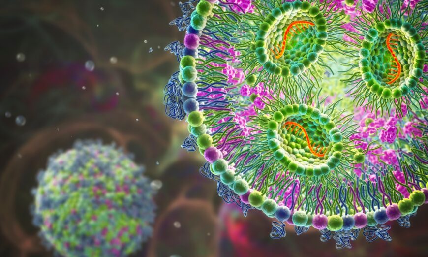
Lipid nanoparticle mRNA vaccine, a type of vaccine used against Covid-19 and influenza. 3D illustration showing cross-section of a lipid nanoparticle carrying mRNA of the virus (orange).
The first author of the article, along with experts from Ireland and the United States, have just followed up their research with a commentary published on December 20, 2022 in the journal Nature Reviews Immunology. Their commentary, “Impact of anti-PEG antibodies induced by SARS-CoV-2 mRNA vaccines,” reinforces the message that antibodies against PEG may be causing health problems. As the authors insist: “larger and longer studies are needed to analyze the longer-term impact of boosting anti-PEG antibodies by LNP mRNA vaccination.”
In other words, the current SARS-CoV-2 vaccines appear to have an anti-PEG antibody problem. Solutions to counter the problem are urgently needed, as millions of people continue to be injected with these problematic products. What is Polyethylene Glycol?
Polyethylene glycol, or PEG, is a hydrophilic polymer. A hydrophilic polymer is a water-soluble substance. Hydrophilic polymers are used in a wide range of products and have many biomedical applications.
PEG is a petroleum-derived compound. It can be found in laxatives and pharmaceutical medications, emulsifiers and surfactants used by the cosmetic industry, and also in food.
PEG is one of the most widely used polymers. PEGylated products have been on the market for over thirty years. mRNA Technology
However, mRNA vaccines are a new technology.
Traditional vaccines contain an attenuated or dead bacteria or virus to prompt the body to generate an immune response. But mRNA injections use messenger RNA—a type of RNA necessary for protein production. In the case of the SARS-CoV-2 mRNA vaccines, this mRNA is injected in order to use the human cells to produce spike proteins found on the virus’s outer membrane.
Using the injected mRNA, the body’s own cells then produce the viral proteins. The immune system recognizes the protein as foreign and produces immune responses (including antibodies) against it. These immune responses then allow the immune system to respond quickly when subsequently exposed to the virus, at least theoretically. PEG in mRNA Injections Lipid,Nanoparticle,Mrna,Vaccine,,A,Type,Of,Vaccine,Used,Against (Kateryna Kon/Shutterstock)
The mRNA in the injections is wrapped in lipid nanoparticles. PEG is incorporated into the vaccines’ lipid nanoparticles in order to stabilize the particles. These nanoparticles help carry the mRNA to human cells.
They also act as an adjuvant—an ingredient that the body recognizes as foreign against which it will mount an immune response.
Lipid nanoparticles in the mRNA injections are “PEGylated.” This means they are chemically attached to PEG molecules, which increase the particles’ stability and viability. Is PEG Safe?
Both the Pfizer/BioNTech and Moderna mRNA injections contain PEG. While PEG is found in many drugs, before the development of mRNA injections it had never been used in approved vaccines before, according to a 2020 article in the journal Science.
PEG is considered non-toxic. The Food and Drug Administration specifies that it is safe for human use depending on the level of exposure.
But this new international commentary is part of a growing body of scientific literature that suggests that many of us will develop anti-PEG antibodies when we are exposed to PEG.
In fact, the American Chemical Society (ACS) paper showed that 53 of 75 people tested—71 percent—had anti-PEG antibodies in their blood before they were exposed to mRNA injections.
There are many unknowns about the role of anti-PEG antibodies, according to these scientists. They hypothesize that one of the reasons the mRNA injections have so many reported side effects—some of which are quite severe—may be connected to anti-PEG antibodies. Anti-PEG Antibodies May Alter Effectiveness
Interestingly, if someone already has high levels of PEG-specific antibodies in the plasma, their immune system may clear the PEGylated nanomedicines in an enhanced way, which could actually limit their efficacy.
Accelerated clearance of PEGylated medicine is one of the concerns the scientists raise. Another is that anti-PEG antibodies could alter vaccine effectiveness, and potentially cause the vaccines to be more inflammatory.
Altered vaccine reactogenicity might include more injection-site pain, redness, and arm swelling, as well as higher fever, body pain, and headaches.
Even more serious vaccine reactions include both short and long-term heart problems, lung blockages, and eye disturbances, to name a few.
“Further research is needed to study the potential links between PEG-specific antibodies, vaccine reactogenicity and enhanced clearance of other PEG-containing medicines,” the scientists insist in their commentary.
This research is particularly important because when vaccination causes severe side effects, people become more hesitant about getting vaccines.
This hesitancy has been well documented in the scientific literature. For example, in a September 2019 article in the journal npj Vaccines, scientists mentioned that vaccines with troublesome side effects “can lead to needle fear, long-term negative attitudes and non-complaint behaviors, which undermine the public health impact of vaccination.” Problems with PEG
Soon after the mRNA injections were widely distributed in the United States and other countries, reports started rolling in about very severe side effects.
A December 2020 article on Science.org linked life-threatening vaccine reactions to the polyethylene glycol in at least eight people.
Anaphylaxis associated with PEG happens within seconds, minutes, or hours of being vaccinated. It often involves a dangerous drop in blood pressure, shortness of breath, and tachycardia.
It may be that people who already have higher levels of anti-PEG antibodies may be at higher risk of having a severe reaction to a vaccine, booster shot, or other medical product that contains PEG. Of the 1,481,226 reports of side effects submitted to VAERS, as of December 9, 2022, there have been 10,240 reports of anaphylaxis following all COVID-19 vaccines. Contaminated With Carcinogens
But the risk of anaphylaxis is only one of the potential health issues with PEG.
The process of making the polymer itself creates byproducts that are known to be toxic to human health. The worst among these may be 1,4-dioxane, a highly toxic compound that the FDA recognizes as “a potential human carcinogen.”
Several animal studies have shown that mammals intentionally exposed to 1,4-dioxane are at greater risk of cancer and other health issues and immune disruption.
Even tiny amounts of 1,4-dioxane may have negative effects on human health. In Europe it is only considered safe for cosmetic products that come in contact with the skin at levels lower than ten parts per million.
PEG can also be contaminated with ethylene oxide, which has been found to increase the risk of miscarriage, preterm birth, and post-term birth.
In one particularly worrisome study, published in 2019 in the journal Laboratory Animals, laboratory mice injected with PEG became so sickly that half of them had to be euthanized.
Since the roll-out of the mRNA vaccines, pathologists have reported seeing a rise in unusually aggressive and fast-growing cancers.
A lifelong promoter of vaccines, the Belgian immunologist Dr. Michel Goldman, experienced this firsthand. Diagnosed with lymphoma, Goldman rushed to get a third Pfizer injection. He believed it would protect him from COVID-19, which could be more severe for him given his compromised immune system.
However, the shot exacerbated his night sweats and left him feeling exhausted. His lymph nodes became even more swollen and tender.
After the third Pfizer injection, a scan showed that he had so many more cancer lesions that it “looked like someone had set off fireworks inside [his] body,” according to an article in The Atlantic. New cancer clusters were blooming in his armpit and along his neck.
The case report about the rapid progression of angioimmunoblastic T cell lymphoma, which was the kind of cancer Goldman was battling, was published in the journal Frontiers in Medicine in September 2021.
While Goldman himself “remains adamant that COVID-19 vaccines are necessary and useful for the vast majority of people,” according to The Atlantic, other scientists disagree.
A team of researchers, led by a senior scientist at the Massachusetts Institute of Technology, believe that the side effects of mRNA vaccines, including cancer, are not nearly as “rare” as the mainstream media suggests.
Their peer-reviewed scientific study, published in June of 2022 in the journal Food and Chemical Toxicology, posited that the mRNA vaccines interfere with innate immunity and causes the immune system to let cancer cells grow unchecked by suppressing the type 1 interferon response.
“The mRNA vaccines potentially cause increased risk to infectious diseases and cancer,” the scientists found.
Dr. Rick Kirschner, a retired naturopathic doctor based in Sandpoint, Idaho who served as president of the Naturopathic Medicine Institute in 2020, said he believes everyone needs to be concerned about the lack of safety and the unusually high reactogenicity of the COVID-19 vaccines.
“It seems to me like the precautionary principle has been thrown out the window for anything related to these mRNA vaccines,” Kirschner told us.
“If something is potentially harmful, you can’t assume it’s safe without adequate testing,” he continued.
“It used to be that you had to prove that something was safe. That’s the precautionary principle. In other words, ‘better safe than sorry,’” Kirschner said. “It sure seems to me like using a known carcinogen and toxin like PEG in this novel way is a great example of throwing caution to the wind.”