Viruses Finally Reveal Their Complex Social Life


New research has uncovered a social world of viruses full of cheating, cooperation and other intrigues, suggesting that viruses make sense only as members of a community.

An illustration shows two hands composed of viral particles lightly touching.
Far from solitary particles, viruses engage in social behaviors within cells and hosts.

Introduction

Ever since viruses came to light in the late 1800s, scientists have set them apart from the rest of life. Viruses were far smaller than cells, and inside their protein shells they carried little more than genes. They could not grow, copy their own genes or do much of anything. Researchers assumed that each virus was a solitary particle drifting alone through the world, able to replicate only if it happened to bump into the right cell that could take it in.

This simplicity was what attracted many scientists to viruses in the first place, said Marco Vignuzzi, a virologist at the Singapore Agency for Science, Research and Technology Infectious Diseases Labs. “We were trying to be reductionist.”

That reductionism paid off. Studies on viruses were crucial to the birth of modern biology. Lacking the complexity of cells, they revealed fundamental rules about how genes work. But viral reductionism came at a cost, Vignuzzi said: By assuming viruses are simple, you blind yourself to the possibility that they might be complicated in ways you don’t know about yet.

For example, if you think of viruses as isolated packages of genes, it would be absurd to imagine them having a social life. But Vignuzzi and a new school of like-minded virologists don’t think it’s absurd at all. In recent decades, they have discovered some strange features of viruses that don’t make sense if viruses are lonely particles. They instead are uncovering a marvelously complex social world of viruses. These sociovirologists, as the researchers sometimes call themselves, believe that viruses make sense only as members of a community.

Granted, the social lives of viruses aren’t quite like those of other species. Viruses don’t post selfies to social media, volunteer at food banks or commit identity theft like humans do. They don’t fight with allies to dominate a troop like baboons; they don’t collect nectar to feed their queen like honeybees; they don’t even congeal into slimy mats for their common defense like some bacteria do. Nevertheless, sociovirologists believe that viruses do cheat, cooperate and interact in other ways with their fellow viruses.

The field of sociovirology is still young and small. The first conference dedicated to the social life of viruses took place in 2022, and the second will take place this June. A grand total of 50 people will be in attendance. Still, sociovirologists argue that the implications of their new field could be profound. Diseases like influenza don’t make sense if we think of viruses in isolation from one another. And if we can decipher the social life of viruses, we might be able to exploit it to fight back against the diseases some of them create.

Under Our Noses

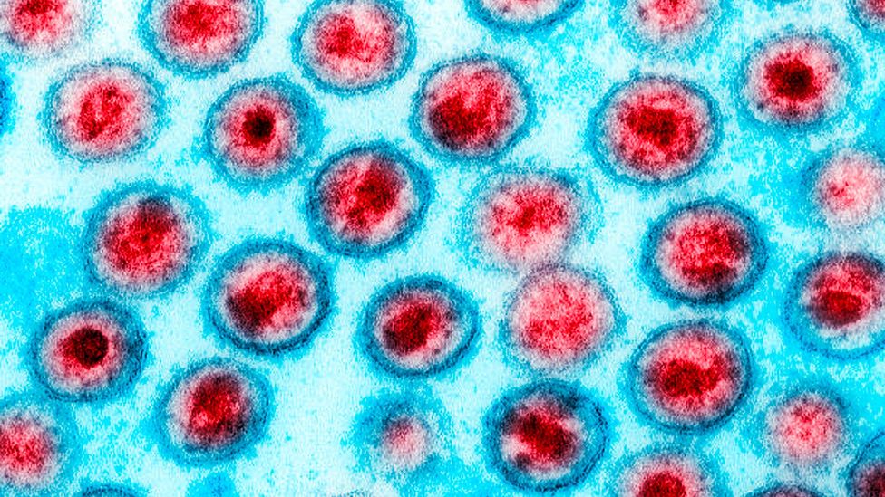
Some of the most important evidence for the social life of viruses has been sitting in plain view for nearly a century. After the discovery of the influenza virus in the early 1930s, scientists figured out how to grow stocks of the virus by injecting it into a chicken egg and letting it multiply inside. The researchers could then use the new viruses to infect lab animals for research or inject them into new eggs to keep growing new viruses.

In the late 1940s, the Danish virologist Preben von Magnus was growing viruses when he noticed something odd. Many of the viruses produced in one egg could not replicate when he injected them into another. By the third cycle of transmission, only one in 10,000 viruses could still replicate. But in the cycles that followed, the defective viruses became rarer and the replicating ones bounced back. Von Magnus suspected that the viruses that couldn’t replicate had not finished developing, and so he called them “incomplete.”

Sam Díaz-Muñoz stands in front of a waterfall.
Sam Díaz-Muñoz at the University of California, Davis, applies social evolution theory originally developed to describe animal behavior, including concepts like cheating and cooperation, to virus-virus interactions.Courtesy of Sam Díaz-Muñoz

In later years, virologists named the boom and bust of incomplete viruses “the von Magnus effect.” To them, it was important — but only as a problem to solve. Since no one had seen incomplete viruses outside of a lab culture, virologists figured they were artificial and came up with ways to get rid of them.

“You have to eliminate these from your lab stocks because you don’t want them to interfere with your experiments,” said Sam Díaz-Muñoz, a virologist at the University of California, Davis, recalling the common view within the field. “Because this is not ‘natural.’”

Researchers in the 1960s observed that incomplete viral genomes were shorter than those of typical viruses. That finding strengthened the view of many virologists that incomplete viruses were defective oddities, lacking the genes needed to replicate. But in the 2010s, inexpensive, powerful gene-sequencing technology made it clear that incomplete viruses were actually abundant inside our own bodies.

In one study, published in 2013, researchers at the University of Pittsburgh swabbed the noses and mouths of people sick with the flu. They pulled out the genetic material from influenza viruses in the samples and discovered that some of the viruses were missing genes. These stunted viruses came into existence when infected cells miscopied the genome of a functional virus, accidentally skipping stretches of genes.

Other studies confirmed this discovery. They also revealed other ways that incomplete viruses can form. Some kinds of viruses carry garbled genomes, for example. In these cases, an infected cell started copying a viral genome only to reverse partway through and then copy the genome backward to its starting point. Other incomplete viruses form when mutations disrupt the sequence of a gene so that it can no longer make a functional protein.

Introduction

These studies demolished the old assumption that von Magnus’ incomplete viruses were only an artifact of lab experiments. “They’re a natural part of virus biology,” Díaz-Muñoz said.

Discovering incomplete viruses in our own bodies has inspired a new surge of scientific interest in them. Influenza is not unique: Many viruses come in incomplete forms. They make up the majority of viruses found in people sick with infections such as respiratory syncytial virus (RSV) and measles.

Scientists have also come up with new names for von Magnus’ incomplete viruses. Some call them “defective interfering particles.” Others call them “nonstandard viral genomes.”

Díaz-Muñoz and colleagues have another name for them: cheaters.

A Viral Grift

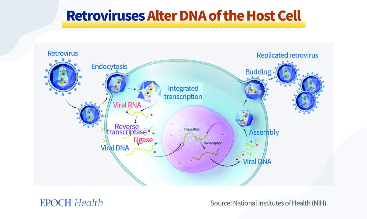
Incomplete viruses can typically get into cells, but once inside, they cannot replicate on their own. They lack some of the genes essential for hijacking their host’s protein-making machinery, such as the one for a gene-copying enzyme known as a polymerase. In order to replicate, they have to cheat. They have to take advantage of their fellow virus.

Fortunately for the cheaters, cells are often infected by more than one viral genome. If a functional virus shows up in a cheater’s cell, it will make polymerases. The cheater can then borrow the other virus’s polymerases to copy its own genes.

In such a cell, the two viruses race to make the most copies of their own genome. The cheater has a profound advantage: It has less genetic material to replicate. The polymerase therefore copies an incomplete genome more quickly than a complete one.

Their edge grows even larger over the course of an infection, as incomplete viruses and functional ones move from cell to cell. “If you’re half as long, that doesn’t mean you get a two-times advantage,” said Asher Leeks, who studies social evolution in viruses as a postdoc at Yale University. “That can mean you get a thousand-times advantage or more.”

Other cheater viruses have working polymerases, but they lack the genes for making protein shells to enclose their genetic material. They replicate by lying in wait for a functional virus to show up; then they sneak their genome into the shells it produces. Some studies suggest that cheater genomes may be able to get inside shells faster than functional ones.

Asher Leeks wears a lab coat.
Asher Leeks’ research into multipartite viruses, which must all be present in a cell to replicate, has shown that what might look like viral cooperation may have evolved from cheating. “In viruses, conflict is dominant,” he said.Nora Pyenson

Introduction

Whichever strategy an incomplete virus uses to replicate, the result is the same. These viruses don’t pay the cost of cooperation, even as they exploit the cooperation of other viruses.

“A cheater does poorly on its own, it does better in relation to another virus, and if there are a lot of cheaters, there’s no one to exploit,” Díaz-Muñoz said. “From an evolutionary perspective, that’s all you need to define cheating.”

The last part of that definition poses a puzzle. If cheaters are so amazingly successful — and, indeed, they are — they ought to drive viruses to extinction. As generations of viruses burst out of old cells and infect new ones, cheaters ought to get more and more common. They should keep replicating until the functional viruses disappear. Without any functional viruses left, the cheaters can’t replicate on their own. The entire population of viruses should get sucked into oblivion.

Of course, viruses such as influenza are clearly escaping this swift extinction, and so there must be more to their social lives than a death spiral of cheating. Carolina López, a virologist at Washington University School of Medicine in St. Louis, believes that some viruses that look like they’re cheating may actually play a more benign role in viral societies. Instead of exploiting their fellow viruses, they cooperate, helping them thrive.

“We think of them as part of a community,” López said, “with everybody playing a critical role.”

Burnout Prevention

López’s initiation into the world of sociovirology started in the early 2000s as she studied Sendai virus, a pathogen that infects mice. Researchers had known for years that two strains of Sendai virus behaved differently. One, called SeV-52, was good at escaping the notice of the immune system, allowing the virus to cause a massive infection. But mice infected with another strain, SeV-Cantell, mounted a swift, powerful defense that helped them quickly recover. The difference, López and her colleagues found, was that SeV-Cantell produced a lot of incomplete viruses.

How were incomplete viruses triggering the mice’s immune systems? After a series of experiments, López and her colleagues established that incomplete viruses cause their host cells to activate an alarm system. The cells produce a signal called interferon, which lets neighboring cells know an invader has arrived. Those cells can prepare defenses against the viruses and prevent the infection from spreading like wildfire through the surrounding tissue.

Carolina López poses for a portrait.
Carolina López hypothesized that while incomplete viruses may cheat inside a given cell, their overall effect — keeping infectious spread in check — may benefit the entire viral community.Matt Miller / Washington University School Of Medicine

This phenomenon wasn’t a quirk of Sendai virus, nor of the mouse immune system. When López and her colleagues turned their attention to RSV), which sickens over 2 million people in the United States every year and causes thousands of deaths, they found that incomplete viruses produced in natural infections also triggered a strong immune response from infected cells.

This effect puzzled López. If incomplete viruses were cheaters, it didn’t make sense for them to provoke a host to cut an infection short. Once the immune system destroyed the functional viruses, the cheaters would be left without any victims to exploit.

Lopez found that her results made sense if she looked at the viruses in a new way. Instead of focusing on the idea that the incomplete viruses were cheating, López began to think about them and the functional viruses as working together toward the shared goal of long-term survival. She realized that if functional viruses replicated uncontrollably, they might overwhelm and kill their current host before transmission to a new host could take place. That would be self-defeating.

“You need some level of immune response for just keeping your host alive long enough for you to move on,” López said.

That’s where the incomplete viruses come in, she said. They might rein in the infection so that their host has a chance to pass viruses onto the next host. In that way, the functional and incomplete viruses might be cooperating. The functional viruses produce the molecular machinery to make new viruses. Meanwhile, the incomplete viruses slow the functional viruses down to avoid burning out their host, which would end the entire community’s infectious run.

In recent years, López and her colleagues have found that incomplete viruses can curb infections in a number of ways. They can trigger cells to respond as if they were under stress from heat or cold, for example. Part of a cell’s stress response shuts down the protein-building factories to save energy. In the process, it also halts the production of more viruses.

In new research published in February, Christopher Brooke reported that an infected cell can produce hundreds of cryptic proteins that are encoded by incomplete viral genomes and new to science.Fred Zwicky

Introduction

Christopher Brooke, a virologist at the University of Illinois Urbana-Champaign, agrees with López that viruses exist in communities. What’s more, he suspects that incomplete viruses have other jobs in cells that he and his fellow scientists have yet to figure out.

Brooke is looking for evidence of these jobs in influenza viruses. A complete influenza virus has eight gene segments, which typically make 12 or more proteins. But when infected cells produce incomplete viruses, they sometimes skip the middle of a gene and stitch the beginning to the end. Despite this drastic change, these altered genes still produce proteins — but new proteins that may have new functions. In a study published in February, Brooke and his colleagues discovered hundreds of these new proteins in flu-infected cells. Because these proteins are new to science, the researchers are trying to figure out what they do. Experiments on one of them suggest that it latches on to polymerase proteins made by intact viruses and blocks them from copying new viral genomes.

For now, however, scientists are largely ignorant of what incomplete viruses accomplish by producing so many strange proteins. “My limited imagination isn’t going to touch a fraction of what’s possible,” Brooke said. “This is raw material for the virus to play with.” But he doubts that the incomplete viruses producing all these strange proteins are cheaters.

“If they really were acting as pure cheaters, I would predict that there would be substantial selective pressure to minimize their production,” Brooke said. “And yet we see them all the time.”

Blurred Lines

Sociovirologists are now trying to figure out just how much cheating and cooperation are going on in the viral world. Scientists who study animal behavior know how hard this can be. An individual may cheat in some situations and cooperate in others. And it’s also possible for a behavior that looks like cooperation to evolve through selfish cheating.

Leeks agrees that incomplete viruses may be productive parts of the viral community. But he thinks it’s always important to consider the possibility that even when they look like they’re cooperating, they are still actually cheating. Evolutionary theory predicts that cheating will often arise in viruses, thanks to their tiny genomes. “In viruses, conflict is dominant,” Leeks said.

Introduction

In fact, cheating can produce adaptations that look like cooperation. One of Leeks’ favorite examples of this hidden conflict is the nanovirus, which infects plants such as parsley and fava beans. Nanoviruses replicate in an astonishing way. They have eight genes in total, but each viral particle has just one of the eight genes. Only when all nanovirus particles, each carrying one of the eight different genes, are infecting the same plant at once can they replicate. The plant cells make proteins from all eight genes, along with new copies of their genes, which then get packaged into new shells.

You might look at nanoviruses and see a textbook case of cooperation. After all, the viruses have to work together for any of them to have a chance to replicate. The arrangement is reminiscent of a beehive’s division of labor, in which the insects split the work of gathering nectar, tending to larvae and scouting new locations for the hive to move to.

But Leeks and his colleagues have charted how nanoviruses — and other so-called multipartite viruses — may have evolved through cheating.

Underneath the façade of cooperation lies viral cheating.

Imagine that the ancestor of nanoviruses started off with all eight genes packaged in one viral genome. The virus then accidentally produced incomplete cheaters that had only one of the genes. That cheater will thrive, as the fully functional viruses copy its gene. And if a second cheat evolves, carrying a different gene, it will get the same benefit of exploiting the intact viruses.

When Leeks and his colleagues built a mathematical model for this evolutionary scenario, they found that viruses can readily break apart into more cheats. They will keep breaking apart until  none of the original viruses that could replicate on their own are left. Nanoviruses may now depend on each other for survival, but only because their ancestors freeloaded off each other. Underneath the façade of cooperation lies viral cheating.

Sorting out the nature of virus societies will take years of research. But solving the mystery may bring a tremendous payoff. Once scientists understand the social behavior of viruses, they may be able to turn viruses against one another.

Turning the Tables

In the 1990s, evolutionary biologists were able to help inform the development of antiviral medicines. When people with HIV took a single antiviral drug, the virus quickly evolved the ability to evade it. But when doctors instead prescribed medicines that combined three antivirals, it became much harder for the viruses to escape them all. The chance that a virus could gain mutations to resist all three drugs was astronomically small. As a result, HIV drug cocktails remain effective even today.

Sociovirologists are now investigating whether evolutionary biology can again help in the fight against viruses. They are looking for vulnerabilities in the way viruses cheat and cooperate, which they can exploit to bring infections to a halt. “We see it as turning the tables on the virus,” Vignuzzi said.

Vignuzzi and his colleagues tested this idea in mice with Zika virus. They engineered incomplete Zika viruses that could ruthlessly exploit functional ones. When they injected these cheaters into infected mice, the population of functional viruses inside the animals quickly collapsed. The French company Meletios Therapeutics has licensed Vignuzzi’s cheater viruses and has been developing them as a potential antiviral drug for a variety of viruses.

At New York University, Ben tenOever and his colleagues are engineering what might be an even more effective cheater from influenza viruses. They’re taking advantage of a quirk of virus biology: Every now and then, the genetic material from two viruses that infect the same cell will end up packaged into one new virus. They wondered if they could create a cheating virus that could readily invade the genome of a functional influenza virus.

Ben tenOever poses for a portrait.
Could incomplete viruses be used in medicine? Ben tenOever’s cutting-edge research has produced a nasal spray of cheater virus that equipped mice to survive a fatal strain of influenza. However, because this cheater virus can spread between animals, the chances of regulators approving such an approach for human medicine seems low.Marcel Indik Photography

Introduction

The NYU team harvested incomplete viruses from influenza-infected cells. From this batch, they identified a super-cheater that was remarkably good at slipping its genes into fully functional influenza viruses. The resulting hybrid virus was bad at replicating, thanks to the cheater’s disruption.

To see how this super-cheater would perform as an antiviral, tenOever and his colleagues packaged it into a nasal spray. They infected mice with a lethal strain of influenza and then squirted the super-cheater into the animals’ noses. The super-cheater virus was so good at exploiting functional viruses and slowing their replication that the mice managed to recover from the flu within a couple weeks. Without help from the super-cheaters, the animals died.

The researchers got even better results when they sprayed the super-cheaters into the noses of mice before they got infected. The super-cheaters lay in wait inside the mice and attacked the functional flu viruses as soon as they arrived.

Then tenOever and his colleagues moved to ferrets for their experiments. Ferrets experience influenza infections more like humans do: In particular, unlike with mice, influenza viruses will readily spread from a sick ferret to a healthy one in an adjacent cage. The scientists found that the nasal spray quickly drove down the number of flu viruses in infected ferrets, just as they saw in mice. However, the scientists got a surprise when they looked at the viruses that the infected ferrets passed to healthy animals. They transmitted not only normal viruses but also super-cheaters stowed away inside their protein shells.

RELATED:


  1. Viruses Can Scatter Their Genes Among Cells and Reassemble
  2. Viruses Have a Secret, Altruistic Social Life
  3. How Many Microbes Does It Take to Make You Sick?
  4. Viruses Would Rather Jump to New Hosts Than Evolve With Them

That finding raises the tantalizing possibility that super-cheaters might be able to stop the spread of a new strain of influenza. If people received sprays of super-cheater viruses, they could rapidly recover from infections. And if they did pass on the new virus strain to others, they would also pass along the super-cheater to stop it. “It’s a pandemic neutralizer,” tenOever said.

That’s true in concept, at least. TenOever would need to run a clinical trial in humans to see if it would work as it does in animals. However, regulators have had qualms about approving such an experiment, he said, as that would not simply be giving people a drug that would work on viruses in their own bodies, but also one that could spread to others, whether they consented to it or not. “That seems to be the kiss of death,” tenOever said, for his hopes of turning the science of social viruses into medicine.

Díaz-Muñoz thinks that it’s right to be cautious about harnessing sociovirology when we still have so much to learn about it. It’s one thing to create medicines from inert molecules. It’s quite another to deploy the social life of viruses. “It is a living, evolving thing,” Díaz-Muñoz said.

Viruses Are the Most Common Cause of Myocarditis in Children


Myocarditis Heart Disease Concept
  • Viral infection is the most common cause of inflammation in the heart muscle, called myocarditis, in children; however, there remains a diverse array of infectious and non-infectious causes of myocarditis that should be considered in diagnosis.
  • Myocarditis caused by a virus is more often seen in children than in adults, and children are more likely to have acute myocarditis (sudden onset) rather than chronic myocarditis, which is more typically seen in adults.
  • The emergence of COVID-19 has led to the description of a new multisystem inflammatory syndrome in children (MIS-C) that can involve the heart muscle and heart arteries in some infected patients.
  • The new scientific statement from the American Heart Association is helpful in guiding the diagnosis and treatment of myocarditis in children. And while not explicitly addressed, since this statement was in development prior to the COVID-19 pandemic, the statement is also useful in informing the clinical care of suspected cases of myocarditis after COVID-19 vaccination and myocarditis after COVID-19 infection, which have been reported primarily in teens and young adults.

Myocarditis in children is a rare yet challenging condition to treat. Diagnosis and treatment includes multiple options, and many cases of myocarditis resolve on their own, according to a new scientific statement from the American Heart Association, “Diagnosis and Management of Myocarditis in Children,” published today in Circulation, the Association’s flagship journal. The scientific statement writing group reviewed the latest research to develop guidance in diagnosis and treatment for myocarditis in children.

Myocarditis is inflammation of the middle layer of the wall of the heart muscle, the myocardium, and it can temporarily or permanently weaken the heart muscle and the heart’s electrical system, which keeps the heart pumping regularly. Approximately 10 to 20 per 100,000 people are diagnosed with myocarditis in the U.S. annually, and in children, the incidence is 1 to 2 per 100,000. Although many cases resolve on their own or with treatment, leading to a full recovery, severe myocarditis can lead to heart failure, abnormal heart rhythms, shock and sudden death. Signs and symptoms of myocarditis include fatigue, shortness of breath, fever, chest pain, and palpitations.

“Myocarditis has distinct characteristics in children and a potential impact on their lifelong health,” said Yuk M. Law, M.D., FAHA, chair of the scientific statement writing group, director of Cardiac Transplant and Heart Failure Service at Seattle Children’s Hospital and professor of pediatrics at the University of Washington School of Medicine in Seattle. “We hope that this statement serves as an educational update as well as a unifying call for much needed research to better understand and treat this important pediatric condition. And, in light of the recently recognized occurrence of myocarditis after COVID-19 infection, as well as the emergence of cases of suspected myocarditis after COVID-19 vaccination, this statement is a resource for clinicians and health care professionals in caring for these patients.”

Direct tissue examination from a biopsy is the standard for proving the presence of myocarditis, which can also identify if viruses are present. However, less invasive testing is widely available now. Additional screening tests for myocarditis may include blood tests to measure for elevated cardiac enzymes that would indicate heart inflammation or injury, including myoglobin, troponin and creatine kinase. Imaging tests include an echocardiogram or a cardiac magnetic resonance imaging (MRI) to determine if there is any visible injury to the heart or abnormalities in how the heart is functioning. Electrocardiography (ECG) screening can assess the heart rhythm and may show signs of injury to the heart as well as signs of pericarditis. Pericarditis is often related to myocarditis and involves swelling and inflammation of the pericardium, a thin, sac-like tissue structure that surrounds the heart to hold it in place and help it function properly.

Key guidance in the statement includes:

  • The incidence of myocarditis in children varies with age, being higher in infants and rising again in young adults.
  • Patients should not participate in competitive sports while active inflammation is present. In addition to normalization of inflammatory and myocardial injury markers, as well as ventricular function and heart failure, 24-hour Holter monitoring and exercise stress testing should be performed in athletes no sooner than 3 to 6 months after diagnosis and before they return to competition.
  • In children, myocarditis is most often the result of a viral infection, and it is most often acute or sudden-onset, rather than chronic myocarditis, which is seen more often in adults.
  • Symptoms in children can range from minimal to showing signs of heart failure, life-threatening arrhythmias, or cardiogenic shock.
  • The most common symptoms of myocarditis in children include fatigue, shortness of breath, abdominal pain, and fever. (Of note, the primary symptom reported in patients with COVID-19 vaccine-associated suspected myocarditis is chest pain.) However, it is important to consider alternative causes of these symptoms since none of them are specific to cardiovascular conditions including myocarditis.   
  • Acute myocarditis can deteriorate rapidly, therefore, close monitoring in an inpatient setting should be considered.
  • The early phase of care should include monitoring for atrial or ventricular arrhythmias. The management of arrhythmia is addressed in the 2017 AHA/ACC/HRS Guideline for Management of Patients with Ventricular Arrhythmias and the Prevention of Sudden Cardiac Death.
  • Acute myocarditis may lead to myocardial injury similar to a heart attack. Early intervention with mechanical circulatory support (MCS) should be considered for high-risk patients and can be lifesaving.
  • Treatment with antiviral and immunotherapies including intravenous immunoglobulin and corticosteroids is common; however, additional evidence-based research is needed to define these treatments more clearly for children.
  • Antiviral therapy should be considered if an active viral infection is found.
  • Myocarditis can also be seen secondary to systemic autoimmune diseases and is associated with rheumatic fever or Kawasaki disease. In these cases, it should be managed according to the guidance for the primary disease.
  • A new condition has emerged during the COVID-19 pandemic – multisystem inflammatory syndrome in children (MIS-C) – that involves the myocardium and coronary arteries in some infected patients. Therapy may consist of antiviral, IVIG (intravenous immunoglobulin), steroid and other anti-inflammatory medications used in atypical Kawasaki disease. 
  • Regular cardiology follow-up including ECG, echocardiography and laboratory tests are recommended initially every 1 to 3 months after onset, then as needed.  

“While our work on this scientific statement preceded the COVID-19 pandemic and the cases of suspected myocarditis after COVID-19 vaccination and after COVID-19 infection being reported in adolescents and young adults, the guidance detailed in this scientific statement can help to advise treatment for these patients as well,” Law said.

Specific to the suspected cases of myocarditis after COVID-19 vaccination in teens and young adults recently reported and continuing to be monitored by the U.S. Centers for Disease Control and Prevention (CDC), the following statement reflects the views of the American Heart Association/American Stroke Association and its science leaders:

  • President Donald M. Lloyd-Jones, M.D., Sc.M., FAHA,
  • Immediate Past President Mitchell S.V. Elkind, M.D., M.S., FAHA, FAAN,
  • President-Elect Michelle A. Albert, M.D., M.P.H., FAHA,
  • Past President Robert A. Harrington, M.D., FAHA,
  • Chief Science and Medical Officer Mariell Jessup, M.D., FAHA,
  • Chief Medical Officer for Prevention Eduardo Sanchez, M.D, M.P.H., FAAFP, and
  • Chair of the Young Hearts Council Shelley Miyamoto, M.D., FAHA.

“The American Heart Association/American Stroke Association recommends all health care professionals be aware of rare adverse events that may be related to a COVID-19 vaccine including myocarditis. Health care professionals should strongly consider inquiring about the timing of any recent COVID vaccination among patients presenting with symptoms related to cardiovascular conditions, as needed, in order to confirm the diagnosis and to provide appropriate treatment quickly. As indicated by the CDC, we agree that cardiologists should be consulted if myocarditis or any heart-related condition is suspected by a primary care clinician.

“This new scientific statement provides a thorough examination of the latest research on the diagnosis, treatment and follow-up for myocarditis in children prior to the COVID-19 era and confirms that myocarditis is a serious yet uncommon condition in children. The effects of COVID-19 infection include its potentially fatal consequences and the potential long-term health effects that are still revealing themselves, such as conditions affecting the heart including myocarditis, the brain, the vascular system and other organs.

“We remain steadfast in our recommendation for all adults and children ages 12 and older in the U.S. to receive a COVID-19 vaccine as soon as they can receive it, as authorized by the U.S. Food and Drug Administration and recommended by the CDC. Overwhelmingly, data continue to indicate that the benefits of COVID-19 vaccination – 91% effective at preventing complications of severe COVID-19 infection including hospitalization and death – far exceed the very rare risks of adverse events, including myocarditis.”

Honey Fights Bacteria and Viruses, Could Be Better Than Antibiotics


Sometimes the therapeutic effects of honey surpass even those of medication.

In this series, we will explore the good and bad sweeteners, uncover the unexpected outcomes of cutting out sugar, and discover the ultimate way to achieve this.

Honey is a sweet food and medicine that nature has gifted us. Its history of medicinal use can be traced back 8,000 years. Sometimes, the therapeutic effects of honey surpass even those of medications, particularly its outstanding antibacterial and antiviral properties.

Honey’s Antibacterial Properties Are Superior to Antibiotics’

“Bacteria quickly learn how to fight off the effects of the antibiotic drugs, but they don’t have honey resistance,” Nural Cokcetin, a research fellow at the Australian Institute for Microbiology and Infection in the science faculty at the University of Technology Sydney, said in an interview with The Epoch Times.

Honey has been used to treat skin problems and wound infections for thousands of years. For instance, ancient Greeks and Egyptians would apply honey topically to the skin to treat wounds and burns. Honey’s effectiveness in treating wounds and eczema was also documented in Persian traditional medicine. Interestingly, the concept of microorganisms such as bacteria and fungi causing wound infections during those historical periods might not have even been known.

The broad-spectrum antibacterial activity of honey has been increasingly recognized and substantiated. In recent years, the prevalence of wound infections caused by antibiotic-resistant bacteria has become a major global health care challenge. Honey can disrupt biofilms that are formed by these resistant bacteria and sometimes penetrate the biofilm structure to eliminate the embedded bacteria. An article published in the journal of the American Society for Microbiology in 2020 reported no documented cases of bacteria developing resistance to honey.

In clinical settings, medical-grade honey has been utilized in various ways. It’s formulated into topical ointments, dressings, and bandages for the treatment of postoperative wound infections, burns, necrotizing fasciitis, nonhealing wounds, boils, venous ulcers, diabetic foot ulcers, eczema, psoriasis, and dandruff. Additionally, it has proven effective in both preventing and treating infections caused by multidrug-resistant microorganisms.

In vitro studies have shown that honey can kill Helicobacter pylori. Furthermore, oral consumption of honey can reduce the duration of bacterial diarrhea.

The antibacterial properties of honey originate from its inherent characteristics and a diverse array of components derived from both nectar-producing plants and bees themselves. (Africa Studio/Shutterstock)

Key Antimicrobial Components

The antibacterial properties of honey originate from its inherent characteristics and a diverse array of components derived from both nectar-producing plants and bees themselves. Additionally, some of these components are formed during the maturation process of honey.

High Viscosity and pH

Honey has a high sugar content, reaching up to 82.5 percent. As a result, it has a viscous and syrupy consistency, exhibiting hygroscopic properties (ability to absorb moisture) and high osmolarity. Bacteria that come into contact with honey undergo dehydration and ultimately perish.

Honey is acidic due to its various organic acids, with a pH value ranging from 3.2 to 4.5. Bacteria can’t survive in honey; their optimal pH typically ranges between 6.5 and 7.5.

Primary Antibacterial Agent: Hydrogen Peroxide

An important enzyme in honey derived from bees facilitates the oxidation of glucose, resulting in the production of hydrogen peroxide.

Hydrogen peroxide is an effective bactericidal agent that can inhibit and kill microorganisms.

Interestingly, water is necessary to induce honey’s hydrogen peroxide production. The maximum level of hydrogen peroxide can be obtained by diluting honey to a concentration of 30 to 50 percent, according to a paper in the International Journal of Microbiology.

Bees Contribute Antibacterial Component Bee Defensin-1

Bee defensin-1 is another natural antibacterial component in honey originating from the honeybee’s hypopharyngeal gland.

Bee defensin-1 is an antimicrobial peptide that can kill various bacteria and microorganisms. Specifically, the defensin protein has been shown to create pores within the bacterial cell membrane, leading to cell death.

Additionally, bee defensin-1 can aid in wound healing by stimulating keratinocytes, the most common type of skin cell.

Unique Antibacterial Component in Manuka Honey: Methylglyoxal

Manuka honey is often mentioned in studies regarding the antibacterial effects of various kinds of honey. As a globally recognized honey with high antibacterial potency, it possesses a unique natural component called methylglyoxal (MGO).

After honeybees gather nectar from the flowers of manuka trees, a natural substance that’s found in the trees undergoes spontaneous dehydration, leading to the formation of MGO in the honey.

“The more of [methylglyoxal] there is, the more antibacterial that honey is,” Dee Carter, a professor from the School of Life and Environmental Sciences at the University of Sydney, told The Epoch Times.

MGO exhibits remarkable antibacterial properties by itself. It can alter the structure of bacterial fimbriae and flagella (appendages), and induce damage to their cell membranes, ultimately resulting in cell death.

However, this substance is sensitive to heat. Heating manuka honey to 98.6 degrees F (37 degrees C) can increase the levels of MGO, but heating it to 122 degrees F (50 degrees C) results in the loss of MGO.

Manuka honey possesses a unique compound derived from the manuka tree called methylglyoxal (MGO), which is an antibacterial compound not found in other types of honey. (HikoPhotography/Shutterstock)
Manuka honey possesses a unique compound derived from the manuka tree called methylglyoxal (MGO), which is an antibacterial compound not found in other types of honey.

Honey’s Antiviral Properties May Reduce COVID-19 Complications

Honey not only possesses antibacterial properties but also exhibits antiviral effects, which help protect the body and boost the immune system. Many individuals find relief from symptoms of cold and respiratory illnesses by consuming honey water, and there’s a valid basis for this remedy.

Combats Influenza and COVID-19

MGO can inhibit the growth of enveloped viruses, including the virus that causes COVID-19.

According to a study published in the Eurasian Journal of Medicine and Oncology in 2020, computer simulations revealed that multiple compounds found in honey can bind to the protease of SARS‑CoV‑2, effectively inhibiting its replication.

MGO in manuka honey can also inhibit the replication of influenza viruses, including drug-resistant strains.

Currently, scientists are researching the potential of MGO for managing and treating COVID-19.

Boosts the Immune System

Honey can activate several immune cells, enabling them to eliminate viruses effectively. For example, oligosaccharides that are present in honey can enhance the activity of natural killer cells. Additionally, certain immune cells can “remember” specific viruses during the antiviral response, offering future protection against the same viral invasions.

Honey can promote the proliferative activity of immune cells, thus increasing their quantity.

Promotes Autophagy

Honey can promote autophagy, which is crucial in combating deadly viruses such as SARS‑CoV‑2, which causes COVID-19. It’s an internal cellular process that involves the degradation and metabolism of cells, allowing for the removal and recycling of unwanted or damaged cells and impurities. This process contributes to the generation of healthier cells and promotes recovery.

Honey’s Anti-Inflammatory and Gut-Health Benefits

Inflammation is a common occurrence in bacterial or viral infections. Left unchecked, it can significantly harm the body, potentially leading to life-threatening complications.

Moreover, honey has been suggested to have a dual role in inflammation control: It can downregulate anti-inflammatory factors, thereby reducing inflammation damage, and stimulating the production of inflammatory mediators, promoting wound healing.

In one of Ms. Cokcetin’s studies, honey could reduce inflammation levels in the intestines. She attributes this effect to the abundant presence of phenolic compounds found in honey.

She explained that honey also contains oligosaccharides and polysaccharides, which are complex sugars. These compounds serve as prebiotics, supporting the growth of beneficial bacteria in the gut and inhibiting the growth of harmful bacteria. Additionally, consuming honey promotes the production of short-chain fatty acids in the intestines.

Choosing Honey With Potent Properties

Generally, raw and darker honey varieties tend to have more robust efficacy.

Commercially available honey found on supermarket shelves differs from raw honey sold by beekeepers or at bazaars, as it’s generally pasteurized. Research has shown that heat processing can lower the antioxidant capacity of honey by 33.4 percent. Alternatively, you can also choose honey processed using high-pressure techniques.

Honey comes in a diverse range of colors, from light yellow to amber, dark red, and even nearly black. These colors reflect the varying composition of honey, such as polyphenols, minerals, and pollen. Numerous studies suggest that dark-colored honey generally contains higher levels of phenolic compounds and exhibits more potent activity.

To obtain manuka honey with exceptional antibacterial properties, it’s recommended to opt for pure honey sourced from New Zealand and Australia.

The manuka tree is exclusive to specific New Zealand and Australian regions, and its annual yield is limited. Certain products labeled as manuka honey may contain blends of other honey types, potentially affecting the honey’s properties. Furthermore, some of these products may even include low-quality ingredients such as sucrose.

Opting for single-origin honey sourced exclusively from the manuka tree and checking for its Unique Manuka Factor (UMF) rating is recommended. A higher UMF rating indicates higher levels of substances related to MGO. Some honey products also specify the amount of MGO in milligrams per kilogram.

However, manuka honey with a higher UMF rating may have a strong flavor that some people find pungent.

Ms. Cokcetin recalled that when she began her research on the antibacterial properties of honey, a professor she worked with said, “We’ve already got this great gift from nature that is a solution to superbugs; we just have to figure out how it works and why it works the way it does.”

The viruses that helped to make you human


A man wearing a helmet shaped like a virus (Credit: Getty Images)

Although they are more commonly associated with causing disease and, at times, devastating pandemics, viruses have also played an important role in human evolution – and without them you wouldn’t be here at all.

The Mabuya lizards that live in the Andes mountains of Colombia are not like other reptiles. While the majority of reptiles lay eggs with hard shells, some Mabuya species give birth to live young. Crucially, the mothers have placentas: specialised organs for feeding the developing young inside their bodies.

Placentas are more commonly associated with mammals like mice and humans: we are placental mammals. But other kinds of animal have also evolved placentas. And in 2001 zoologists Martha Patricia Ramírez-Pinilla and Adriana Jerez of the Industrial University of Santander in Bucaramanga, Colombia, revealed that Mabuya lizards have extremely advanced placentas, not too dissimilar from ours.

While this was surprising enough for a reptile, which typically lay leathery eggs, the real revelation came 16 years later, when Ramírez-Pinilla teamed up with geneticist Thierry Heidmann of Gustave Roussy in Paris, France and his colleagues. They found that the lizards have a gene that is essential for the formation of the placenta, and that gene came from a virus.

Within the last 25 million years, the ancestors of the lizards were infected by a virus that incorporated some of its own DNA into their genome. But instead of being harmed, the lizards somehow co-opted the viral DNA and used it to develop their first placentas. Thanks to the virus, the lizards evolved a new organ.

“The genomic acquisition coincided with the shift from non-placental to placental lizard,” says Heidmann.

The unusual thing about this story, however, is that it is not unusual. About a tenth of the human genome comes from viruses, and that viral DNA has played crucial roles in our evolution. Some of it was the source of the mammalian placenta. Other bits are involved in our immune response against disease and in the formation of new genes. Without viruses, humans could not have evolved.There are four known retroviruses that infect humans, including HIV-1 – viruses like this have helped to shape our evolution (Credit: Getty Images)

There are four known retroviruses that infect humans, including HIV-1 – viruses like this have helped to shape our evolution

Viruses are so simple that many biologists do not regard them as fully alive. Each virus is essentially a microscopic package of genetic material. They can only reproduce by infecting living cells: they subvert the cell’s machinery to make copies of themselves. In doing so they often make their hosts ill.

Viruses that insert their own genetic material into the host’s genome are called retroviruses. Their nature was first understood in the 1960s and 1970s, although some had been isolated decades before. Following a suggestion in 1964 that some viruses might copy their own genetic material into the DNA of their hosts, researchers identified DNA of viral origin in the genomes of chickens.

Despite being a large and diverse group of viruses, only four retroviruses are currently known to infect humans. All were discovered in the 1980s: human T-lymphotropic virus 1 (HTLV-1), which causes a form of cancer, along with the closely related HTLV-2; and human immunodeficiency virus (HIV) types 1 and 2, which cause Aids.

If a retrovirus infects a cell in a person’s lungs or skin, that may be bad news for the person, but it has limited consequences for our species’ evolution as this DNA is not passed onto the next generation. However, sometimes a retrovirus gets into the germline: the cells that give rise to eggs and sperm, where viral DNA can be passed onto our offspring. These pieces of viral DNA are called endogenous retroviruses or ERVs. It is these heritable bits of viral DNA that can change the course of evolution.

ERVs everywhere

The sheer scale of human ERVs was revealed when the first draft of the human genome was published in 2001. “It turned out there was a huge amount of viral sequence,” says Heidmann. About 8% of the human genome consists of ERVs.

Some of them are truly ancient. One study in 2013 identified an ERV on the human chromosome 17 that is at least 104 million years old and probably older. That means it is the result of a virus infecting a mammal deep in the era when dinosaurs ruled the Earth. The ERV is only found in mammals with placentas, so it may have integrated soon after placental mammals split from their marsupial cousins

ERVs aren’t confined to mammals and reptiles, either. “All vertebrates have endogenous retroviruses,” says molecular virologist Nicole Grandi at the University of Cagliari in Italy.

Most human ERVs are not unique to our species but can also be found in at least some other primates, such as chimpanzees. This means they got into primate genomes millions of years ago, long before our own species evolved, and we then inherited them from our ape ancestors.

Some ERVs help the immune system to target cancerous cells

Curiously, there is no evidence of new ERVs entering the human genome in the last few thousand years. The only retroviruses our species currently has to deal with are HTLV and HIV, says Grandi, and neither has been reported to infect germline cells. “Currently we cannot see active endogenisation in humans,” says Grandi.

This is in stark contrast to some other species. Koalas are currently being invaded by koala retrovirus (KoRV), DNA from which is found in some populations of koalas but not others. Koala geneticists are therefore able to watch “a genome invasion in real time“.

Origin of the placenta

Human ERVs were originally thought of as inactive “fossil sequences” or part of the genome’s “junk DNA”. However, in common with a lot of supposed junk DNA, it turns out that many human ERVs are active. (Learn about the crucial role junk DNA may play in this article by David Cox.)

The most studied ERVs in the human genome are called HERV-W, first described in 1999. They encode proteins called syncytins, which are found in the placenta. Just like in the Mabuya lizards, these viral genes are essential for the placenta to form.

This link between viruses and the placenta makes sense when you consider what syncytins actually do. These proteins have the ability to fuse two or more cells into one. When they were viral proteins, they were used by the virus to fuse with the outer membrane of a cell and thus infect it. This fusion ability has been co-opted by the placenta. By fusing cells from the mother and cells from the embryo, the placenta can transfer nutrients to the embryo and extract wastes.

And it’s not just humans. Similar syncytin proteins are found in other apes like gorillas. More recent studies have shown that retroviruses have infected mammals repeatedly throughout their evolutionary history, so different groups of mammals often have different syncytins derived from different retroviruses.

“We hypothesise that in fact there was a founding ERV capture 150 million years ago, which led to the emergence of placental mammals,” says Heidmann. Since then, repeated infections seem to have overwritten that original ERV, so it cannot be found in any living mammal. The Mabuya lizard study was important because it demonstrated that the lizards only acquired placentas after first acquiring the ERV from a virus – suggesting the same thing happened in the ancestor of all placental mammals. “It gave the demonstration of the link between acquisition of a placenta and acquisition of a syncytin,” says Heidmann.A few koala populations, such as those on Kangaroo Island, a free of the koala retrovirus while others are being invaded by the disease (Credit: Getty Images)

A few koala populations, such as those on Kangaroo Island, a free of the koala retrovirus while others are being invaded by the disease

Beyond the Genome

It has been 20 years since the Human Genome Project was “completed”, but this enormous effort to sequence and map the human “book of life” was only just the beginning. Far from closing the question of what makes our bodies tick and why they do so differently, research on the human genome has revealed a far more complex picture than anyone could have imagined. Beyond the Genome examines just how far our understanding of our genetics has come in the past two decades.

The story of syncytins and the placenta is one of the most dramatic examples of viral DNA affecting evolution. It is particularly noteworthy because a complete viral gene has survived in the human genome and encodes a protein. Many other ERVs do not encode proteins, but they still have functions.

Some play a role in stem cells: the multipurpose cells found in developing embryos. Some stem cells are pluripotent, meaning they can develop into any type of cell in the body, from neurons to muscle fibres.

A family of retroviruses called HERV-H are essential for pluripotency. However, they do not code for proteins. Instead, the HERV-H sequences are copied onto molecules called RNAs, and these keep the cell pluripotent. “If they are suppressed, then the cell morphology changes and it loses the ability to maintain its undifferentiated state,” says virologist Christine Kozak of the National Institute of Allergy and Infectious Diseases in Bethesda, Maryland.

Other ERVs regulate the activity of genes, and therefore control bodily processes. For example, our bodies use an enzyme called amylase to break down carbohydrates like starch in our food. “We have amylase in the pancreas and we have amylase in the mouth in the saliva,” says Grandi. The amylase gene is activated in the salivary gland by a DNA sequence called a promoter – which comes from an ERV.

The viruses that keep us healthy

Unsurprisingly, given that ERVs come from viruses, many scientists are interested in their role in health and disease. One such example was described in 2022 by researchers led by Cédric Feschotte, a molecular biologist and geneticist at Cornell University in Ithaca, New York. The team was trying to find an instance in humans of a phenomenon already well-known in other animals – sometimes, ERV genes code for proteins that can be co-opted by the immune system and used to fight off other viruses. The target viruses can be closely related to the virus that gave rise to the ERV in the first place, or only distantly related. Feschotte says antiviral proteins from ERVs have been studied in mice, chickens and cats. “But to my knowledge there was no examples of that in the human genome,” he says.

https://emp.bbc.com/emp/SMPj/2.51.0/iframe.html

Could ancient viruses emerge once again?

The team scanned the known ERVs in the human genome and identified hundreds of sequences that could potentially encode antiviral proteins. Then they zeroed in on one gene called Suppressyn, which encodes a protein similar to those that make up the outer envelopes of viruses. The Suppressyn protein blocks retroviruses from entering cells, because it binds to receptors on the cell outer membrane that the viruses would themselves use to get into the cell. Feschotte compares it to inserting a broken key into a lock, preventing anyone from unlocking the door.

Suppressyn is mostly found in the placenta and developing embryo. This suggests its original use was to prevent retroviruses from infecting embryos, which have very weak immune systems. “It’s protective of the germline rather than of the organism as a whole,” says Feschotte.

But he believes that ERVs probably do a lot more in our immune system. “We have 1,500 candidates,” he says. “That’s a lot of genes.” While many geneticists still think of ERVs as inert or defective, that’s misleading. “They are decaying but they still make RNA and they still make a lot of proteins,” says Feschotte. “We need to take a good look at that.” And the picture is still emerging – a study published in April 2023 found that some ERVs help the immune system to target cancerous cells.

Do ERVs make us ill?

But while they can protect us from disease, it would not be surprising if some ERVs might also be responsible for producing negative health affects in humans too. “There’s really a lot of interest right now in the possibility that human ERVs can be associated with disease,” says Kozak. “At this point there’s a lot of suggestive evidence but no smoking gun.”

Humans are truly a mosaic species – around 8% of our genome comes from viruses

For Feschotte, it’s key to think through exactly what the ERVs are doing – and we haven’t always got it right. “Ever since endogenous retroviruses were discovered, people were trying to tie those to cancer,” he says. That’s because the first ones discovered in animals were causing cancer. Funders “poured tonnes of money” at ERV research in the hope that it would uncover mechanisms of cancer and thus potential treatments. “Many people came back empty-handed.”

The key point is that human ERVs are not capable of forming viruses, which could then infect other cells. “In the mouse there is plenty, and in the chicken there is plenty,” says Feschotte. “They cause all kinds of disease.” But human ERVs have been brought under a great level of control by the rest of the genome, so do not cause viral infections.

“It’s a lot more subtle and probably about gene regulation or dysregulation, I think,” says Feschotte. Because ERVs are so widely distributed in the human genome, they can coordinate the activities of multiple genes that are separated by huge expanses of sequence. Many bodily processes need genes to be turned on and off in precise sequences, and ERVs play a big role in controlling that. “Now we’re revisiting the role of these things in disease, but through different mechanisms.”

The role of ERVs in disease is shrouded for now. But what is clear is that they are an engine of evolution. By inserting new bits of DNA throughout our genome, viruses have spurred massive changes to our genetic make-up. Once ERVs are in place, they can trigger the duplication or deletion of swathes of DNA – and if the changes are beneficial, they spread. No animal, including humans, would exist in its current form without them.

The final lesson is that humans are truly a mosaic species. Many of us have some DNA, around 2% of our genome, from Neanderthals. Some populations also have some DNA from another extinct hominin group, the Denisovans. And all of us get around 8% of our genome from viruses.

“If you think about the human gene catalogue, it’s kind of almost an existential question,” says Feschotte. Around 20,000 protein-coding genes are known, and a comparable quantity of our DNA comes from viruses. “It’s kind of mind blowing.”

Viruses Can Exploit Built-In Antibody Bias to Reinfect Hosts


A massive crowd of people shouts as one, “WE ARE ALL INDIVIDUALS!” Then, a lone person in the crowd sheepishly mutters, “I’m not.”

The scene above, one of the best from the Monty Python troupe, brings to mind a phenomenon known as the immunodominant public antibody response. It is a widely shared immune response to a pathogen. In different people, the same epitopes on viral proteins end up being targeted by the same antibodies—public antibodies.

The phenomenon, which appears to reflect our immune systems’ tendency to be efficient, can sometimes leave us vulnerable. That is, it may cause us to produce antibodies that repeatedly target the same epitopes, even if the antibodies are not protective. Or, it can give viruses, such as SARS-CoV-2, an easy way to evade the immune response. For example, a virus could mutate just a few target residues and gain the ability to outmaneuver antibodies that are widely shared by many people.

A better understanding of the phenomenon is now possible thanks to a new study led by investigators from Brigham and Women’s Hospital. According to this study, the generation of antibodies is far from random because of germline-encoded amino acid–binding (GRAB) motifs. GRAB motifs represent a germline-encoded component of the architecture of the antibody repertoire that predisposes antibodies to recognize particular structures and thus influences epitope selection and composition.

The study—”Germline-encoded amino acid–binding motifs drive immunodominant public antibody responses”—was published in Science. “Our research may help explain a lot of the patterns we’ve seen during the COVID-19 pandemic, especially in terms of re-infection,” said corresponding author Stephen J. Elledge, PhD, the Gregor Mendel professor of genetics at the Brigham and Harvard Medical School. “Our findings could help inform immune predictions and may change the way people think about immune strategies.”

Before the study, there were hints, but no clear evidence, that people’s immune systems didn’t target sites on a viral protein at random. In isolated examples, investigators had seen recurrent antibody responses across individuals—people recreating antibodies to home in on the same viral protein location (known as an epitope). But the study by Elledge and colleagues helps explain the extent and underlying mechanisms of this phenomenon.

The team used a tool the Elledge lab developed in 2015 called VirScan, which can detect thousands of viral epitopes—sites on viruses that antibodies recognize and bind to—and give a snapshot of a person’s immunological history from a single drop of blood. For the new study, the researchers used VirScan to analyze 569 blood samples from participants in the United States, Peru, and France. They found that recognition of public epitopes—viral regions recurrently targeted by antibodies—was a general feature of the human antibody response.

“By mapping 376 immunodominant ‘public epitopes’ at high resolution and characterizing several of their cognate antibodies, we concluded that germline-encoded sequences in antibodies drive recurrent recognition,” the article’s authors wrote. “Systematic analysis of antibody-antigen structures uncovered 18 human and 21 partially overlapping mouse GRAB motifs within heavy and light V gene segments that in case studies proved critical for public epitope recognition.”

GRAB motifs correspond to antibody regions that are particularly good at picking out one specific amino acid. They help explain why human antibodies tend to focus on regions where these amino acids are available for binding, and thus repeatedly bind the same spots. A small number of mutations can help a virus avoid detection by these shared antibodies, allowing the virus to reinfect populations that were previously immune.

“We find an underlying architecture in the immune system that causes people, no matter where in the world they live, to make essentially the same antibodies that give the virus a very small number of targets to evade in order to reinfect people and continue to expand and further evolve,” said lead author Ellen L. Shrock, PhD, of the Elledge lab.

Interestingly, the team noted that nonhuman species produce antibodies that recognize different public epitopes from those that humans recognize. And, while it is more likely for a person to produce antibodies against a public epitope, some people do produce rarer antibodies, which may more effectively protect them from reinfection. These insights could have important implications for treatments developed against COVID-19, such as monoclonal antibodies, as well as for vaccine design.

“The more unique antibodies may be a lot harder to evade,” Elledge said. “[This] is important to consider as we think about the design of better therapies and vaccines.”

Enzyme That Protects Against Viruses May Promote Tumor Mutation


According to a study led by investigators at Weill Cornell Medicine, an enzyme that defends human cells against viruses can help drive cancer evolution towards greater malignancy by causing myriad mutations in cancer cells. The finding suggests that the enzyme may be a potential target for future cancer treatments.

In the new study, published in Cancer Research, scientists used a preclinical model of bladder cancer to investigate the role of the enzyme called APOBEC3G in promoting the disease and found that it significantly increased the number of mutations in tumor cells, boosting the genetic diversity of bladder tumors and hastening mortality.

“Our findings suggest that APOBEC3G is a big contributor to bladder cancer evolution and should be considered as a target for future treatment strategies,” said study senior author Dr Bishoy M. Faltas, assistant professor of cell and developmental biology at Weill Cornell Medicine, and an oncologist who specializes in urothelial cancers at NewYork-Presbyterian/Weill Cornell Medical Center.

The APOBEC3 family of enzymes is capable of mutating RNA or DNA—by chemically modifying a cytosine nucleotide (letter “C” in the genetic code). This can result in an erroneous nucleotide at that position. The normal roles of these enzymes, including APOBEC3G, are to fight retroviruses like HIV—they attempt to hobble viral replication by mutating the cytosines in the viral genome.

The inherent hazardousness of these enzymes suggests that mechanisms must be in place to prevent them from harming cellular DNA. However, starting about a decade ago, researchers using new DNA-sequencing techniques began to find extensive APOBEC3-type mutations in cellular DNA in the context of cancer. In a 2016 study of human bladder tumor samples, Dr Faltas, who is also director of bladder cancer research at the Englander Institute for Precision Medicine and a member of the Sandra and Edward Meyer Cancer Center, found that a high proportion of the mutations in these tumors were APOBEC3-related—and that these mutations appeared to have a role in helping tumors evade the effects of chemotherapy.

Such findings point to the possibility that cancers generally harness APOBEC3s to mutate their genomes. This could help them not only acquire all the mutations needed for cancerous growth but also boost their ability to diversify and “evolve” thereafter—enabling further growth and spread despite immune defenses, drug treatments, and other adverse factors.

In the new study, Dr Faltas and his team, including first author  Dr Weisi Liu, a postdoctoral research associate, addressed the specific role of APOBEC3G in bladder cancer with direct cause-and-effect experiments.

APOBEC3G is a human enzyme not found in mice, so the team knocked out the gene for the sole APOBEC3-type enzyme in mice, replacing it with the gene for human APOBEC3G. The researchers observed that when these APOBEC3G mice were exposed to a bladder cancer-promoting chemical that mimics the carcinogens in cigarette smoke, they became much more likely to develop this form of cancer (76% developed cancer) compared with mice whose APOBEC gene was knocked out and not replaced (53% developed cancer). Moreover, during a 30-week observation period, all the knockout-only mice survived, whereas nearly a third of the APOBEC3G mice succumbed to cancer.

To their surprise, the researchers found that APOBEC3G in the mouse cells was present in the nucleus, where cellular DNA is kept using an ‘optical sectioning’ microscopy technique. Previously, this protein had been thought to reside only outside the nucleus. They also found that the bladder tumors of the APOBEC3G mice had about twice the number of mutations compared to the tumors in knockout-only mice.

Identifying the specific mutational signature of APOBEC3G and mapping it in the tumor genomes, the team found ample evidence that the enzyme had caused a greater mutational burden and genomic diversity in the tumors, likely accounting for the greater malignancy and mortality in the APOBEC3G mice. “We saw a distinct mutational signature caused by APOBEC3G in these tumors that is different from signatures caused by other members of the APOBEC3 family” said Dr Liu.

Lastly, the researchers looked for APOBEC3G’s mutational signature in a widely used human tumor DNA database, The Cancer Genome Atlas, and found that these mutations appear to be common in bladder cancers and are linked to worse outcomes.

“These findings will inform future efforts to restrict or steer tumor evolution by targeting APOBEC3 enzymes with drugs,” said Dr Faltas.

8 Percent of Human DNA Comes From Viruses, May Be Good for Immunity


Endogenous retroviruses account for up to 8 percent of human DNA. (ESB Professional/Shutterstock)

Endogenous retroviruses account for up to 8 percent of human DNA.

The concept that microbes such as those in the gut and skin contribute to human health is not new. In fact, scientists have also discovered that hundreds of thousands of DNA fragments in the human body are derived from viruses; these fragments were embedded in the human body long ago and passed down from generation to generation; and these DNA fragments and the viruses from which they are derived are known as “endogenous retroviruses”.

What do these viral DNA do to the human body? It was not until recent years that people discovered that these foreign genetic materials may also play a role in boosting human immunity.

Yuanyu Jeng, a former attending physician at the Department of Infectious Disease of Taipei Veterans General Hospital, explained this topic in detail.

A Study in the Science Journal: Fragments of Viral DNA Protect Against Viral Infection

A study published in Science in October 2022 pointed out that a protein called Suppressyn can protect human cells from RD114 virus infection.

Suppressyn can be produced by the human body itself, but scientists have found through in-depth analysis that the DNA that produces Suppressyn in the human body was introduced by a virus. They believe that the process of virus invasion occurred in our ancestors. The virus can be passed from generation to generation since it infects germ cells, to begin with. So today, humans generally possess this viral gene.

Therefore, everyone is born with viral genes inherited from their ancestors. Studies have now found that about 8 percent of the genetic information in human DNA is composed of endogenous retroviruses.

How Does Retroviral Invasion Alter Cellular DNA?

The human body can produce ribonucleic acid (RNA) with reference to the genetic information of DNA, thereby producing the essential substance that constitutes the human body—proteins. “Transcription” refers to the process by which the human body makes RNA with reference to DNA.

Naturally, the DNA of the human body needs to be protected, so scientists used to believe that the process of transcribing DNA into RNA is unidirectional and irreversible.

However, scientists were taken by surprise by the discovery of “retroviruses”, as these viruses can use human cells with reference to their own RNA, make DNA fragments with viruses, and place them in the DNA of cells. In other words, it is possible for the DNA in the cells of an organism to be invaded and altered.

Simply put, retroviruses can insert their own genetic information into the DNA of human cells, thereby changing their DNA.

The HIV virus that causes AIDS and the hepatitis B virus that causes hepatitis are common retroviruses.

Epoch Times Photo

Can Viruses That Alter Human DNA Turn People Into ‘Mutants’?

After the human DNA is altered, will people be transformed into “mutants”, “cyborgs” or “zombies” as seen in movies?

Of course not.

For example, the hepatitis B virus only alters liver cells, while HIV targets helper T cells. The DNA of other cells in the body is not altered. In other words, the DNA of the patient’s offspring will not be affected by the viruses.

However, if a retrovirus infects germ cells, such as spermatogonia or oogonia, and alters their DNA, the alterations can be passed on to the patient’s offspring and then passed down from generation to generation.

Thus, all of us are born with viral DNA that we inherited from our ancestors who were infected with the virus. They are named “endogenous” retroviruses, as the DNA information of these viruses is “naturally” present in our bodies.

Tracing back to the origin, the endogenous retroviral fragments in human DNA were acquired from external infection by our ancestors. Other animals, including pigs, monkeys, and orangutans, also contain endogenous retrovirus information in their DNA.

Can Viral Genes Inherited From Our Ancestors Still Cause Infection?

When it was first discovered that there were fragments of foreign viruses in human DNA, scientists thought that they might just be “fossils” left over from ancient viruses and were non-functional. Basically, humans or other animals survive after overcoming these viral infections, but they cannot get rid of the foreign DNA, so they remain in the DNA like “residues”.

Human endogenous retroviruses have not been found to be pathogenic or infectious, but it has been confirmed that human cells can produce a viral entity called human endogenous retrovirus K.

Although this is the only complete virus discovered so far, endogenous retroviruses actually account for up to 8 percent of human DNA; other parts of retroviruses, such as RNA fragments and protein particles, can also be made from human cells.

Looking at it from another perspective, in the process of entering human cells and passing on from generation to generation, some of the DNA of these viruses have become incomplete, some have become inactive, and some are even beneficial to humans.

Endogenous Retroviruses Have Both Good and Bad Effects on the Human Body

As an example, the body produces a protein called syncytin that stabilizes the placental structure. Without syncytin, pregnant women will experience placenta-related conditions and diseases, such as miscarriage, fetal growth restriction, and preeclampsia.

Studies have found that the gene that encodes syncytin is brought by an endogenous retrovirus, and syncytin has been a crucial stability factor for human reproduction.

However, some diseases, including multiple sclerosis, schizophrenia, and bipolar disorder, may be associated with syncytin abnormalities in the nervous system.

Studies also found that some other endogenous retroviral components may also be related to autoimmune diseases, including Type 1 diabetes and rheumatoid arthritis.

It is conceivable that the viral genes inherited by human beings were originally “invaders” to our ancestors. After being passed down from generation to generation, the immune system gradually tolerates the existence of these genes.

Then, is it possible for the immune system to regard these viral genes as foreign “invaders” and launch an attack, thereby causing autoimmune diseases? In this regard, current studies have found a preliminary association.

However, just like the principle of “mutual promotion and mutual restraint” taught by Taoism, there are always pros and cons to everything. 

If the downside of a viral gene embedded in human DNA is autoimmune diseases, what is the upside? Well, it allows the immune system to respond earlier by simulating an environment in the body similar to a viral invasion.

Endogenous Retroviruses Use Ordinary Cells to Fight Viruses

Speaking of the benefits of endogenous retroviral genes to the human body, the aforementioned Science study has found that a protein derived from endogenous retroviral genes, Suppressyn, has antiviral properties.

The study also found that placental and embryonic stem cells have high levels of Suppressyn gene expression, which makes the Suppressyn protein. In other words, the human body already has the ability to fight against viruses when it is still in the embryonic stage, which was never thought of in the past.

So, how does Suppressyn fight viral infections?

The RD114 virus detected by the researchers is a common virus in feline species, such as domestic cats. The virus can bind to the ASCT2 receptor on the surface of human cells and infect the cells. Suppressyn can interfere with the binding process of RD114 virus and ASCT2 receptor, thus preventing the virus from infecting the cells.

Of course, Suppressyn’s antiviral properties are just an example. There are more than 100,000 DNA fragments of endogenous retroviruses in the human body, and they help the immune system to fight against viruses in more than one way.

For instance, virus invasion will stimulate the body to produce antibodies with high binding capacity. It is one of the functions of the acquired immune system.

Why is it called “acquired” immunity? This is because when the human body is exposed to a pathogen, the immune system needs to identify the characteristics of the pathogen before it can make antibodies through “acquired learning”. Hence, it takes time for the human body to make antibodies, and antibodies can only fight against one specific target.

However, after the virus invades the human body, it also produces a substance called double-stranded RNA, which can trigger and activate the innate immune system and enhance antiviral capabilities. The innate immune response has a broad effect as the process does not require the identification of specific characteristics of the virus. Innate immunity can quickly fight viral infections when acquired immunity fails.

Studies found that some cells will activate the endogenous retroviral genes, express double-stranded RNA, and call on the immune system to make a stronger response when foreign viruses invade. Such findings were seen in cell experiments with influenza A.

The endogenous retroviruses of human DNA also have other ways of fighting viruses, such as the “receptor interference” described below.

There are many complex steps in the process by which a virus invades a cell, replicates in large numbers, and then leaves the cell. Human cells can use the endogenous retroviral genes to produce a variety of virus-like components that can occupy the positions where viruses need to bind, such as the cellular entry receptors or the exit pathways. There are also components that can interfere with viral activity, such as the process of viral replication.

In fact, every cell in the human body has a complete set of DNA information, including DNA fragments of all endogenous retroviruses. In theory, many cells in the body have the potential to utilize these viral DNAs. This is equivalent to launching an “all-out anti-virus defense” with ordinary cells of the human body, instead of relying only on immune cells.

Endogenous Retroviruses May Be Useful in Detecting Cancer

Fragments of endogenous retroviruses in human DNA may have another application.

It has been found that cancer cells are particularly prone to activating the expression of these endogenous retroviral genes. It is uncertain whether the cells deteriorate and become cancerous because these genes are activated abnormally, or whether the cancer cells have these genes activated.

 In any case, there is one more identifiable difference between cancer cells and normal cells that we may be able to utilize. This is because the expression products of these genes are ancient viral components, and on the basis of existing cancer immunotherapy, they can induce the immune system to target the endogenous retroviral genes that are activated in cancer cells. It is a promising approach to cancer treatment.

How are viruses discovered and identified in the first place?


The earthshaking Etienne De Harven interview by Celia Farber

The question I’ve been asking since 1987—

If the experts are going to claim a particular virus causes a particular disease—how do they know that virus exists in the first place?

For example, the supposedly new coronavirus in China. For example, Ebola. For example, HIV. For example, the coronavirus supposedly causing SARS (2003). How do researchers know these viruses exist?

“Well, of course they know. They must.”

That is not a satisfactory answer—even though most people would offer it.

The question can become very interesting, when you stop and consider researchers working away in biowar labs fiddling with viruses. How do they know they’re tweaking viruses that actually exist?

On a more mundane frontier, when scientists tell us they’re rushing to develop a vaccine against a virus that is harming the population, how do they know that virus exists to begin with?

I came to this question when I was researching HIV in 1987. I began to think about it seriously in 1990. During all these years, I’ve reached out to independent researchers, and I’ve tried to stitch together their answers. I can’t say it’s been a smooth trip.

But I have found some answers; and I have certainly found some fake mainstream assertions, which glitter like baubles on plastic branches of 99-cent store Xmas trees.

Here are a few clues. You need to take a tissue sample from a live human being. You need to filter that sample correctly so you arrive at a much smaller sample you believe might contain a virus. You need to put a drop of that sample under an electron microscope and observe what looks like a virus.

How much virus? How many identical particles of virus? Opinions differ on this. It could be one definite virus, one particle. It could be many, many identical particles.

Sidebar: If you’re trying to prove this virus is actually causing DISEASE in a person, you have to go further. You have to show the very same virus is active and replicating at a very high rate in the person’s body, and his immune system isn’t defeating it. Beyond noticing the patient is sick, how do you test for all THAT? I’m still looking for a definitive technical answer—if there is one.

All right, let’s get back to the electron microscope. Let’s say you’ve observed many identical particles of what looks like a virus in the electron microscope photograph, called an EM. You can then say, “Found it.” But you need to be sure. You need to figure out that this virus isn’t just something that ordinarily lives in the human body like a couch potato and does nothing—a passive endogenous virus. No. You want to show this virus comes from the outside as an invader—an exogenous virus. And how do you perfectly make that differentiation every time? Another question that might have no precise formula as an answer.

Big question: CAN WE BE SURE ALL VIRUSES THAT ARE SAID TO EXIST AND SAID TO CAUSE EPIDEMICS ARE ACTUALLY FOUND AND OBSERVED AND IDENTIFIED ON ELECTRON MICROSCOPE PHOTOGRAPHS? CAN WE AT LEAST SAY THAT?

No.

In which case, the researchers have been, at least some of the time, up the creek without a paddle. They’ve jumped the gun. They’ve bolted out of the starting gate too soon. They’ve laid their money down on a horse that may not even be in the race. They’ve written a check no one can cash. They’re talking about lockdowns and quarantines without having proved their favorite virus of the moment exists. Sure, people on the back end will make big money from these unwarranted presumptions, but money is not science. It might control science, but it ISN’T science.

All right. I’ve now set the stage for an excerpt from an interview, a profound interview with a late mainstream master who, in the face of fake science, suddenly was characterized as a rebel, Etienne De Harven. The interview was conducted several years ago by the brilliant reporter, Celia Farber. You can find the whole interview here. I strongly suggest you read it sixteen times. Yes, it gets technical. You’ll also notice names of elite scientists you haven’t run across. Learn the meaning of the words you’ve never seen before. Dig in. This isn’t television-type brush-off conversation. This isn’t a YouTube throwaway.

I have another reason for exposing readers to this interview—it’s what a conversation about serious scientific issues looks like…this is what trying to bridge the gap between researchers, honest reporters, and the public looks like. There should be hundreds and thousands of such print-interviews taking place, laid before readers. They can handle it. Dumbing down people is partly an illusion: they can wake up. They WILL wake up if they’re sufficiently interested.

Etienne De Harven’s background: president of the Electron Microscopy Society of America; researcher, Memorial Sloan-Kettering Cancer Center; Cornell professor of cell biology; professor of pathology, University of Toronto; recognized pioneer in the field of electron microscopy.

The interview focuses on HIV; whether it was ever found and isolated. The implications and questions spread out to any and all viruses.

DE HARVEN: Unacceptably frustrated by the total lack of success in all attempts to demonstrate virus particles in human cancer by EM, the “impresarios” of the cancer/virus “dream” (Gallo, Fauci, and others) totally engaged in the molecular approach.

Consequently, they invented molecular markers to compensate for the missing viral particles…This would have been acceptable if the specificity of these new molecular markers would have been clearly established. Unfortunately, this was not the case. The most misleading molecular marker was probably the first one, i.e. the enzyme [called] reverse transcriptase (RT). Following Temin and Baltimore 1970 papers in “Science”, the RT enzymatic activity has been, most abusively, used as a specific retroviral marker. Both Temin and Baltimore demonstrated RT activity in samples of supposedly “purified” retrovirus.

Embarrassingly, they both omitted to verify the “purity” of their samples by EM. Some of their samples were simply purchased from a commercial company… True, the label on the vials read “pure retrovirus”… However, it was known that these commercial “pure retrovirus” were heavily contaminated by cellular debris!

And since it is also known that all cells contain RT (see Varmus), cellular debris are most likely carrying similar RT enzymes.

Temin and Baltimore did not, therefore, prove that RT is a specific molecular marker for retroviruses. It would have been so simple to check, by EM, the degree of “purity” of the samples they used. This would have, most probably, shown important cell debris contamination, and would have obliged Temin and Baltimore to be much more cautious in the interpretation of their results. In 1975, the members of the Nobel Committee, most regrettably, failed to scrutinize this “purity” problem…

In 1983, at Pasteur Institute in Paris, reliance on the RT marker was a key element in the claimed “isolation” of a new retrovirus [HIV]. Still, Montagnier himself recognized “We did not purify”… He dangerously omitted to consider the misleading interference of cell debris, just as Temin and Baltimore did in 1970.

But a paper on the discovery of a new retrovirus looks much better if it contains at least… one EM picture! So, members of Montagnier’s team spent hours at the TEM [transmission electron microscope], looking at their mixed cell cultures, and they found the virus!

See Fig. 2 in their “historic” 1983 “Science” paper! It is, by the way, a good quality EM picture. It shows unquestionable retroviral particles, budding at the surface of a cell. But the legend of this Fig. 2 states that this cell is a cord blood lymphocyte. Indeed, cord blood lymphocytes were admixed to these complex cell cultures (why?)

Montagnier and his co-workers should have known that human embryonic tissues, and the placenta in particular, are very rich in endogenous retroviruses (HERVs), and that cord blood lymphocytes should therefore be expected to carry the same endogenous retroviruses (under the TEM, endogenous and exogenous viruses, looking identical, cannot be distinguished.)

The budding of these particles has perhaps been stimulated by some of the growth factors also present in these cell cultures. An essential control would have been to repeat the experiment using lymphocytes from the peripheral blood instead of from cord blood. This control is unfortunately missing.

In short, I would frankly state that the Pasteur 1983 paper (whose 30th anniversary has just been celebrated in a “grand messe” of official HIV retro-virology!) contributed very little in AIDS research because its conclusion (i.e. “the isolation of a new retrovirus”) is based on 1) the use of a non specific RT molecular marker, and 2) is falsely supported by EM pictures of, most probably, endogenous human retroviruses.

More details and appropriate references on this analysis can be found in my 2010 paper published in the Journal of American Physicians and Surgeons [— “Human Endogenous Retroviruses and AIDS Research: Confusion, Consensus, or Science?”] (jpands.org/vol15no3/deharven.pdf).

CELIA FARBER: When antibody and VL [viral load] tests became widespread as diagnostic tools for “HIV infection” over the ensuing decades, what happened with EM inside of HIV science and literature? It is my understanding that nobody has ever found HIV in human blood, on EM. Is this an accurate way to say it?

DE HARVEN: In my views, Western Blot [antibody] tests lost all credibility after the publication of Eleni Papadopulos’s et al. (1993) paper, and antibody tests (“Elisa”) [lost credibility] after Christine Johnson’s report (1996). The notion of a “Viral load” (VL), however, brought a new parameter in AIDS diagnosis (Ho,1996). It called attention to the actual number of HIV particles supposedly present in the blood plasma of AIDS patients, PCR technologies [tests] being presumed to offer a way to quantify that number.

If such a viremia (i.e. presence of virus particles in the blood) is indeed present in AIDS patients, it reminisces the retroviral viremia well known in leukemic mice. In such case, retroviral particles should be readily demonstrable, by TEM, of appropriately prepared patient plasma samples. Unfortunately, it has never been possible to demonstrate by TEM one single retroviral particle in the blood plasma of any AIDS patient, even if one selects patients presenting with a so-called “high viral load.”

I was apparently the first researcher to make that statement, during the opening session of President T. Mbeki’s major AIDS conference, in Pretoria, SA, in May 2000. My statement to that effect has never been refuted.

CELIA FARBER: How come?

DE HARVEN: That question must be answered because “something” is measured by PCR technologies in the blood of many AIDS patients. Actually, what is being measured is definitely not the number of retroviral particles (phantom-like, i.e. EM invisible!). In fact, what is being PCR identified, amplified, and supposedly quantified is the number of genomic nucleotide sequences that are extremely similar to sequences known to be part of the retroviral genome. Most regrettably, these sequences were misinterpreted as an indication as a certain number of … HIV particles! This did a lot to consolidate the quasi-religious dogma of HIV as the cause of AIDS, a dogma that has been sharply criticized, a few years ago, by David Rasnick who wrote, authoritatively, about “The AIDS Blunder”…

This interpretation would have been acceptable only if retroviral particles would have been readily demonstrated, by EM, in the blood plasma of these patients; but, since this is not the case, another explanation for the presence of these nucleotide sequences has to be founded.

I presented at the RA conference in Oakland, CA, in 2009, and further developed in my 2010 JAPS paper such a much needed explanation for the presence of these retroviral-like nucleotide sequences. My explanation is based on the well known, variable amounts of circulating DNA in the blood of severely ill patients, and on the fact that we all carry [irrelevant] retroviral-like sequences in our DNA, as endogenous, defective retroviruses, i.e. HERVs (HERVs, for “Human endogenous retroviruses”) (See “Virus in all of us”, R. Lower at al., 1996 PNAS paper).

No surprise, therefore, that these nucleotide sequences are recognized by PCR [tests] in the blood of many AIDS patients, who are indeed severely ill. As already demonstrated in 2008 in Robin Weiss laboratory, HERVs can interfere as confounding factors in the search for novel retrovirus in chronic human diseases…

CELIA FARBER: …Paint a picture for us. The story of the [HIV] virus, the “new deadly virus,” what happens first: What steps did they [—] Montagnier, on one hand, Gallo on the other [—] take to “find” the new entity? Then once they ‘found’ it, what shape was it in? It was not an entity, a thing, with a body, right? It was not coherent. Can we say that? So it lived where? It was seen only through the technologies developed to find it, Elisa, WB [both are antibody tests]? Later PCR/VL [tests]? But what happened back THEN when they tried to see it on EM? Why didn’t everybody look for it on EM? Too expensive?

DE HARVEN: No, EM is not cheap but not that expensive! And its cost has certainly nothing to do with the fact that it has barely been used for the past 30 years in AIDS research! It has not been used because “They” knew it was not going to show anything of retroviral significance in samples coming directly from AIDS patients. And since AIDS had become big business, the stocks of involved giant pharmaceutical companies could not be jeopardized! It had to be saved at all cost, even at the cost of trusting non specific molecular markers… Fear is good business, and viruses generate fear most efficiently… So, the HIV flag has to be maximally agitated. In worldwide medias, with thousands of computer-generated, colorful caricatures of an idealistic retrovirus… By contrast, the medias have been dominated by the most rigorous censorship when it comes to inform the public about views of rethinking dissidents. This total censorship put a safety lock on any information that could jeopardize the colossal, entirely HIV derived profits of the major pharmaceutical companies.

But I am glad we have Internet!

Daring to say that HIV does not exist amounts to some sort of a capitalistic crime…

Yes, the HIV dogma is probably the darkest page in the history of modern medicine.

CELIA FARBER: Etienne, if you could sum up: Does HIV exist? If so, where and how and as what?

If you could examine 1,000 HIV positive people’s blood under EM, what would you expect to find? If you don’t find HIV on EM in human blood, can any argument be made that the virus is “hiding” and so forth, or that the drugs suppressed the virus to undetectable levels? This is what the defenders of the orthodoxy seem to be saying about the results seen in the Nushawn Williams case.

DE HARVEN: This is the main question! Questioning the very existence of HIV is not something that should be debated only between specialized retro-virologists. It is an essential question that concerns all of us.

CELIA FARBER: Why?

DE HARVEN: Simply because 100% of AIDS research funding is based on the dogmatically postulated existence of HIV. If HIV does not exist, it would follow that AIDS research is the most appalling case of total misappropriation of public research funds! And it would also follow that the monumental amounts of money, so far exclusively devoted to HIV research, would be much better used in other directions. Could you imagine what world we would live in, today, if the total amount of money wasted over the past 30 years on HIV research had been, instead, used for feeding starving Africans, for clean water supply equipment, for public hygiene infrastructures, and for public health education? This would happen only if HIV research is totally stopped! And for this, the scientific and public health organizations have to face the fact that, indeed, HIV does not exist!

…we all have to, courageously, face the fact that the very existence of an exogenous HIV has never been scientifically verified.

—end of interview excerpt—

Again, you can read the whole interview here.

De Harven unmasks HIV research. How many other unproven viruses have likewise been prematurely massaged into existence and prominence? How many times have researchers pulled “special markers” like rabbits out of hats—spuriously claiming these markers establish the existence of otherwise never-observed viruses?

And therefore, when these researchers state they have published the genetic sequences of these viruses—what are they really sequencing? Harmless and passive endogenous viruses that wouldn’t hurt a fly and prefer to lie around in the body for the whole course of a lifetime watching television?

And when someone steps forward, and claims a new and never-before-seen virus is actually a manmade weapon, and he knows this from studying its genetic sequence—is he right, or is he looking at the sequence of an irrelevant microbe that has been rudely coaxed from its long languishing snooze in the warmth of the human body?

Vitamin D: The Pac-Man of Viruses, Bacterial Infections, and Cancer Cells


Vitamin D, also called the Sunshine Vitamin, is actually a steroid with hormone-like activity. (Shutterstock)

Vitamin D, also called the Sunshine Vitamin, is actually a steroid with hormone-like activity. (Shutterstock)

In a fascinating study conducted at the University of Copenhagen, researchers found that vitamin D is essential in order to activate the body’s immune system. Without it — or with insufficient levels — the immune system’s killer T cells can’t fight off serious infections and instead remain unheroically dormant. But with sufficient levels of vitamin D, these T cells spring into action, do what they’re designed to do, and gobble up viruses, harmful bacteria, and can even destroy cancer cells.

According to Professor Carsten Geisler (Department of International Health, Immunology and Microbiology): “When a T cell is exposed to a foreign pathogen, it extends a signaling device or ‘antenna’ known as a vitamin D receptor, with which it searches for vitamin D. This means that the T cell must have vitamin D or activation of the cell will cease. If the T cells cannot find enough vitamin D in the blood, they won’t even begin to mobilize.”

With an estimated 42% of the world’s population deficient in vitamin D, many experts consider this deficiency to be a global health problem. But it’s  a problem that can be easily remedied.

It begins with knowing what your current vitamin D levels are.

Checking Your Vitamin D Levels—How Much Is Enough?

Knowing your vitamin D levels is critical, especially if you have cancer. Fortunately, a simple blood test is all you’ll need. Your doctor can recommend the test, or you can order your own from companies such as LabCorp, Private MD Labs, and Life Extension.

You’ll want your vitamin D levels to be a minimum of 60-80 ng/mL on this test. Anything below 25 is dangerously deficient.

Risk Factors

Obesity. Because vitamin D is fat-soluble, people with higher amounts of body fat will store vitamin D in fat cells, causing lower amounts of vitamin D to circulate in the bloodstream. Those who are obese typically require higher amounts of vitamin D to correct a deficiency.

Ethnicity. African Americans are of particular risk for vitamin D deficiency. A study from the Medical University of South Carolina states that this class of people is 90% more likely to be deficient in vitamin D and that daily doses of 4,000IU may be necessary to combat the deficiency. According to a study published in the Journal of Investigative Medicine, if you’re black AND obese, you are 70% more likely to be deficient in Vitamin D.

Age. For those aged 50 and older, vitamin D deficiency can be a problem. A variety of reasons may account for this, such as excessive time spent indoors, reduced appetite and malabsorption of nutrients. In addition, our skin becomes thinner as we age, affecting the body’s ability to synthesize vitamin D.

The Ideal Form of Vitamin D

Vitamin D, also called the Sunshine Vitamin, is actually a steroid with hormone-like activity. It regulates the functions of over 200 genes and, in addition to its phenomenal immune system boosting properties, is absolutely essential for strong bones.

It only takes about 20 minutes in the midday sun (from 10AM—2PM) for the body to absorb UVB rays through the skin and, via a chemical reaction process, turn it into one of the best immune system defenses on the planet. Studies show that vitamin D derived from the sun may circulate for double the time as vitamin D from food or supplements.

You would think that since the sun shines on all of us, there wouldn’t be a lack of vitamin D, but for many reasons, this simply isn’t the case. For those who are housebound or who live at latitudes too far from the equator, getting enough natural vitamin D can be problematic. And for those who use sunscreens, they’re missing out on a chance for the body to produce this precious steroid, as sunscreen blocks the body’s ability to produce vitamin D.

The Shadow Rule: you make more vitamin D when you are taller than your shadow.

You’re fooling yourself if you think that by sitting indoors near a sunny window or driving in the car on a sunny day is enough to increase your levels of vitamin D. Window glass blocks UVB ultraviolet light. You really need to be outdoors, exposing as much skin as possible.

If you’re curious as to how much vitamin D potential you have in the area where you live, check this chart from National Oceanic and Atmospheric Administration (NOAA): https://gml.noaa.gov/grad/solcalc/azel.html

Enjoy Vitamin D-rich Foods

You can fortify your diet by increasing dietary sources of vitamin D. These include egg yolks, beef liver, salmon, herring, sardines, cod liver oil, and mushrooms.

When eating fatty fish like salmon, be sure to opt for wild-caught as wild-caught salmon (on average) contains anywhere from 988-1,300 IU of vitamin D per 3.5-ounce serving. Farmed salmon disappoints with only about one-quarter of the amount of vitamin D.

If you’re not a fish eater, consider cod liver oil. It was used for centuries as a preventative measure against vitamin D deficiency. Those who lived in northern climates may be familiar with the advantages of relying on cod liver oil as a vitamin D supplement when sunlight is scarce in wintertime.

A word about eggs — vitamins, minerals, and fat are concentrated in the yolk, and the protein in eggs is found mainly in the whites. One typical egg yolk contains approximately 37 IU of vitamin D.

Vitamin D Mushroom Hack

Mushrooms are a great source of vitamin D. Like humans, mushrooms can synthesize this vitamin when exposed to UV light. Researchers in Virginia have documented that sliced or chopped mushrooms when exposed to natural sunlight for only 15 minutes recorded a significant increase in Vitamin D levels. In some cases, daily Vitamin D requirements of 600 IU were exceeded by just three sliced white button mushrooms! (Bruce Hudson, Fitness and Health)

Simply place sliced mushrooms in a pan and place in the sunlight for about 15 minutes before adding to a fresh salad or your favorite recipe. Be sure to soak up the sunlight yourself while you’re waiting!

Tan-Through Clothing

Ideally, you’ll want to have at least 40% of bare skin exposed to the sun, with NO sunscreens or lotions. If you’re too modest to bare up to 40% of your body to the sunlight, consider tan-through clothing options like this line of swimwear and shirts: https://www.tanthrough.com/.

Vitamin D Supplements

Vitamin D supplements are available in two forms: D2 (made from plants) and D3 (found in animal foods). Vitamin D3 is the type that is naturally produced in the human body and is widely considered to be the optimal form of supplementation.

Look for quality brands—never skimp on this important vitamin. We recommend those such as Bio-D-Mulsion Forte by Biotics Research, Nordic Naturals Arctic Cod Liver Oil, and Thorne Vitamin D/K2. We also recommend that you check with your personal healthcare practitioner to determine if supplementation is right for you.

The Good that Viruses Do


The surprising and beneficial contributions that viruses bring to life

Paris, 1917. Hospitalized soldiers were dying from dysentery as Shigella bacteria overwhelmed their guts. Nothing could be done for them. Antibiotics wouldn’t be discovered for another decade.

Experimenting with Shigella cultured from the ill, microbiologist Félix d’Hérelle uncovered a difference between samples from patients who survived and those who succumbed. In survivors, an entity too small to be seen through his microscope was killing the bacteria. He called the attackers bacteriophages, or bacteria eaters.

D’Hérelle recognized that the mysterious phages offered a way to fight bacterial infections. In 1919, he isolated phages from Salmonella bacteria that were causing a typhoid outbreak in chickens and used them to cure the birds. A few months later he thought he would risk treating a boy with a dire case of dysentery. First, however, d’Hérelle and his team drank a concoction of phages they’d isolated from another dysentery patient. When no one felt the worse for wear, they gave it to the boy.

He recovered.

Advances in microscopy later revealed what phages really are: viruses that infect bacteria and single-celled microbes known as archaea while ignoring plants and animals.

Endeavors like d’Hérelle’s have helped show humanity that viruses can provide medical and research benefits.

#NotAllViruses

Although some two hundred kinds of viruses are known to infect, sicken, or kill us, as the emergence of SARS-CoV-2 has most recently hammered home, that’s only one part of the picture. Viruses also keep us alive. They form part of the body’s microbiome and safeguard our health. They can be harnessed to treat illness, deliver vaccines, and diagnose infections. They’re wielded as research tools to illuminate biology and disease and develop new drugs. We can thank snippets of viral genomes, incorporated into our DNA tens of millions of years ago, for how our reproductive and nervous systems work.

“We have these amazing approved therapies, yet we’re just scratching the surface of what viruses can do to modify and treat diseases.”

Protovirus components likely even contributed to the emergence of life on Earth, and viruses continue to drive evolution today. They form a crucial part of the global ecosystem that allows us to survive.

“We can’t generalize viruses as being harmful,” says Mohammadsharif (Sharif) Tabebordbar, PhD ’16, who led the development of an experimental gene therapy that uses modified virus components.

“Despite the devastating effects of viral diseases, the viruses that count most in our lives are crucial not in disease but in health and in all aspects of life,” says Eugene Koonin, an expert on the genetics of evolution and viruses at the National Institutes of Health’s National Center for Biotechnology Information.

While scientists and physicians across the HMS community study ways to combat the viruses that plague us, other colleagues are uncovering and exploiting viruses’ potential for good. Some, like Connie Cepko, the Bullard Professor of Genetics and Neuroscience in the Blavatnik Institute at HMS, do both.

“We love viruses for all of our work,” says Cepko. Her lab members have devised virus-based tools to map circuits in the brain, prolong vision in mouse models of inherited blindness, and test for SARS-CoV-2.

“Viruses are useful in a ton of ways in research and the clinic,” agrees Timothy Lu, MD ’10, an associate professor of biological engineering and electrical engineering and computer science at MIT and CEO at Senti Biosciences. “We have these amazing, approved therapies, yet we’re just scratching the surface of what viruses can do to modify and treat diseases.”

The old is new again

Strangely, the idea of phages as treatments has never overcome initial skepticism. Outside of areas such as eastern Europe, the medical community discarded them when antibiotics emerged mid-century. Today, however, phage-based therapies are gaining traction.

That’s partly because phages kill bacteria in a different way from antibiotics, offering a potential lifeline as antibiotic resistance plays a role in the deaths of 5 million people each year worldwide. Phages also offer pinpoint targeting, since most phages evolved to infect one or a few strains of bacteria or archaea. Identifying the bacterium causing a patient’s illness and finding a phage that kills it could wipe out the troublemaker and leave beneficial bacteria unharmed.

In the past few years, doctors operating under compassionate use allowances have saved a small number of people from life-threatening bacterial infections that defied all other treatments. The number of phage therapy clinical trials is ticking upward. In 2021, the U.S. FDA and the National Institute of Allergy and Infectious Diseases awarded $2.5 million in grants to groups developing phage-based therapies.

As an MD-PhD student, Lu gravitated toward phage engineering after learning about the problem of antibiotic resistance. “I thought it was crazy we didn’t have a solution,” he recalls. In advisor James Collins’s lab at Boston University, Lu “got really intrigued by phages. They had this weird vibe of being an old technology but with this whole new toolset.”

portrait of Timothy Lu
Timothy Lu
 

Collins and Lu showed that phages can break up biofilms, stubborn webs of bacteria and extracellular matrix that immune cells and antibiotics have difficulty penetrating. Modifying the phages to deliver genes into bacteria to enhance the activity of antibiotics and adding enzymes from other phages that “chew up” biofilm matrices produced even better results. Harking back to d’Hérelle, in 2019 Lu and colleagues at Massachusetts General Hospital built an intestine organoid and showed how a phage they’d isolated from Shigella vanquished infection.

Phages’ potential is vast, but finding the right ones isn’t easy. Each case requires combing through meager phage libraries or sampling places where the bacterium lives, such as in sewage. Often, the hunt fails or yields phages that are difficult to work with. Synthetic biologists like Lu would love to prompt one type of phage to open multiple bacterial doors, but there’s limited space inside phage “bodies” to cram genetic keys. Though these hurdles and more lie ahead, Lu and others in the field are optimistic that they’re surmountable.

Not in my human

Another group of researchers is interested in viruses that infect humans without causing disease and then fend off more dangerous viruses and bacteria. GB virus C, an asymptomatic blood-borne virus, slows progression to AIDS in people with HIV and lowers the risk that infection with Ebola virus will prove deadly. Mouse studies suggest that certain innocuous herpesviruses and cytomegaloviruses prevent infection by Listeria and Yersinia pestis, which causes bubonic plague. Even harmful viruses can harbor disease-combating strategies for scientists to adapt. The hepatitis A virus can protect against hepatitis C, and researchers have used lymphoma-associated viruses to cure type 1 diabetes in mice.

Proponents lament that human viruses remain underexplored as infection fighters. “Medicine may benefit from taking mutualistic viruses more seriously,” reads a 2011 article in Nature Reviews Microbiology titled “The good viruses.”

Subscribe to Harvard Medicine magazine

Oncology has had better luck. Records spanning millennia tell of unusual cases where infection with what we now know to be viruses that cause diseases such as influenza, chicken pox, and measles temporarily beat backpeople’s cancers. After a century of attempts with sometimes disastrous results, researchers in the past decade have succeeded in safely wielding viruses to cure or curb cancers or sensitize tumors to other therapies.

Oncolytic viruses—from onco, meaning mass or tumor, and lysis, to break apart—work by killing cancer cells and by helping the immune system do so. Unlike chemotherapy, radiation, and surgery, oncolytic viruses go after cancer cells while largely sparing healthy cells. Researchers defang the viruses so they don’t cause disease themselves and can engineer them to deliver molecules that draw immune cells to the tumors.

The only oncolytic virus approved in the United States, called T-VEC, earned FDA clearance in 2015. It’s a modified herpesvirus injected into late-stage melanomas in the skin and lymph nodes. Dozens of other candidates are being tested for treatment of numerous cancers. They may work solo, in tandem with conventional treatments, or when combined with advanced immunotherapies such as checkpoint inhibitors. The main challenge, researchers say, is stopping the immune system from destroying the viruses before they do their work.

“When I started my PhD, clinicians thought we were nuts for wanting to put viruses into people,” says Lu. “Now it’s a totally different world. People have seen that this type of engineering can transform patients’ lives.”

Your package has been delivered

Cepko went to college in the ’70s and fell in love—with viruses.

“I appreciated their wiliness, their different life cycles, their ways of escaping host surveillance,” she says. “I thought they were super interesting and valuable to study.”

All that viral variety serves one goal: to latch onto cells, inject them with DNA or RNA, and turn those cells into virus copy machines. As doctors and scientists sought new and better ways to get treatments into patients’ cells, particularly newfangled gene therapies, Cepko joined them in asking why not take advantage of viruses’ innate skills.

Now,after decades of trial and error, viruses have become indispensable medical delivery vehicles.

“I appreciated their wiliness, their different life cycles, their ways of escaping host surveillance. I thought they were super interesting.”

“Gene therapy would be way, way, way behind if we didn’t use viruses and viral vectors,” says Cepko.

Researchers first disarm the virus by removing some or all of its genome from inside the virus’s protein shell, or capsid. They might tweak capsid proteins to generate less of an unwanted immune attack or to home in on certain cell types. Then they fill the hollowed-out capsid with whatever they want to insert into cells, be it a healthy copy of a defective gene, genome editing machinery, a drug, or a vaccine.

The FDA issued its first gene therapy approval in 2017. Additional approvals have followed, and clinical trials have shot up into the thousands. So far allthe gene therapies licensed worldwide, which have saved the lives of tens of thousands ofadults and children with otherwise untreatable and often terminal diseases, use viral vectors in some way.

Hollowed-out adenoviruses and adeno-associated viruses, or AAVs, are the most popular choices for injecting healthy genes into the body. That’s how approved gene therapies for spinal muscular atrophy and a form of inherited vision loss known as retinal dystrophy work. Cepko’s lab has engineered AAVs to deliver gene cocktails into the eye to mop up inflammatory, oxidative, and metabolic damage caused by hereditary retinal degeneration. The therapy has successfully prolonged vision in mouse models. This spring, the lab partnered with a biotech company that secured the retinal dystrophy approval to take Cepko’s therapy forward.

portrait of Connie Cepko
Connie Cepko
 

“We haven’t treated people yet, so I can’t really say, but I would be very proud if we could help anybody retain their vision for any amount of time,” she says.

When researchers want to change the genomes of cells permanently, such as to teach bone marrow stem cells to make nonsickled hemoglobin, they turn to modified lentiviruses and retroviruses: viruses that don’t just inject free-floating genetic material into the cell nuclei they infect but actually integrate their genes into a host cell’s DNA. These treatments take place outside the body. In the FDA-approved CAR-T cell therapies that revolutionized cancer treatment in recent years, clinicians retrieve T cells from the blood of patients with particular types of cancer and send the cells to a lab, where technicians use viral vectors to deliver a gene that helps the cells attack that cancer. The augmented T cells are then multiplied and reinfused into the patient. Similar strategies are being explored for blood disorders, HIV/AIDS, and dozens of other conditions.

Although promising, viral vector-based gene therapies still present challenges. Capsid proteins sometimes instigate immune response. As with phage capsids, AAV capsids have limited cargo space. Viral vectors introduced systemically accumulate in the liver, with only a small percentage reaching the tissues that need treatment. The low uptakemeans patients must receive massive doses to get enough vectors where they’re supposed to go, which can cause fatal liver toxicity. Finally, costs are astronomical—up to $2 million per treatment—raising concerns about equitable access.

Tabebordbar and collaborators may at least have solved the liver issue for the treatment of genetic muscle diseases.

Personal inspiration

Tabebordbar grew up watching his father slowly lose muscle strength and coordination from a rare genetic disease. The decline motivated Tabebordbar to become a scientist and pursue treatments for muscle-wasting conditions. His PhD advisor, Amy Wagers, the Forst Family Professor of Stem Cell and Regenerative Biology at Harvard and HMS, inspired him to seek solutions in gene therapy.

The breakthrough came when Tabebord­bar was a postdoctoral fellow in the lab of Pardis Sabeti, MD ’06, a professor of organismic and evolutionary biology and of immunology and infectious diseases at Harvard and a member of the Broad Institute of MIT and Harvard. Tabebordbar and a pan-Harvard team including Sabeti and Wagers generated more than 5 million slightly differing AAV capsid variants and introduced them into animal models. They identified the 30,000 capsid types that reached muscle tissue, evolved those into new variations, and identified a family of capsids that zeroed in on muscle and mostly bypassed the liver. The vectors, dubbed MyoAAV, worked at one-hundredth the doses currently given to people. The team published the results in September 2021 in Cell.

The work gives researchers a protocol for developing liver-sparing AAV capsids that reach tissues other than muscle. Sabeti’s group has already attempted one for the central nervous system, which Cepko is testing in the retina. Tabebordbar, meanwhile, spun off a company to take MyoAAV to human trials and now serves as its chief scientific officer. Although his father’s disease has progressed too far for him to benefit, Tabebordbarhopes the therapy will prove safe and effective for thousands of others with genetic muscle disorders.

“It’s exciting to engineer these viruses to develop technologies that can help humanity,” he says.

Viral vectors also have become a hot topic for researchers developing vaccines. Half a dozen have been approved worldwide, all targeting SARS-CoV-2 or Ebola virus. Among them isJanssen Pharmaceuticals’ COVID-19 vaccine, the development of which drew upon years of work by Dan Barouch, MD ’99, the William Bosworth Castle Professor of Medicine at HMS and Beth Israel Deaconess Medical Center. Barouch had been working on an AAV-based vaccine for HIV, then redirected the project when the new pandemic struck.

“It’s exciting to engineer these viruses to develop technologies that can help humanity.”

Researchers are testing other viral vector-based vaccines for intractable pathogens, including Zika virus and malaria parasites. Bacteriophages may aid in this arena, too. A team including Richard Sidman, MD ’53, the HMS Bullard Professor of Neuropathology, Emeritus, published a proof-of-concept study in PNAS in 2021 showing the promise of using phages to deliver COVID-19 vaccines.

Long-term tenants

Astronomer Carl Sagan used to say, “We are made of star stuff.” Recent years have shown that we are also made of microbes. Some estimates hold that bacteria in and on our bodies outnumber our own cells 10 to 1. Now scientists say we may harbor a tenfold greater number of viruses.

The rise of metagenomic sequencing in the past fifteen years has allowed scientists to identify a good chunk of the viruses that comprise our virome, says NIH’s Koonin. Understanding what those viruses are doing should follow. Early studies suggest that while some cause damage or await opportunities to do so, others do us favors, including modulating our microbiomes and fighting off invaders. Such findings have led to speculation that manipulating the virome could help treat gastrointestinal and mood disorders.

Other viruses go deeper, down to our DNA. Every so often in the far recesses of history as humans and our evolutionary forebears evolved, a retrovirus infected an egg or sperm cell; that cell became a fertilized embryo that developed to term with viral DNA incorporated everywhere; and the resulting offspring went on to have offspring of their own, passing the DNA through subsequent generations. According to Koonin, about 50 percent of our genome once belonged to viruses and related mobile genetic elements.

Most of the viral gene remnants, or endogenous retroviruses, studied so far have proven either inert or latent, lurking until they reawaken and contribute to disease. An unknown number, however, were evolutionarily repurposed for our benefit. Mounting evidence suggests that endogenous retroviruses spurred the rise of placental mammals. Genes derived from viruses make proteins that form a foundational layer in the placenta and regulate a hormone that controls birth timing in primates. In the nervous system, endogenous retroviruses appear to contribute to brain development, long-term memory formation, and neuronal communication. Our once-viral genes also may influence immune function, embryonic development, and “probably a number of things we are not fully aware of yet,” says Koonin. More will surely resolve as researchers peer closer.

Evolution engines

The viruses that threaten humans and the animals and plants we’re familiar with barely register among the estimated 1023-1031 virus particles that dominate our planet, most of which stay busy infecting other microbes. Their effects on cellular life run deep.

Viruses cull microbes around the world in incredible numbers every day, creating sediment that sustains food chains, providing nutrients for photosynthetic marine organisms that produce half the world’s oxygen, and helping power carbon, nitrogen, and phosphorus cycles.

“Viruses basically regulate ecology and biogeochemistry on a global level,” says Koonin.

The so-called arms race, in which viruses spur hosts to develop antiviral defenses that then incite viruses to overcome those defenses and so on, represents one of the most powerful drivers of evolution on Earth, Koonin says, as do viruses’ abilities to transfer genes among one another and from host to host.

Although it seems intuitive that if viruses need cells to replicate, then they must have evolved after cells, observations and computational analyses have led a group of researchers including Koonin to the conclusion that certain building blocks of viruses predated the development of cellular life. Protoviral elements in the primordial soup formed RNA and DNA and ultimately laid the path for the evolution of cells, Koonin explains. Later, cells provided proteins for structures such as capsids; viral elements began to co-opt cells for replication; and modern viruses arrived.

“The emergence of complex life would not have been possible without contributions from genetic parasites in general and viruses in particular,” Koonin says.

“Viruses basically regulate ecology and biogeochemistry on a global level.”

From the origins of life to technologies that preserve it, learning about viruses can transform people’s opinions on what once seemed like straightforward agents of disease and death.

“When I hear the word virus, I’m not scared anymore,” says Tabebordbar. “I ask, ‘Okay, what type of virus? What does it do? What are the implications?’ ”

Even as their lives and careers warp around a pandemic virus, he and colleagues worldwide continue to unveil the good that viruses can do.