The World’s First Space Telescope


50 years ago, astronomers launched the Orbiting Astronomical Observatory, whose descendants include the Hubble, Spitzer and James Webb telescopes

The World's First Space Telescope
Credit: NASA

In July 1958, an astronomer at the University of Wisconsin–Madison named Arthur “Art” Code received a telegram from the fledgling Space Science Board of the National Academy of Sciences. The agency wanted to know what he and his colleagues would do if given the opportunity to launch into Earth’s orbit an instrument weighing up to 100 pounds.

Code, newly-minted director of the University’s Washburn Observatory, had something in mind. His department was already well known for pioneering a technique for measuring the light emitted by celestial objects, called photoelectric photometry, and Code had joined the university with the intent of adapting it to the burgeoning field of space astronomy.

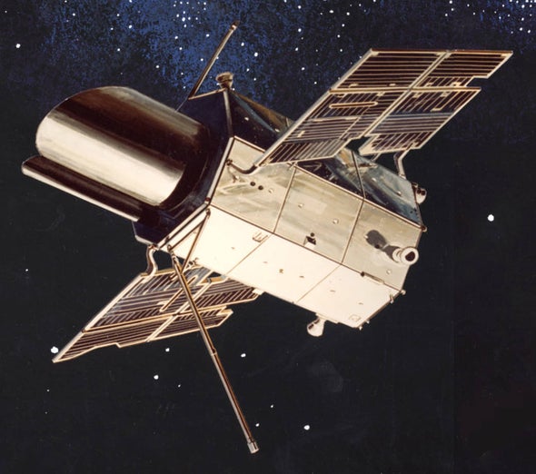
He founded the Space Astronomy Laboratory at UW–Madison and, with his colleagues, proposed to launch a small telescope equipped with a photoelectric photometer, designed to measure the ultraviolet (UV) energy output of stars—a task impossible from Earth’s surface. Fifty years ago, on December 7, 1968, that idea culminated in NASA’s launch of the first successful space-based observatory: the Orbiting Astronomical Observatory, or OAO-2.

With it was born the era of America’s Great Observatories, bearing the Hubble, Spitzer, Chandra and Compton space telescopes, a time during which our understanding of the universe repeatedly deepened and transformed. Today, dwindling political appetite and lean funding threaten our progress. Contemporary projects like the James Webb Space Telescope flounder, and federal budgets omit promising projects like the Wide Field Infrared Survey Telescope (WFIRST).

In celebrating the half century since OAO-2’s launch, we are reminded that major scientific achievements like it become part of the public trust, and to make good on the public trust, we must repay our debt to history by investing in our future. Advances like those made by Hubble are possible only through sustained, publicly-funded research.

These first investments originated in the late 1950s, during the space race between the U.S. the USSR. They led to economic gains in the private sector, technological and scientific innovations, and the birth of new fields of exploration.

Astronomer Lyman Spitzer, considered the father of the Hubble Space Telescope, first posited the idea of space-based observing seriously in a 1946 RAND Corporation study. By leaving Earth’s atmosphere, he argued, astronomers could point telescopes at and follow nearly anything in the sky, from comets to galaxy clusters, and measure light in a broader range of the electromagnetic spectrum.

When Code pitched Wisconsin’s idea to the Space Board, the result was NASA funding to create part of the scientific payload for OAO. The agency went to work planning a spacecraft that could support these astronomical instruments. The Cook Electric Company in Chicago and Grumman Aircraft Engineering Corporation in New York won contracts to help pull it off.

The payload, named the Wisconsin Experiment Package (WEP), bundled five telescopes equipped with photoelectric photometers and two scanning spectrophotometers, all with UV capabilities. The Massachusetts Institute of Technology created a package of X-ray and gamma detectors.

Scientists and engineers had to make the instruments on OAO both programmable and capable of operating autonomously between ground contacts. Because repairs were impossible once in orbit, they designed redundant systems and operating modes. Scientists also had to innovate systems for handling complex observations, transmitting data to Earth digitally (still a novelty in those days), and for processing data before they landed in the hands of astronomers.

The first effort, OAO-1, suffered a fatal power failure after launch in 1966, and the scientific instruments were never turned on. But NASA reinvested, and OAO-2 launched with a new WEP from Wisconsin, and this time a complementary instrument from the Smithsonian Astrophysical Observatory, called Celescope, that used television camera technology to produce images of celestial objects emitting UV light. Expected to operate just one year, OAO-2 continued to make observations for four years.


Numerous “guest” astronomers received access to the instruments during the extended mission. Such collaborations ultimately led to the creation of the Space Telescope Science Institute, which Code helped organize as acting director in 1981.

And the data yielded many scientific firsts, including a modern understanding of stellar physics, surprise insights into stellar explosions called novae, and exploration of a comet that had far-reaching implications for theories of planet formation and evolution.

To be responsible beneficiaries of such insights, we must remember that just as we are yesterday’s future, the firsts of tomorrow depend on today. We honor that public trust only by continuing to fund James Webb, WFIRST, and other projects not yet conceived.

In the forward of a 1971 volume publishing OAO-2’s scientific results, NASA’s Chief of Astronomy Nancy G. Roman wrote: “The performance of this satellite has completely vindicated the early planners and has rewarded … the entire astronomical community with many exciting new discoveries and much important data to aid in the unravelling of the secrets of the stars.”

Let’s keep unraveling these stellar secrets.

Invasion of the Nostril Ticks.


Tony Goldberg had been back from Uganda for only about a day when he felt a distressingly familiar itch in his nose. A veterinary epidemiologist at the University of Wisconsin, Madison, he had just spent a few weeks in Kibale National Park studying chimpanzees and how the diseases they carry might make the jump to humans. Now, he realized, he might have brought one of their parasites home with him.

There was only one way to be sure. Goldberg quickly gathered the necessary supplies—a pair of forceps, a flashlight, and a mirror—and steeled his resolve. Using the mirror to steer his hand, he poked the instrument into his irritated nostril, latched onto a suspicious lump, and quickly yanked it out, careful not to snag any nose hairs in the process. There it was: an adolescent tick. At that point, Goldberg knew, it had likely been living in his nostril for several days.

This was not Goldberg’s first nostril tick, and it’s unlikely to be his last. (On the whole, he says, the experience is “not pleasant but not as bad as you might think.”) He’s seen lots of chimpanzees with nostril ticks during his time in the field, so he’s not surprised a few of the parasites have taken advantage of his presence to burrow into the nose of a closely related primate. This particular tick, however, presented a unique opportunity. Because he found it when he was already back in his lab, Goldberg says, “I was in a position to preserve it for DNA analysis. It was just lucky that the timing was right.”

The nostril tick belonged to the genus Amblyomma, species of which are known to carry diseases that can infect mammals ranging from cows to people. But for now, that’s all Goldberg knows. “Its genetic sequence didn’t match anything in any known databases. So it could be a known species of tick that hasn’t been genetically characterized yet, or a completely new species,” he says. Goldberg reports his analysis in the latest issue of The American Journal of Tropical Medicine and Hygiene.

“It’s fun to welcome Tony to that small, elite club of publishers on ticks in the nose,” says Gary Aronsen, an anthropologist at Yale University who is one of the few other scientists to have written about a close encounter with a nostril tick. (He sneezed his out during a layover in Amsterdam and brought it home with him in a chewing gum wrapper, though he wasn’t able to sequence its DNA.) Picking up parasites like these is “part of the glory and glamour of fieldwork.”

Although researchers know very little about nostril ticks, including which other species they infest and if they carry any diseases, Goldberg speculates that his might be adapted to live in noses of chimpanzees. Chimps are fastidious groomers, so any parasite that wants to hang around for a while needs to fly under the radar. “I can’t think of a better way to do that than hide in an anatomic site that is difficult to access with the fingers,” Goldberg says. “There are several of those—some of which we won’t discuss—but the nostril certainly counts.” (In case you’re wondering, yes, chimps do pick their noses, but it doesn’t seem to dislodge the ticks.)

Because most ticks need to feed on at least three different hosts in their lifetimes, they are exceptionally good at transmitting disease. Species-jumping nostril ticks are “yet another example of how nature provides opportunities for pathogen spillover,” says tick biologist Thomas Mather of the University of Rhode Island, Kingston. Still, the thought of nostril ticks spreading throughout North America isn’t keeping him up at night. “I’m not looking at this as a likely pathway for the introduction of exotic ticks. How many ticks are going to be in a person or two’s nose?”

Nearly a year and a half after removing his own nostril tick, Goldberg hasn’t suffered any ill effects. But the parasite remains a mysterious creature, and for now, the only thing to do is wait for more specimens to turn up. He hopes his paper will raise awareness among his fellow field scientists. Soon, he suspects, “somebody somewhere will come up with another nose tick and will advance the field to the next level.”

Sleep ‘boosts brain cell numbers’


Scientists believe they have discovered a new reason why we need to sleep – it replenishes a type of brain cell.

Sleep ramps up the production of cells that go on to make an insulating material known as myelin which protects our brain’s circuitry.

_69607872_synapses,_artwork-spl-1

The findings, so far in mice, could lead to insights about sleep’s role in brain repair and growth as well as the disease MS, says the Wisconsin team.

The work is in the Journal of Neuroscience.

Dr Chiara Cirelli and colleagues from the University of Wisconsin found that the production rate of the myelin making cells, immature oligodendrocytes, doubled as mice slept.

The increase was most marked during the type of sleep that is associated with dreaming – REM or rapid eye movement sleep – and was driven by genes.

In contrast, the genes involved in cell death and stress responses were turned on when the mice were forced to stay awake.

Precisely why we need to sleep has baffled scientists for centuries. It’s obvious that we need to sleep to feel rested and for our mind to function well – but the biological processes that go on as we slumber have only started to be uncovered relatively recently.

Growth and repair

Dr Cirelli said: “For a long time, sleep researchers focused on how the activity of nerve cells differs when animals are awake versus when they are asleep.

_69630898_matthew

“Now it is clear that the way other supporting cells in the nervous system operate also changes significantly depending on whether the animal is asleep or awake.”

The researchers say their findings suggest that sleep loss might aggravate some symptoms of multiple sclerosis (MS), a disease that damages myelin.

In MS, the body’s immune system attacks and destroys the myelin coating of nerves in the brain and spinal cord.

Future studies could look at whether or not sleep affects the symptoms of MS, says Dr Cirelli.

Her team is also interested in testing whether lack of sleep, especially during adolescence, may have long-term consequences for the brain.

Sleep appears necessary for our nervous systems to work properly, says the US National Institute of Neurological Disorders and Stroke (NINDS).

Deep sleep coincides with the release of growth hormone in children and young adults. Many of the body’s cells also show increased production and reduced breakdown of proteins during deep sleep.

Since proteins are the building blocks needed for cell growth and for repair of damage from factors like stress and ultraviolet rays, deep sleep may truly be “beauty sleep”, says NINDS.

Source:BBC

Words prompt us to notice what our subconscious sees.


It’s a case of hear no object, see no object. Hearing the name of an object appears to influence whether or not we see it, suggesting that hearing and vision might be even more intertwined than previously thought.

Studies of how the brain files away concepts suggest that words and images are tightly coupled. What is not clear, says Gary Lupyan of the University of Wisconsin in Madison, is whether language and vision work together to help you interpret what you’re seeing, or whether words can actually change what you see.

Lupyan and Emily Ward of Yale University used a technique called continuous flash suppression (CFS) on 20 volunteers to test whether a spoken prompt could make them detect an image that they were not consciously aware they were seeing.

CFS works by displaying different images to the right and left eyes: one eye might be shown a simple shape or an animal, for example, while the other is shown visual “noise” in the form of bright, randomly flickering shapes. The noise monopolises the brain, leaving so little processing power for the other image that the person does not consciously register it, making it effectively invisible.

Wheels of perception

In a series of CFS experiments, the researchers asked volunteers whether or not they could see a specific object, such as a dog. Sometimes it was displayed, sometimes not. When it was not displayed or when the image was of another animal such as a zebra or kangaroo, the volunteers typically reported seeing nothing. But when a dog was displayed and the question mentioned a dog, the volunteers were significantly more likely to become aware of it. “If you hear a word, that greases the wheels of perception,” says Lupyan: the visual system becomes primed for anything to do with dogs.

In a similar experiment, the team found that volunteers were more likely to detect specific shapes if asked about them. For example, asking “Do you see a square?” made it more likely than that they would see a hidden square but not a hidden circle.

James McClelland of Stanford University in California, who was not involved in the work, thinks it is an important study. It suggests that sight and language are intertwined, he says.

Lupyan now wants to study how the language we speak influences the ability of certain terms to help us spot images. For instance, breeds might be categorised differently in different languages and might not all become visible when volunteers hear their language’s word for “dog”. He also thinks textures or smells linked to an image might have a similar effect on whether we perceive it as words.

Source: http://www.newscientist.com

Brain cells give insight into Down’s syndrome.


downs

Brain cells have been grown from skin cells of adults with Down’s syndrome in research that could shed new light on the condition.

US scientists found a reduction in connections among the brain cells and possible faults in genes that protect the body from ageing.

The research in the Proceedings of the National Academy of Sciences gives an insight into early brain development.

Down’s syndrome results from an extra copy of one chromosome.

This generally causes some level of learning disability and a range of distinctive physical features.

A team led by Anita Bhattacharyya, a neuroscientist at the Waisman Center at the University of Wisconsin-Madison, grew brain cells from skin cells of two individuals with Down’s syndrome.

This involved reprogramming skin cells to transform them into a type of stem cell that could be turned into any cell in the body.

Brain cells were then grown in the lab, providing a way to look at early brain development in Down’s syndrome.

One significant finding was a reduction in connections among the neurons, said Dr Bhattacharyya.

“They communicate less, are quieter. This is new, but it fits with what little we know about the Down syndrome brain.”

Continue reading the main story

“Start Quote

It seems to be another step forward, giving us insight into the effects of having three copies of chromosome 21”

Carol BoysChief executive, Down’s Syndrome Association

Brain cells communicate through connections known as synapses. The brain cells in Down’s syndrome individuals had only about 60% of the usual number of synapses and synaptic activity.

“This is enough to make a difference,” added Dr Bhattacharyya. “Even if they recovered these synapses later on, you have missed this critical window of time during early development.”

The researchers looked at genes that were affected in the stem cells and neurons from two individuals with Down’s syndrome.

They found that genes on the extra chromosome, chromosome 21, were increased greatly, particularly genes that responded to damage from free radicals, which may play a role in ageing.

This could explain why people with Down’s syndrome appear to age quickly, although this remains to be tested, said the University of Wisconsin-Madison team.

Commenting on the study, Carol Boys, chief executive of the UK Down’s Syndrome Association, said it was interesting work from an established, well-known team.

“It seems to be another step forward, giving us insight into the effects of having three copies of chromosome 21,” she said.

“We are learning more all the time about the mechanisms that cause certain aspects of the condition Down’s syndrome and this may ultimately result in the development of therapies for treatment.”

Source: BBC

Ask NIH to Stop Funding Cruel UW Experiment on Cats.


For decades, experimenters at the University of Wisconsin-Madison (UW) have been conducting cruel and useless taxpayer-funded “sound localization” studies in which cats have steel coils implanted in their eyes, holes drilled into their skulls, and electrodes implanted in their brains. Sometimes, cats used in this experiment have had their ears cut off or are intentionally deafened by having a toxic chemical applied to their inner ear. The cats are then deprived of food for several days in order to coerce them to look in the direction of sounds during experimental sessions in which their heads are immobilized by a bolt screwed into their skulls.

Internal UW documents and photographs obtained by PETA in response to a successful lawsuit detail the miserable life and death of one of the cats, a gentle tabby named Double Trouble, who was abused and killed in this barbaric experiment. In one instance, Double Trouble woke up while experimenters were cutting into her head. Following a series of invasive surgeries, she developed infections, became lethargic and depressed, started to twitch, and suffered paralysis in half her face. After UW deemed the experiment a failure, the experimenters killed and decapitated Double Trouble so that they could examine her brain.

Experimenters have justified the use of 30 cats like Double Trouble per year in this cruel project not by saying that it would enhance human health but by stating that they needed to “keep up a productive publication record that ensures our constant funding.”

Shockingly, the federal government continues to support this irrelevant and deadly project and has provided UW with more than $3 million in grant money to abuse animals—even though researchers at other institutions around the world are already using modern methods with human volunteers to investigate how the brain locates and processes sound.

PETA has called on federal officials to investigate the circumstances surrounding Double Trouble’s horrendous treatment and take disciplinary action against UW for likely violations of federal animal welfare laws. You can help our efforts by contacting the National Institutes of Health and urging the agency to cut funding for this crude and deadly project.

Sign the petition:

https://secure.peta.org/site/Advocacy?cmd=display&page=UserAction&id=4317&utm_campaign=0912%20Ask%20NIH%20to%20Stop%20Funding%20Cruel%20UW%20Experiment%20on%20Cats%20Post&utm_source=PETA%20Facebook&utm_medium=Promo

Source: PETA