Soft Tissue Sarcoma: What You Need to Know About this Rare Cancer


Soft Tissue Sarcomas account for <1% of all adult tumours and 15% of paediatric tumours. They are a complex, heterogeneous group of tumours that can occur in any part of the body, making them diagnostically challenging. 

Sarcomas can spread to other parts of your body, while aggressively destroying the soft tissues or bones.

The treatment of Soft Tissue Sarcoma depends on the type, stage, size, and other factors, as well as whether your body is suited for chemotherapy or radiotherapy. 

Let’s get a deeper insight into this rare disease.

What Is Soft Tissue and What Is Soft Tissue Sarcoma?

How-cancer-cells-grow

Our soft tissues comprise muscles, tendons, ligaments, fat, blood vessels and other tissues. They connect and surround the organs, while supporting the other structures of our body.

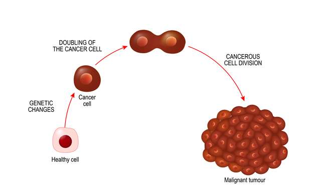
Tumours in the soft tissues can be benign ( non- cancerous) or malignant ( cancerous). The malignant soft tissue tumours are called soft tissue sarcomas.

Sarcoma is a type of cancer that begins in the tissues and can be found anywhere in the body. However, they are most commonly found in the head, arms, neck, abdomen, and legs. 

What Are the Types of Soft Tissue Sarcoma?

Despite having more than 30 types, Soft Tissue Sarcoma is broadly categorised based on the origin tissue. They are:

  1. Muscle tissue
  2. Fibrous tissue
  3. Peripheral nerve tissue
  4. Joint tissue
  5. Blood and lymph vessels

The most commonly identified types are Undifferentiated Pleomorphic Sarcoma (UPS), Gastrointestinal Stromal Tumour (GIST), Liposarcoma, Leiomyosarcoma, Ewing’s Sarcoma, and Synovial Sarcoma. However, in India, the two main types are Ewing’s Sarcoma and Synovial Sarcoma.

What Is Ewing’s Sarcoma?

Ewing’s sarcoma is a rare cancerous disease. It is a tumour in the bone or soft tissues. It occurs in common areas like the pelvis, the femur, the humerus, the ribs, and collarbone. Symptoms include lumps in their legs and arm, which grows over weeks or months.

Ewing’s sarcoma occurs in teenagers and young adults, with a male/female ratio of 1:6. Due to its complex nature, it is often difficult to diagnose Learn more about Ewing Sarcoma here.

What Is Synovial Sarcoma?

Synovial Sarcoma is one of the rarest types of cancer. It usually starts in the thighs, knees, feet, or forearms. It is usually diagnosed after a lump or some related pain. 

Synovial Sarcoma has a male to female ratio of 1.2:1. It can be caused due to multiple risk factors, but mostly due to inherited conditions.

What Are the Risk Factors of Soft Tissue Sarcoma?

Risk-factors-for-Soft-tissue-sarcoma

The exact cause of what causes most soft tissue sarcomas is still under research. However, there have been certain risk factors identified with it. Most of these risk factors are inherited conditions due to gene mutations, and affects the genes in the cells of the soft tissue.

Medical research states the following as risk factors for developing soft tissue sarcoma:

  1. Radiation during treatment of other cancers: Patients might develop sarcoma during treatment for breast cancer or lymphoma. However, it constitutes less than 5% of all sarcomas. The time between radiation exposure and diagnosis of sarcoma is approximately 10 years.
  2. Family Cancer Syndrome: They are disorders caused due to gene mutations. People are usually born with it, and it may increase their chances of developing soft tissue sarcoma. The main types of family cancer syndromes that may cause soft tissue sarcoma are:
    •  Neurofibromatosis, also called von Recklinghausen disease. It forms in the nerves under the skin. 5% of people with Neurofibromatosis will develop sarcoma.
    • Gardner Syndrome, that causes polyps in the colon and intestine. It increases the risk of colon cancer as well, and may also cause problems outside the colon.
    • Li-Fraumeni Syndrome, in which the susceptibility to developing cancers of the breastbrain, or blood is high. People with this syndrome are highly sensitive to radiation, and may eventually develop sarcoma in a new part of the body while being treated.
    • Retinoblastoma, a type of eye cancer prevalent in children.
    • Werner Syndrome, a condition that causes children to have age-related medical conditions like cataracts, skin changes, arteriosclerosis (clogged arteries) and an increased risk of soft tissue sarcoma.
  3. Exposure to cancer-causing chemicals, particularly vinyl chloride, presents a higher risk of developing a sarcoma. Exposure to polycyclic hydrocarbons, asbestos, and dioxin can also up the risk.
  4. Damaged Lymph System, caused due to lymph (clear fluid that contains cells from the immune system) buildup caused by damaged lymph nodes during radiation therapy. It can result in swelling and is also known as lymphedema.

What Are the Symptoms of Soft Tissue Sarcoma?

Symptoms-of-soft-tissue-sarcoma

Soft Tissue Sarcoma may not present any initial sign or symptom due to its complex nature. However, here are some of the symptoms you need to keep an eye out for:

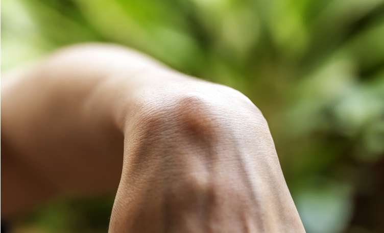
  1. A new lump that is growing in size
  2. Chronic, degenerative pain in the abdomen
  3. Blood in your vomit or stool
  4. Black, sticky bowel (due to internal bleeding in the stomach)

However, lumps and bumps in the body necessarily do not mean Sarcoma. It is best to consult a medical expert if you’re experiencing any of the above.

How Is Soft Tissue Sarcoma Diagnosed?

Diagnosing-Soft-Tissue-Sarcoma

There are a number of tests that are used to identify Soft Tissue Sarcomas. It usually begins with the medical expert determining if the tumour is cancerous or benign and how much it has spread in the body. 

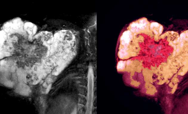
After reviewing the symptoms, the lump specifically, a computed tomography (CT) scan or magnetic resonance imaging (MRI) scan is done to obtain a detailed view of the tumour. 

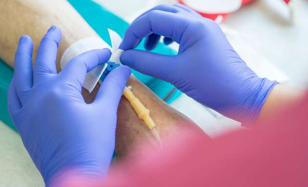
To confirm the diagnosis and determine the type of tumour, a biopsy is done. A biopsy sample can be obtained by withdrawing bits of tissue from the lump with a needle. In some cases, the tissue sample may be obtained during surgery. The tissue sample is examined under the microscope by a pathologist. In addition to making the diagnosis, this specialist can determine how active the sarcoma is by estimating the number of dividing cells (mitoses) in the specimen. Cancers with a large number of mitoses have a worse prognosis and may need aggressive treatment.

If the diagnosis of malignant soft tissue tumour( Sarcoma) is established, the next step is to determine the cancer’s stage—a measure of how much it has spread. The stage is based on:

  • the tumour’s size
  • the tumour’s grade (how rapidly the cells are dividing and how abnormal they look under the microscope)
  • whether or not cancer cells are in nearby lymph nodes
  • whether or not the cancer has spread beyond its original site to other parts of the body. A positron emission tomography (PET- CT) scan may be done to identify a spread of tumour( Staging).  

PET scan  provides a more accurate picture of where cancer is located. Because PET looks at the entire body, it can be useful when your doctor thinks the cancer may have spread to the other parts of the body.

Treatment will be advised only after the stage, type, and other important factors have been determined.

What Are the Stages of Soft Tissue Sarcoma?

Doctors first determine the type of Sarcoma you have. It is then graded based on how your cancer might behave or spread to the other parts of your body ; followed by the stage it is on.

Sarcomas are graded on a scale of G1 to G3, with G1 being low-grade and almost similar to normal cells, G2, or medium-grade, and G3 or high-grade tumour which may spread readily to other parts of your body.

The general stages of a Soft tissue sarcoma are:

Stage 1: The tumour is small and low grade

Stage 2: The tumour is small but of a higher grade

Stage 3: The tumour is large and of a higher grade

Stage 4: The cancer has spread to other parts of the body

Tumour sizes are often measured in centimeters (cm) or inches. Common food items that can be used to show tumour size in cm include: a pea (1 cm), a peanut (2 cm), a grape (3 cm), a walnut (4 cm), a lime (5 cm or 2 inches), an egg (6 cm), a peach (7 cm), and a grapefruit (10 cm or 4 inches).

Treatment Options for Soft Tissue Sarcoma

Treatment-for-soft-tissue-sarcoma

There are different methods to treat the different types of soft tissue sarcoma. It is often a combination of therapies best suited for the patient. However, the four standard types of treatment are:

  • Surgery
  • Radiation therapy
  • Chemotherapy
  • Isolated regional therapy

The Prognosis for Soft Tissue Sarcoma

In general, people with localised Soft Tissue Sarcomas have a very good prognosis with a high rate of cure. The main feature of an excellent prognosis is a tumour that is completely removed by surgery and hasn’t spread beyond the margins of the tumour. Children tend to have a better prognosis than adults for both localised tumours and those that have spread.

Preventing Soft Tissue Sarcoma

There is no known way to prevent sarcomas. However, because HIV infection seems to increase the risk of some sarcomas, you should avoid behaviours that can lead to HIV infection.

If your occupation exposes you to substances that can cause soft tissue sarcomas, use proper protective equipment to reduce your exposure.

With advancements in medical science and continual research and development to treat cancers, soft tissue sarcoma, despite being a rare type of cancer, may be treated if detected early on. Overall, the 5-year survival rate for soft tissue sarcoma is more than 65%.

It is important to understand that not everyone with a risk factor will develop soft tissue sarcoma, especially due to its rare nature. However, it is best to consult your doctor if you observe any visible lumps or unmanageable pain.

Genetic Map Helps Pinpoint Genes That Cause Sarcoma


Sarcomas are rare cancers arising in bone, muscle, fat, or cartilage and account for about 20% of the cancers diagnosed in people under the age of 20. Now, researchers from Omico, the Garvan Institute of Medical Research, and UNSW Sydney report several genes that cause sarcoma have been identified in the first comprehensive genetic map of sarcomas.

Their new study is published in Science in an article titled, “Heritable defects in telomere and mitotic function selectively predispose to sarcomas.”

“Cancer genetics has to date focused on epithelial malignancies, identifying multiple histotype-specific pathways underlying cancer susceptibility,” wrote the researchers. “Sarcomas are rare malignancies predominantly derived from embryonic mesoderm. To identify pathways specific to mesenchymal cancers, we performed whole-genome germline sequencing on 1,644 sporadic cases and 3,205 matched healthy elderly controls.”

The researchers found that one in 14 individuals diagnosed with sarcoma carries a clinically important gene. The researchers also identified a previously unrecognized genetic pathway specific to sarcomas.

“The findings uncovered by this research are so important because by understanding how individuals develop sarcomas, we move closer to earlier detection and better treatments,” explained Mandy Ballinger, PhD, lead author of the paper, and group leader of the genetic cancer risk group at Garvan.

“Cancer is fundamentally a genetic disease, and genomics is the key to unlocking its secrets. This international collaboration has developed new methods for mapping the genetic basis for cancer and identified new heritable pathways that increase cancer risk. These findings fill important gaps in the missing heritability of cancer,” said David Thomas, head of the Genomic Cancer Medicine Laboratory at Garvan and CEO of Omico, a non-profit nationwide network of genomic cancer research and treatment centers.

The map and findings pave the way for people with a family history of sarcoma to test for their genetic risk of developing the disease.

Persistent opioid use common among adolescents, young adults after sarcoma treatment


Adolescents and young adults with sarcoma prescribed opioids for pain often continued using those substances after treatment completion, according to study results published in Cancer.

Approximately two-thirds of adolescents and young adults (AYAs) received prescriptions for opioids during cancer treatment. Nearly one-quarter of AYAs who used opioids during sarcoma treatment met criteria for new persistent opioid use after completing cancer therapy.

Opioid use among younger patients.
Data derived from Beauchemin MP, et al. Cancer. 2022;doi:10.1002/cncr.34238.

Opioid use was expectedly high, given that most of these patients undergo intense multimodal treatment,” Melissa P. Beauchemin, PhD, RN, CPNP-PC, CPON, director of the NCI-Community Oncology Research Program at Herbert Irving Comprehensive Cancer Center at Columbia University and assistant professor at Columbia University School of Nursing, told Healio. “In our study, Medicaid insurance, bone tumors compared with soft tissue sarcomas, and concurrent lorazepam use were associated with new persistent use.”

Background and methodology

AYAs with sarcoma often develop acute and chronic pain due to their disease and the treatments they receive.

Prior research showed high risk for persistent opioid use after cancer therapy among previously opioid-naive older adults, according to study background.

Melissa P. Beauchemin, PhD, RN, CPNP-PC, CPON

Melissa P. Beauchemin

Beauchemin and colleagues aimed to describe patterns of new persistent opioid use among AYAs treated for sarcoma. They defined persistent opioid users as previously opioid-naive patients who filled at least one opioid prescription during active therapy and at least two opioid prescriptions in the year after therapy completion.

Researchers also assessed the association between insurance type and opioid use.

Beauchemin and colleagues used the IBM Marketscan Database to identify patients aged 10 to 26 years diagnosed with sarcoma between 2008 and 2016 who had not used opioids in the period 365 days to 31 days prior to their first treatment.

Their analysis included 938 patients (55.6% men; 62% aged younger than 18 years) with an International Classification of Diseases code for sarcoma. All patients received anticancer therapy — chemotherapy, surgery and/or radiation — within 30 days of the initial diagnosis code and had continuous insurance coverage (78% commercial, 22% Medicaid) for more than 12 months prior to diagnosis and more than 12 months after their final therapy.

Twenty-nine percent of patients had a mental health diagnosis before or during the treatment period.

Key findings

A majority (64%) of patients received opioid prescriptions during sarcoma treatment.

Half (49%) of patients used opioids only during treatment.

Nearly one-quarter (23%) of those who used opioids during treatment received at least two prescriptions in the year following therapy.

Multivariable analysis showed non-soft tissue sarcoma type (Ewing sarcoma, OR = 3.23; 95% CI, 1.81-5.78; osteosarcoma, OR = 2.05; 95% CI, 1.36-3.09) and insurance type (Medicaid vs. commercial, OR = 1.74; 95% CI, 1.15-2.64) appeared associated with increased likelihood of persistent opioid use.

“Medicaid insurance may be a proxy for other social determinants of poorer health, including access to quality health care, education level and social support networks,” Beauchemin told Healio.

Patients with a mental health diagnosis exhibited a higher likelihood of persistent opioid use than those without (21% vs. 12%; P < .01). Concomitant use of lorazepam — often used as an antiemetic for chemotherapy-induced nausea — also appeared significantly associated with persistent opioid use (OR = 3.38; 95% CI, 2.26-5.05).

Implications

The results highlight an urgent need for age-appropriate education and guidance for AYAs, as well as development of novel pain management strategies to limit long-term negative effects of opioid use, researchers concluded.

Future studies should focus on managing pain among AYAs undergoing treatment while developing ways to limit or discontinue opioid use safely and early to reduce misuse, Beauchemin said.

“Psychosocial screening and targeted interventions should be developed for AYAs, including close monitoring and provision of anticipatory guidance for AYAs and their family members,” Beauchemin told Healio. “Generations of new knowledge around opioid use in AYAs, and potential strategies to mitigate long-term misuse, will help to inform development of clinical practice guidelines to support evidence-based clinical decision-making.”

Uterine Cancer Incidence and Deaths on the Rise in US


The incidence of uterine cancer and deaths from the disease are on the increase, with black women disproportionately affected, warn researchers at the Centers for Disease Control and Prevention (CDC). They call for greater awareness of the symptoms to allow early detection and treatment.

Uterine cancer “is one of the few cancers with increasing incidence and mortality in the United States,” the CDC notes. This reflects, in part, increases in the prevalence of overweight and obesity since the 1980s.

It is the fourth most common cancer diagnosed in US women and is the seventh most common cause of death.

The findings were published online December 7 in the Morbidity and Mortality Weekly Report.

S. Jane Henley, MSPH, from the National Center for Chronic Disease Prevention and Health Promotion, CDC, and colleagues studied the official incidence and mortality rates for uterine cancer from 1999 to 2015/6.

They found that rates of the disease have been increasing by approximately 0.7% per year, with uterine cancer deaths rising by an average of slightly more than 1.0% per year.

Worryingly, in comparision with other groups, black women were more likely to be diagnosed with harder-to-treat forms of the disease and with later-stage disease, in particular in comparision with white women.

“Multifactorial efforts at individual, community, clinical, and systems levels to help women achieve and maintain a healthy weight and obtain sufficient physical activity might reduce the risk for developing uterine cancer,” the authors write.

“Promoting awareness among women and health care providers of the need for timely evaluation of abnormal vaginal bleeding can increase the chance that uterine cancer is detected early and treated appropriately,” they add.

Study Details

The team gathered incidence data from the CDC’s National Program of Cancer Registries and the National Cancer Institute’s Surveillance, Epidemiology, and End Results Program.

In addition, they obtained mortality data from the National Vital Statistics System, which covered 98% of the overall US population for the period 1999 to 2015/2016.

Uterine cancers were classified by histologic site and stage at diagnosis. Individuals were classified as white, black, non-Hispanic American, Indian/Alaska Native (AI/AN), non-Hispanic Asian/Pacific Islander (API), or Hispanic.

The researchers found that in 2015, there were 53,911 new, confirmed cases of uterine cancer, which occurred at a rate of 27 cases per 100,000 women. The rates were highest among white and black women (27 per 100,000 each).

The most commonly reported form the disease was endometrioid carcinoma, which occurred in 68% of women. The proportion was much lower in black women, at 47%. Black women who were more likely to have other carcinomas, carcinosarcomas, and sarcomas.

In non-black women, uterine cancers were diagnosed at the localized stage in 66% to 69% of cases. In black women, that rate was 55%.

Black women are also more likely to be diagnosed with disease of distant stage than other groups, at 16% vs 8% to 10%. This was particularly the case for sarcoma.

Sarcomas were more likely to be diagnosed at the distant stage (36%) than carcinosarcomas (22%), other carcinomas (18%), and endometrioid carcinomas (3%).

The incidence rate of uterine cancers increased between 1999 and 2015 by 12%, or an average of 0.7% per year.

The increase was far higher among AI/AN (53%), black (46%), API (38%), and Hispanic (32%) women than among white women (9%).

In 2016, there were 10,733 deaths due to uterine cancer, at five deaths per 100,000 women. The rate was highest among black women, at nine per 100,000 women.

The rate of uterine cancer deaths increased between 1999 and 2016 by 21%, or 1.1% per year on average.

The increases were higher in API (52%), Hispanic (33%) and black (29%) women than white women (18%). Rates of uterine cancer deaths remained stable in AI/AN women.

Obesity a Contributing Factor

The researchers say that one contributing factor in the increase in incidence could be “excess body weight,” inasmuch as overweight or obese women are two to four times more likely to develop endometrial cancer than women of healthy weight.

“During 2013-2016, approximately 40% of women in the United States had obesity, including 56% of black women and 49% of Hispanic women,” they add.

The team points out that, “as with other cancers, the odds of surviving uterine cancer are much higher when it is detected at an early stage, when treatment is more effective.” The rate of survival is 90% for patients with localized cancers, vs <30% for patients with distant cancers.

“This report found that black women were more likely to receive a diagnosis at distant stage and with more aggressive histologic types than were other women, which might in part account for the higher death rate among black women,” the investigators write.

Carbon Ion Radiotherapy Promising for Inoperable Spinal Tumors.


Carbon ion radiotherapy (CIRT) is a promising option for patients with spinal tumors that cannot be surgically removed, say researchers from Japan. This form of radiation is not currently available in the United States but is being tested in Germany.

CIRT can control cancer growth and prolong life in this “challenging” patient population, they report online today in the journal Cancer.

“In Japan, there are 3 working facilities providing [CIRT],” Reiko Imai, MD, PhD, from the Research Center Hospital for Charged Particle Therapy, National Institute of Radiological Sciences in Chiba, told Medscape Medical News. “The access is open for patients with unresectable sarcomas considered to be radioresistant tumors.”

Safe and Effective, Preserves QoL

Dr. Imai and colleagues carried out a retrospective review on data on 47 patients (24 men and 23 women) with 48 medically unresectable spinal sarcomas, excluding sacral tumors, who received CIRT between 1996 and 2011. Most of the patients (88%) had tumors located less than 5 mm from the spinal cord.

The median age of the patients was 54 years; they were enrolled in phase 1/2 and phase 2 clinical trials of CIRT for bone and soft tissue sarcoma. The applied dose ranged from 52.8 gray equivalents (GyE) to 70.4 GyE (median, 64.0 GyE) in 16 fixed fractions during a 4-week period.

The median follow-up was 25 months, and the median survival was 44 months (range, 5.2 to 148 months).

The researchers say CIRT yielded a 5-year local tumor control rate of 79% and overall and progression-free survival rates of 52% and 48%, respectively. None of the 15 patients with tumors measuring less than 100 cm3had a local recurrence.

No fatal toxicities occurred from treatment. One patient had a grade 3 late skin reaction, and 1 had a grade 4 late skin reaction. Seven patients suffered vertebral body compression, which was salvaged by surgical intervention. One patient had a grade 3 late spinal cord reaction.

Twenty-two of the 28 patients who were alive at last follow-up could walk without supportive devices.

Overall, these findings indicate that CIRT is “both safe and feasible,” the investigators say.

“In this analysis, we would like to emphasize that [CIRT] has the potential to overcome sarcomas and to preserve patients’ quality of life, even if the patients are not candidates for surgery,” Dr. Imai told Medscape Medical News.

“A String of Impressive Papers”

In a telephone interview with Medscape Medical News, David Kirsch, MD, PhD, who specializes in treating sarcoma but who was not involved in the study, noted that this group from Chiba, Japan, has been conducting CIRT trials since 1996 for medically inoperable bone and soft tissue sarcomas and has published “a string of impressive papers.” This includes a study reported by Medscape Medical News in 2002.

Dr. Kirsch is an associate professor of radiation oncology at Duke University School of Medicine in Durham, North Carolina. He thinks CIRT is “an important type of radiation; it has high linear energy transfer [LET] and the potential to kill cells by different mechanisms. These [new] results are good, and I think it’s an important modality to test head to head with standard radiation therapy or proton radiation therapy.”

CIRT is not currently available in the United States. “It would be really nice to have a carbon ion facility in the US and to do a randomized study to figure out if high LET radiation is really killing tumor cells that low LET radiation can’t; that’s kind of the theory,” Dr. Kirsch said.

The problem is cost. “To put up a carbon ion facility is about double the cost of proton therapy, which is also really expensive, although there are certain academic centers [in the United States] that are talking about building a carbon ion center,” Dr. Kirsch said.

He noted that a group in Germany is also testing CIRT against proton therapy in a randomized study.

Source: Medscape.com