Deep learning tool uses MRI to enhance brain tumor diagnosis


Jointly developed by investigators of the Vall d’Hebron Institute of Oncology’s (VHIO) Radiomics Group and the Bellvitge University Hospital’s Neuro-Radiology Unit, the Diagnosis in Susceptibility Contrast Enhancing Regions for Neuroncology (DISCERN) is an open access, deep learning tool based on the training of patterns using artificial intelligence models from information of standard magnetic resonance imaging (MRI).

Published in the journal Cell Reports Medicine, results of a VHIO-led study demonstrate the feasibility and accuracy of DISCERN as an enabler of accurate brain tumor diagnosis from perfusion MRI, outperforming conventional methods. 

Glioblastoma multiforme, brain metastasis from solid tumors, and primary central nervous system lymphoma account for up to 70% of all malignant brain cancers. Given that each of these malignancies require a distinct therapeutic approach, differential diagnosis between these different malignancies represents an unmet clinical need. “The non-invasive differential diagnosis of brain tumors is currently based on the assessment of magnetic resonance imaging before and after contrast administration. However, a definitive diagnosis often requires neurosurgical interventions that compromise the quality of life of patients,” said Raquel Perez-Lopez, Head of VHIO’s Radiomics Group and corresponding author of this present study. 

Validating this deep learning tool in more than 500 additional cases, results of this present study demonstrate 78% accuracy in the classification of these tumors, surpassing conventional methodsRaquel Perez-Lopez

“This work is the result of more than five years’ research focused on the identification of innovative magnetic resonance perfusion imaging biomarkers to enable the differential diagnosis of brain tumors. This present study integrates insights generated by other previous research projects on artificial intelligence, resulting in the development of software that automates presurgical diagnostic classification with very good precision, while facilitating its clinical applicability with a user-friendly interface for clinicians,” added Albert Pons-Escoda, a Clinical Neuroradiologist and Investigator of the Neuroradiology at the Bellvitge University Hospital, and co-author of this study. 

This novel deep learning tool leverages the full spatial and temporal information of conventional MRI to identify behavioral patterns on imaging specific to each tumor. “Deep learning teaches the machine the characteristics of each tumor detected by magnetic resonance imaging of already diagnosed patients. For example, if we show the machine thousands of images of dogs and cats, it will learn the distinct and defining characteristics of both species, and when it sees a new image, it can differentiate between the two,” added Alonso García-Ruiz, a PhD Student of VHIO’s Radiomics Group and first author of this study.

In this case, the learning units are voxels which represent the minimum measurement of volume to study MRI scans and are equivalent to pixels, but three-dimensional display elements. The investigators trained DISCERN to learn the characteristics of these three most common brain malignancies on 50,000 voxels from 40 diagnosed patients. “Validating this deep learning tool in more than 500 additional cases, results of this present study demonstrate 78% accuracy in the classification of these tumors, surpassing conventional methods,” observed Perez-Lopez 

“DISCERN is a computerized diagnostic support tool that facilitates the classification of brain tumors to help guide medical decision making by multidisciplinary teams regarding the need for and type of surgery required to confirm diagnosis,” said Carles Majós, Clinical Neuroradiologist and Investigator of the Neuro-Rdaiology at the Bellvitge University Hospital, and a co-author of this study. 

To enhance study reproducibility and accelerate its adoption in clinical studies, the proposed method developed by VHIO’s Radiomics Group, in close collaboration with investigators of HUB’s Neuro-Radiology Unit, has been implemented on the user-friendly, open access DISCERN application. For demonstrative and research purposes, the DISCERN app can be accessed here

This study was carried out in collaboration with  colleagues at the Neuro-Radiology Unit of the Bellvitge Biomedical Research Institute – IDIBELL and Clínic Hospital (Barcelona, Spain), and HT Medica (Andalucia, Spain),  and Radiation Medicine and Applied Sciences and Bioengineering Departments of the University of California (San Diego, USA). This work has been carried out thanks to the support received from the CRIS Cancer Foundation, FERO Foundation, and “la Caixa” Foundation, which fund research of VHIO’s Radiomics Group through various initiatives. 

Source: Vall d’Hebron Institute of Oncology

MRI shows cardiac diagnostic value


Cardiovascular magnetic resonance (CMR) imaging has become faster, simpler and more widely available in recent years because it has evolved to deliver effective assessment and diagnosis of a range of heart conditions with expanding guideline indications.

Report: Mark Nicholls

‘MRI is the reference test for anatomical imaging of the heart, for quantifying chamber sizes and function,’ explains Professor Sven Plein, British Heart Foundation Professor of Cardiovascular Imaging and Professor of Cardiology at the University of Leeds, United Kingdom. 

Over recent years, with his Leeds team he has conducted extensive research on MRI – significantly in the cardiac arena – and in particular on the cardiovascular effects of diabetes using MRI and in developing quantitative MRI methods. ‘Cardiovascular MRI,’ he points out, ‘is a non-invasive and safe imaging test with the main benefit of providing high tissue contrast, spatial detail and comprehensive tissue characterisation. Recent developments in parametric mapping and automated perfusion analysis are continuing to move MRI from visual analysis to quantitative description of pathology.’

MRI vs CT

Photo

Whilst MRI offers a number of benefits over CT, he emphasises that the two modalities should be regarded as complementary rather than competing. ‘CT is the method of choice for imaging the coronary arteries, while MRI is most powerful for functional imaging of the myocardium,’ Plein adds. ‘Of course, CT exposes patients to a small dose of X-ray radiation, while MRI doesn’t, and MRI contrast agents are generally safer than CT contrast agents. In practice, however, both tests are increasingly used to replace invasive methods for different indications.

‘In congenital heart disease, MRI is the “go-to” test to figure out complicated anatomy, and plan and assess the effectiveness of complex surgery. In children in particular, we need to avoid repeated exposure to X-rays, so would always aim to use MRI as the first line test. Increasingly, cardiologists are using MRI not just to take anatomical pictures, but also to assess tissue composition and tissue characteristics of heart muscle.’

Photo

Plein outlines the benefits of parametric mapping as an important area of development and one that delivers a quantitative colour map output that encodes a range of tissue properties such as T1, T2 and T2* times. ‘We can map areas and create normal values for these parameters. Increasingly, these quantitative methods are used to describe the composition of the cardiac muscle and its content in terms of fibrotic tissue, fat and water content as well as abnormal loading with, for example, iron in thalassaemia patients.’ 

After several years of development, myocardial blood flow can now also be measured quantitatively with automated in line methods via MRI. That development – which is important in ischaemia detection – will shift the approach from the subjective observation of an experienced cardiologist assessing dynamic images of heart muscle enhanced by contrast, to a numeric quantification of blood flow from a colour-coded map.

Stress perfusion imaging

In daily practice MRI is becoming an indispensable test for patients with congenital heart disease, cardiomyopathy and heart failure, ischaemic heart disease and many moreSven Plein

‘Techniques are now coming onto the market where software generates a colour map that shows the amount of blood flow through the heart muscle at rest and during stress – stress perfusion imaging – and whether there are any abnormalities,’ Plein continues. ‘These will soon be commercially available, offering another exciting addition to the growing quantitative MRI methodology.’

Magnetic resonance imaging continues to help improve understanding of heart disease. ‘MRI provides detailed information about cardiac and vascular function, presence of scar or fibrosis in the heart muscle, fat content, blood flow and much more,’ he points out. ‘In daily practice MRI is becoming an indispensable test for patients with congenital heart disease, cardiomyopathy and heart failure, ischaemic heart disease and many more. In research, MRI methods are in development to image the microstructure of the heart, visualise complex 4-dimensional flow, and heart metabolism. This helps us better understand the mechanism of heart failure and other conditions.’

Advanced MRI detects brain changes after Covid-19.


Researchers at Linköping University have examined the brains of 16 patients previously hospitalised for Covid-19 with persisting symptoms. They have found differences in brain tissue structure between patients with persisting symptoms after Covid-19 and healthy people.

Their findings, published in the journal Brain Communications, can bring insights into the underlying mechanisms of persisting neurological problems after Covid-19. 

Several previous studies of persisting problems after Covid have involved MRI brain scanning. Although researchers have found differences compared with healthy brains, these differences are not specific to Covid-19. 

“It can be frustrating for me as a doctor when I understand that the patients have problems, but I can’t find an explanation because there’s nothing in the MRI scan to explain it. To me, this underlines the importance of trying other examination technologies to understand what’s happening in the brain in patients with persisting symptoms after Covid-19,” says Ida Blystad, neuroradiologist in the Department of Radiology at Linköping University Hospital and researcher affiliated with the Department of Health, Medicine and Caring Sciences at Linköping University and the Centre for Medical Image Science and Visualization (CMIV). 

In their current study, the researchers have therefore added a new type of MR imaging called advanced diffusion MRI. They were particularly interested in the brain’s white matter. This consists mainly of nerve axons and is very important for transporting signals between the different parts of the brain and the rest of the body.

“Diffusion MRI is a very sensitive technology that allows changes in how the nerve axons are organised to be detected. This is one of the reasons why we wanted to use diffusion MRI to study the effects of Covid-19 on the brain that other imaging technologies might not pick up,” says Deneb Boito, doctoral student at the Department of Biomedical Engineering at Linköping University. 

To get an idea of what diffusion MRI is, we can imagine a big city at night. Car headlights and rear lights shine like red and white strings of pearls on the most trafficked roads. We cannot see the road itself, but we understand that it is there, as the cars can easily move about right there. Similarly, doctors and researchers can get an insight into how the brain is constructed on a microscopic level through diffusion MRI. This technology builds on the fact that there is water everywhere in the brain moving in the tissue according to the law of least resistance. Water molecules move more easily along the neural pathways. By measuring the movement of water molecules through the neural pathways, researchers can indirectly infer the structure of neural pathways, just as we can indirectly understand that there is a motorway where there are many cars driving.

To me, these findings are a sign that we must investigate long-term effects of Covid-19 in the brain using more advanced MRI technology than conventional MRIIda Blystad

Healthcare usages of diffusion MRI include diagnosing stroke and planning brain surgery. In their current study, the researchers used a more advanced version of diffusion MRI. They examined 16 men who had been hospitalised for severe Covid-19 and who are participating in the Linköping Covid-19 Study (LinCos) at the Department of Rehabilitation Medicine in Linköping. They still had persisting symptoms after seven months. This group was compared with a group of healthy individuals without post-Covid symptoms who had not been hospitalised for Covid. The participants’ brains were examined with both conventional MRI and diffusion MRI. 

“The two groups differ when it comes to brain white matter structure. This can be one of the causes of the neurological problems experienced by the group that had suffered from severe Covid-19. It’s a result that’s in line with other studies that have shown changes to the brain’s white matter. However, having examined only a small group of patients, we are cautious about drawing any major conclusions. With this technology, we’re not measuring the function of the brain, but its microstructure. To me, these findings are a sign that we must investigate long-term effects of Covid-19 in the brain using more advanced MRI technology than conventional MRI,” says Ida Blystad.

Years after the first outbreak and spread of coronavirus Sars-CoV-2, its impact can still be felt in everyday life. Keep up-to-date with the latest research news, political developments, and background information on Covid-19.

There are several issues that the researchers want to study further. It appears, for instance, that white matter in different parts of the brain is affected in different ways, although it is too early to draw any conclusions as to what these differences mean. An upcoming study will investigate whether changes detected with diffusion MRI are in any way connected to brain activity, and how different parts of the brain communicate with each other through the brain white matter in patients suffering from post-Covid fatigue. 

Another question is what happens over time. The MRI scan provides an image of the brain at that particular moment. As the participants were examined on one occasion only, it is not possible to know whether the differences between the two groups will disappear over time or whether they are permanent.

MRI Helps Researchers Link Enlarged Spaces in Infant Brains to Higher Risk of Autism, Sleep Disorders


Researchers Dea Garic, PhD, and Mark Shen, PhD, both at the UNC School of Medicine’s Department of Psychiatry, discovered that infants with abnormally enlarged perivascular spaces have a 2.2 times greater chance of developing autism compared to infants with the same genetic risk. Their research also indicated that enlarged perivascular spaces in infancy are associated with sleep problems seven to 10 years after diagnosis.

“These results suggest that perivascular spaces could serve as an early marker for autism,” said Garic, assistant professor of psychiatry and a member of the Carolina Institute for Developmental Disabilities (CIDD).

The researchers studied infants at increased likelihood for developing autism, because they had an older sibling with autism. They followed these infants from 6-24 months of age, before the age of autism diagnosis. Their study, published in JAMA Network Open, found that thirty% of infants who later developed autism had enlarged perivascular spaces by 12 months. By 24 months of age, nearly half of the infants diagnosed with autism had enlarged perivascular spaces.

Starting 10 years ago, there has been a resurgence of research on the important functions of CSF in regulating brain health and development. Shen’s lab was the first to report that excessive volume of CSF was evident at 6 months of age in infants who would later develop autism. The current study showed that excessive CSF volume at 6 months was linked to enlarged perivascular spaces at 24 months.

Every six hours, the brain expels a wave of CSF that flows through perivascular spaces to remove potentially harmful neuroinflammatory proteins, such as amyloid beta, from building up in the brain. The CSF cleansing process is especially efficient when we are asleep, as the majority of CSF circulation and clearance occurs during sleep.

Disrupted sleep, however, can reduce CSF clearance from perivascular spaces, leading to dilation or enlargement, but this has previously only been studied in animal studies or in human studies of adults. This is the first study of its kind in children.

Shen, senior author of the JAMA Network Open paper, and Garic hypothesized that CSF abnormalities in infancy would be related to later sleep problems, based on  Shen’s earlier research. The current sleep analysis revealed children who had enlarged perivascular spaces at two years of age had higher rates of sleep disturbances at school age.

“Since autism is so highly linked with sleep problems, we were in this unique position to examine CSF dynamics and sleep,” said Garic, who is first author of the paper. “It was really striking to observe such a strong association separated by such a long period of time over childhood. But it really shows how perivascular spaces not only have an effect early in life, but they can have long term effects, too.”

The research was done in conjunction with the Infant Brain Imaging Study (IBIS), a nationwide network of researchers investigating brain development, autism, and related developmental disabilities. The network consists of five universities, of which the University of North Carolina-Chapel Hill is the lead site.

For their study, Garic and Shen analyzed 870 MRIs from IBIS to measure excessive CSF volume and enlarged perivascular spaces. MRIs were obtained from babies during natural sleep at six, 12, and 24 months of age to observe changes over time.

The infant brain undergoes rapid development over this period. Previously, measurement of perivascular spaces was only thought to be clinically relevant for disorders of aging in older adults, such as in dementia. These findings suggest that younger populations may need to be considered and monitored for these types of brain abnormalities.

“Our findings were striking, given that neuroradiologists typically view enlarged perivascular spaces as a sign of neurodegeneration in adults, but this study reported it in toddlers,” said Garic. “This is an important aspect of brain development in the first years of life that should be monitored.”

Garic and Shen hypothesize that excess CSF volume is stagnant, or clogged, and not circulating through the brain as efficiently as it should. For their next research endeavor, the researchers are planning to once again use MRIs to measure CSF in a sleeping infant’s brain, but this time focusing on the physiology and speed of CSF flow throughout the brain.

The research team is also working with other collaborators to quantify the size of perivascular spaces and the severity of behavioral outcomes. The team also plans to extend their research to neurogenetic syndromes associated with autism, such as Fragile X syndrome and Down syndrome.

“Collectively our research has shown that CSF abnormalities in the first year of life could have downstream effects on a variety of outcomes, including later autism diagnosis, sleep problems, neuroinflammation, and possibly, other developmental disabilities,” said Shen.

An Elderly Woman With Ventricular Tachycardia


A woman in her 90s presented to the emergency department with symptoms of fever, hemoptysis, and syncopal episodes. She had a history of breast cancer and pulmonary nodules with biopsy findings negative for metastatic disease but positive for granulomatous inflammation. Initial vital signs included blood pressure of 103/66 mm Hg, heart rate of 190 bpm, and temperature of 38.7 °C. Electrocardiogram showed monomorphic ventricular tachycardia with a morphology suggesting right ventricular (RV) origin (Figure 1). In addition to receiving intravenous amiodarone hydrochloride, she had correction of mild hypokalemia (3.1 mEq/L [reference range, 3.5-5.2 mEq/L]; to convert to mmol/L, multiply by 1.0). Successful arrhythmia termination occurred shortly thereafter, and her vital signs remained stable. Chest computed tomographic angiography excluded pulmonary embolism but showed pulmonary nodules and mediastinal lymphadenopathy that had progressed compared with her previous computed tomography. An echocardiogram showed severe RV and left ventricular (LV) systolic dysfunction with multiple areas of hypokinesis.

Patient electrocardiogram showing monomorphic regular wide complex tachycardia with a left bundle branch block morphology, with a precordial transition at V3 and inferior axis suggesting right ventricular origin.

Patient electrocardiogram showing monomorphic regular wide complex tachycardia with a left bundle branch block morphology, with a precordial transition at V3 and inferior axis suggesting right ventricular origin.

Discussion

Diagnosis

Cardiac sarcoidosis

What to Do Next

A. Cardiac magnetic resonance imaging

Discussion

The patient underwent cardiac magnetic resonance imaging, which demonstrated areas of nonischemic near-transmural late gadolinium enhancement (LGE) of the interventricular septum and epicardium to midmyocardium of the LV basal to midanterior and basal inferior walls extending to the RV walls and septal papillary muscle (Figure 2A). However, T2 mapping revealed only discrete evidence of edema of a small region of the midanterior LV wall. Taken together, these findings raised the possibility of end-stage cardiac sarcoidosis (CS).

Figure 2.  

A, Cardiac magnetic resonance imaging using phase-sensitive inversion recovery with motion correction demonstrates multiple areas of subepicardial, midmyocardial, and transmural and near-transmural late gadolinium enhancement in the left ventricle (pink arrowhead) and extending to the right ventricular wall and septal papillary muscle (blue arrowhead). B, Selected matched contiguous short-axis rubidium-82 (top row) and fluorodexoyglucose F 18 (FDG) positron emission tomographic images (bottom row) demonstrate reduced perfusion in the midseptum with marked FDG uptake in the septum and inferior wall, sparing the lateral wall.

A, Cardiac magnetic resonance imaging using phase-sensitive inversion recovery with motion correction demonstrates multiple areas of subepicardial, midmyocardial, and transmural and near-transmural late gadolinium enhancement in the left ventricle (pink arrowhead) and extending to the right ventricular wall and septal papillary muscle (blue arrowhead). B, Selected matched contiguous short-axis rubidium-82 (top row) and fluorodexoyglucose F 18 (FDG) positron emission tomographic images (bottom row) demonstrate reduced perfusion in the midseptum with marked FDG uptake in the septum and inferior wall, sparing the lateral wall.

Cardiac fluorodeoxyglucose F 18 (FDG) and rubidium-82 positron emission tomography (PET) for CS was obtained with a strict high-fat, low-carbohydrate diet, followed by 12 hours of fasting prior to the examination per American Society of Nuclear Cardiology recommendations.1 Because the patient was hospitalized, dietary compliance was ensured. Furthermore, FDG uptake was segmental and the LV blood pool maximum standard uptake value was only 1.1, demonstrating adequate suppression of normal myocardial glucose utilization. There was an extensive FDG uptake (Figure 2B), with maximum standard uptake value of 10.4 in the LV septum. There was also extracardiac FDG uptake in hilar and mediastinal lymph nodes and in multiple pulmonary nodules. Rubidium-82 PET showed only mildly reduced perfusion in the basal to apical septal wall.

This case highlights multiple learning points in the use of multimodality imaging for evaluation of patients with CS and understanding its complex pathophysiology. First, the utility of T2 mapping for detecting myocardial edema, a potential surrogate for active inflammation, needs to be better understood, as this can be challenging and in this case underrepresented the extensive positive FDG uptake seen on cardiac PET. Despite extensive LV and RV LGE and depressed biventricular systolic function, substantial myocardial inflammation in this patient is consistent with active rather than end-stage CS. Second, determining individual patient prognosis and assessment of immunosuppression response in patients with CS using cardiac imaging remains challenging. It has been demonstrated that patients without LGE are at an extremely low risk of arrhythmic events.2,3 However, although there is increased risk of arrhythmic events in patients with cardiac involvement identified by LGE, only a fraction of these patients experience arrhythmic events. Patients with both RV and LV LGE are at elevated risk of cardiovascular events compared with patients with only LV enhancement.24 Last, while CS is extremely rare after 70 years of age, as the oldest patient reported in the literature was 80 years old,5 this patient presented with CS in her 90s, reminding us that CS remains on the differential diagnosis in patients with sustained ventricular arrythmias regardless of age.

Patient Outcome

Heart failure guideline–directed medical treatment, oral amiodarone, and immunosuppressive therapy consisting of a high, tapering dose of prednisone with mycophenolate mofetil were started. The patient remained stable with no more arrythmias. After shared decision discussions with the patient, a cardiac defibrillator was not implanted given the patient’s age and out of respect for her wishes. She will undergo follow-up FDG-PET to monitor her response to therapy and disease activity.

MR May Detect Brain Changes Before Symptoms of Dementia Appear


According to a study in Radiology, an advanced MR technique shows promise in helping understand early structural changes in the brain before symptoms of dementia become apparent. The research indicates the structural integrity of the brain’s white matter, as measured with an advanced MR technique, is lower in cognitively normal people who carry a genetic mutation associated with Alzheimer’s disease than it is in non-carriers. Researchers said the findings show the promise of widely available imaging techniques in helping to understand early structural changes in the brain before symptoms of dementia become apparent.

People who carry the autosomal dominant Alzheimer disease (ADAD) mutation have a higher risk of Alzheimer’s disease. The mutation is linked to a buildup of abnormal protein called amyloid-beta in the brain that affects both the gray matter and the signal-carrying white matter.

“It’s thought that the amyloid deposition in the gray matter could disrupt its function, and as a result the white matter won’t function correctly or could even atrophy,” said study lead author Jeffrey W. Prescott, M.D., Ph.D., neuroradiologist at the MetroHealth Medical Center in Cleveland.

An earlier study by Dr. Prescott and colleagues on patients with sporadic Alzheimer’s disease, which comprises 99% of cases, found that white matter structural connectivity, as measured with the MR technique diffusion tensor imaging (DTI), degraded significantly as patients developed more amyloid burden.

“The current work extends these results by showing that similar findings are detectable in asymptomatic at-risk patients,” said Jeffrey R. Petrella, M.D., professor of radiology at Duke University and senior author on both studies.

In the new study, Dr. Prescott and colleagues used data from the Dominantly Inherited Alzheimer Network (DIAN) to compare ADAD mutation carriers with non-carriers to see if there were changes in structural connectivity that could be related to the mutation.

The study participants included 30 mutation carriers, mean age 34 years, and 38 non-carriers, mean age 37. The participants all had normal cognition when they underwent structural brain MR and DTI.

Analysis showed that mutation carriers had lower structural connectivity in the frontoparietal control network, which connects areas mainly in the parietal and frontal lobes, two regions known to be involved with Alzheimer’s disease. Among mutation carriers, there was a correlation between expected years until onset of symptoms and white matter structural connectivity in the frontoparietal control network, even when controlling for amyloid plaque burden.

“This suggests that DTI measures of network integrity may serve as a surrogate for the brain’s resilience to pathologic attack,” Dr. Petrella said.

“We used a network measurement called global efficiency, in which a decreased efficiency can be taken as a breakdown in the organization of the network,” Dr. Prescott added. “The results show that for mutation carriers, global efficiency would decrease significantly as they approach the estimated age of symptom onset.”

The study findings support a potential role for imaging-based identification of structural changes of the brain in people at genetic risk for early-onset Alzheimer’s disease in understanding how genes influence the disease process that leads to dementia.

“This shows the potential of MR as an evaluation tool in patients who are deemed at-risk for Alzheimer’s disease before they develop symptoms,” Dr. Prescott said. “Use of these advanced MR techniques could help further refine identification of at-risk patients and risk measurements.”

The findings also point to a role for imaging in studying therapeutic drugs to treat Alzheimer’s disease. While the majority of trials so far have been performed with patients who already have Alzheimer’s disease or cognitive impairment, earlier identification and treatment of patients at risk represents a more promising avenue for preventing or at least delaying the onset of dementia.”

“One potential clinical use of this study tool would be to add quantitative information to risk factors like family history and use that to help identify patients early, when they may benefit from treatment,” Dr. Prescott said. “But until we have an effective treatment, we will have to wait for that to be implemented.”

The researchers hope to do a follow-up using advanced imaging and updated data from the DIAN network to evaluate the progression of Alzheimer’s disease in the study participants.

Scientists use High-Resolution MRI to Create New Map of Young Human Brain Cortex


Using high-resolution MRI, scientists at the UNC School of Medicine have mapped the surface of the cortex of the young human brain with unprecedented resolution, revealing the development of key functional regions from two months before birth to two years after.

Published in the Proceedings of the National Academy of Sciences, the new cortical development mapping represents a valuable resource for further research on brain development and offers a powerful new approach to the study of brain-development conditions such as autism and schizophrenia.

“These results provide an important reference for exploring and understanding the dynamics of early brain development,” said study senior author Gang Li, PhD, associate professor of radiology at the UNC School of Medicine.

The study’s first author was Ying Huang, a PhD candidate in Li’s laboratory.

The cortex is a sheet of brain cells that wraps around much of the rest of the brain. The most evolutionarily advanced brain region, it is proportionately larger in humans than in other mammals, and is responsible for higher, distinctively human functions including language abilities and abstract reasoning.

The third trimester of pregnancy through the first two years of life is the most dynamic period in cortical development. The cortex thickens markedly during this interval, and grows at an even faster pace in terms of surface area, by forming complicated cortical folds.

Disruptions to cortical thickening and expansion in this phase have been linked to autism and schizophrenia. However, neuroscientists haven’t had as detailed an understanding of this developmental phase as they would like. In particular, they’ve had a need for more comprehensive, high-resolution mapping, across the fetal-to-toddler age range, that divides or “parcellates” the developing cortex into distinct regions with their own growth rates – especially surface area growth rates.

In the study, Li and colleagues performed just such a mapping. They first gathered a set of 1,037 high-quality magnetic resonance imaging (MRI) scans of infants in the third-trimester-to-two-year age interval. The scans came from two other research projects, the UNC/UMN Baby Connectome Project  (BCP) and the Developing Human Connectome Project. The team analyzed the scan data using state-of-the-art, computer-based image-processing methods, essentially dividing the cortical surface into a virtual mesh containing thousands of tiny circular areas, and calculating the surface expansion rate for each of these areas.

The analysis didn’t start with assumptions about the locations of brain structures or functional regions, but this regionalization of the brain became evident anyway from the resulting maps, based solely on the different rates at which areas of the surface expanded. In all, the researchers defined 18 distinct regions, which they found correlated well with what is already known about the developing cortex’s functional regions.

“All these regions show dramatic expansion in surface area during this developmental window, with each region having a distinct trajectory,” Li said.

The maps revealed that each region tended to have the same developmental path as its counterpart in the cortex’s opposite hemisphere. Sex differences were apparent too. Even when controlling for sex differences in overall surface area – male brains having greater area – there remained differences in multiple regions. For example, the medial prefrontal region in the left hemisphere, which is believed to host important functions such as attention and working memory, became proportionately larger in males early in the second year of postnatal life.

The analysis also showed that the patterns of cortical surface area expansion in this early period of life were very different from the patterns of cortical thickness development, suggesting that these two measures of brain development involve distinct mechanisms.

All in all, Li said, the mapping provides fundamental new insights into brain development.

He and his team now plan to extend this approach with MRI scan datasets that start at earlier ages and end at older ones. They also hope eventually to study scan datasets covering children who have autism-spectrum or other neurodevelopmental conditions. Such analyses might offer not only clues to the origins of these conditions, but also the identification of early signs or biomarkers, which in the future could be used to administer early and more effective treatments.

MRI Monitors Liver Fat Response to Bariatric Surgery


Quantitative chemical shift-encoded magnetic resonance imaging (CSE-MRI) is an effective, noninvasive way to monitor liver fat levels over time after bariatric surgery, a study published today in Radiology reports.

Common in obese people, nonalcoholic fatty liver disease (NAFLD) can progress to fibrosis, cirrhosis, and cancer. Although weight loss can reduce the extent of NAFLD, biopsy has been the only possible method of assessing the long-term impact of weight loss on hepatic steatosis.

B. Dustin Pooler, MD, an adjunct assistant professor at the University of Wisconsin School of Medicine and Public Health and a radiologist at Madison Radiologists, S.C., in Madison, Wisconsin, and colleagues used the noninvasive MRI technique to measure average liver proton density fat fraction (PDFF) in 50 patients before bariatric surgery and at several points during the year afterward. They compared this MRI biomarker with postsurgical changes in body mass index (BMI), weight, and waist circumference.

The study cohort, which included 43 women and seven men and was recruited from 2010 to 2015 from medical centers at the University of California San Diego and the University of Wisconsin-Madison, had a mean age of 51.0 years and a mean BMI of 44.9 kg/m2.

Presurgical preparation entailed a very low-calorie diet of 600 to 900 calories per day, started at a mean of 2.6 weeks before surgery. Bariatric procedures included gastric banding (n = 2), gastric bypass (n = 28), gastric sleeve (n = 19), and gastric plication (n = 1). PDFF was assessed presurgically before and after the diet, and along with anthropomorphic measurements was assessed at 1, 3, and 6 months after surgery.

Liver fat content began to normalize early during the low-calorie diet and soon after surgery, suggesting metabolic disturbances of NAFLD begin to correct rapidly in response to the caloric deficit of the presurgical diet even before substantial weight loss.

Overall, 32 (64%) of 50 study participants saw their liver fat normalized and an estimated 90% reached that goal by 42 weeks. Six to 10 months postsurgery, patients’ mean PDFF had returned to the normal range of < 5%, falling from 18.1% to 4.9%, with a mean estimated time to normalization of 22.5 weeks.

Mean BMI fell to 34.5 kg/m2 (mean decrease 10.4), and the cohort’s final mean weight of 91.6 kg represented am overall reduction of 29.9 kg. The final mean waist circumference of 110.9 cm represented a mean decrease of 21.3 cm. All reductions were statistically significant (P < .0001)

Rohit Loomba, MD, director of the NAFLD Research Center at the University of California San Diego and chair of the NAFLD Special Interest Group for the American Association for the Study of Liver Diseases, told Medscape Medical News the findings are an important contribution to the field of liver study.

“Previously the MOZART trial conducted by our group demonstrated the role of MRI-PDFF in assessing treatment response in [nonalcoholic steatohepatitis; NASH] trials. This excellent study clearly extends those findings to examine longitudinal changes in liver fat by MRI-PDFF specifically after bariatric surgery,” said Loomba, who was not involved in the research.

Loomba noted that further studies are needed to examine longitudinal changes that might suggest progressive NAFLD, including biomarkers of fibrosis and inflammation.

Earlier this year, Duke University researchers confirmed MRI’s utility for detecting NAFLD, but concluded it was not sufficiently accurate to replace biopsy in distinguishing between NAFLD and its more advanced relative, nonalcoholic steatohepatitis.

Interestingly, whereas initial PDFF level strongly predicted both rate of liver fat change and time to normalization, body anthropometrics did not predict either outcome. “Decreases in liver fat content were only weakly correlated with starting weight and the amount of overall weight loss, suggesting possible utility in monitoring liver fat with MRI following bariatric surgery, independent of monitoring weight loss,” Pooler and colleagues write.

They suggest that PDFF measurements could also help identify appropriate candidates for bariatric surgery as a result of this robust association between liver fat reduction and pretreatment steatosis levels.

According to the authors, their study is the first longitudinal analysis of changes in liver fat over time in a bariatric surgery population.

Here’s What to Expect Before, During, and After an MRI


MRIs sound like a nightmare—but they don’t have to be.
MRI-Machine

Getting an MRI scan probably doesn’t top your list of ways to spend your free time, unless you like lying in tubes that make loud and mystifying noises. Can’t relate!

Unfortunately, sometimes getting an MRI (which stands for magnetic resonance imaging) is just a medically necessary evil. In that case, you’ll have to schlep over to your local radiology clinic or hospital to spend some quality time inside a machine that lets doctors see what’s up inside your body. If the thought sends shivers down your spine, there’s some good news: MRIs often aren’t as scary as they seem.

In case you’re not familiar with the test, an MRI uses a magnetic field and radio waves to make detailed pictures of your insides.

When you’re inside an MRI machine, its magnetic field temporarily realigns hydrogen atoms in your body, according to the Mayo Clinic. Radio waves make these atoms create very faint signals—and those are used to make cross-sectional images. Those images are layered on top of each other to give doctors a really good view of the inside of your body that they can see from different angles.

Doctors will often turn to an MRI when they suspect you have an injury or illness that an X-ray, CT scan, or ultrasound won’t pick up, Mina Makary, M.D., chief diagnostic radiology resident at The Ohio State University Wexner Medical Center, tells SELF. “It provides excellent anatomical detail of the soft tissues, which is helpful for the evaluation of specific conditions,” she explains.

There is a huge range of issues an MRI can spot, including disk abnormalities in your spine, joint problems, tumors in various organs like your kidneys and ovaries, structural problems in your heart, and brain injuries, according to the Mayo Clinic.

You don’t usually have to do a ton of preparation before you get an MRI.

In most cases, you’ll make an appointment to have your scan done and just show up with zero prep work, Kerry L. Thomas, M.D., a radiologist at Moffitt Cancer Center, tells SELF. But if you’re undergoing a pelvic or abdominal MRI, your doctor may ask you to avoid eating or drinking for a few hours beforehand. Skipping food and beverages for a bit will improve the image quality by causing less movement in your gastrointestinal tract, Bachir Taouli, M.D., a professor of radiology and director of body MRI at the Icahn School of Medicine at Mount Sinai, New York, tells SELF.

There are a few things that can mess with your test, which is why it’s so important to be upfront and honest about your health history.

If you have tattoos, the Mayo Clinic advises asking your doctor whether they might impact your test results, since some darker inks can contain metal. “The most important part of having an MRI is that you do not have any metal on for your scan,” Dr. Thomas says. “The machine is very a strong magnet, and metals can cause problems.”

It’s also important to tell your doctor if you’re pregnant or think you may be pregnant. Medical experts don’t understand the effects of magnetic fields on fetuses, and your doctor may recommend using an alternative test or postponing the MRI until after you give birth, the Mayo Clinic says.

Once you arrive at the appointment, you’ll need to remove all metal you might be wearing, like rings, earrings, or glasses and fill out a checklist to make sure you don’t have metal inside your body, like an artificial heart valve, pacemaker, or cochlear implants. Your doctor may also ask if you have a copper IUD (sold under the brand name ParaGard), since copper is a metal. While it’s safe to get an MRI when you have a copper IUD, the prescribing information recommends doing it at what’s known as 1.5 Tesla (the unit used to measure MRI strength), which isn’t as powerful as the 3.0 Tesla often used for MRIs, Dr. Taouli says. This is to avoid the (very minimal) chance of the magnet affecting the metal in the IUD.

Depending on why you’re having your MRI, you may need an injection of a contrasting agent beforehand.

In some cases, your doctor will want to perform an MRI with contrast, which means you’ll be injected with a contrasting agent like gadolinium right before your MRI. Gadolinium lights up when you get a scan and can help doctors get a better look at your brain, heart, and blood vessels. This can aid them in making a diagnosis of things like cancer or an inflammatory condition like multiple sclerosis, Suresh Mukherji, M.D., chairman of the department of radiology at Michigan State University, tells SELF. The American College of Radiology notes that the use of contrast agents is “not completely devoid of risk,” pointing out that some people may have side effects ranging from minor discomfort to “rare severe life-threatening situations.” According to the ACR, the adverse event rate for gadolinium-based contrast media (GBCM) ranges from 0.07 percent to 2.4 percent, which includes mild reactions (like coldness or warmth, headache, nausea) to more severe allergic-like reactions.

The ACR notes that millions of MRIs are done with contrast every year without issues. Allergic-like reactions and severe life-threatening anaphylactic reactions are uncommon, but can happen in less than 1 percent of cases, according to the ACR.

Some people also worry about after-effects. The ACR notes that residual gadolinium was recently found in the brain tissue of people who received multiple gadolinium-based contrasts in the past. The Food and Drug Administration also released a safety alert stating that the brain can retain gadolinium deposits, but also said it found no evidence that this is harmful. Ultimately, the FDA says that the benefits of an MRI with contrast exceed the potential risks.

If you’re nervous about having an MRI with contrast, talk to your doctor about why they requested this particular test and whether you have other options.

Once you change into a gown, it’s time to get into the MRI machine.

The machine will typically be long and tube-shaped with one or two open ends, though newer “open” MRI machines may not be closed on the sides. An MRI technician will ask you to lie down on a table and will often hand you a headset to put on before the actual test gets started. “Patients are given a headset to allow for communication during the MRI scan,” Dr. Taouli explains.

When it’s time for your test to begin, the technician will go behind a partition and the platform you’re lying on will move into the MRI machine. The table you’re on might move you around to allow for better imaging, but you’ll typically need to keep your body as motionless as possible during your exam. “It is critical to lie still during an MRI examination as any movement can disrupt the images being formed, and the exam will need to be repeated,” Dr. Makary says. The only exception is during a functional MRI, when the technician might ask you to perform small tasks like tapping your thumb against your fingers to see how your brain works, according to the Mayo Clinic.

It’s probably going to be loud and you might feel kind of claustrophobic, but there are some things you can do to make getting an MRI as comfortable as possible.

While there’s some variation depending on your injury or illness, MRIs can take anywhere from 15 minutes to over an hour, according to the Mayo Clinic. That’s a lot of time to be amped up with anxiety, so there are a few steps you can take to stay calm.

During your MRI, you’ll hear really loud noises like thumping and tapping as the machine goes to work. If you already know that’s going to freak you out, you can ask for earplugs. The MRI technician may also be able to play music through the headset you’re wearing, so you can ask if this is a possibility when setting up your scan. You might also want to ask if you’ll be able to use an “open” MRI machine rather than one closed at the sides, or at least one that’s newer and might be roomier than past models.

Even though the inside of newer MRI machines aren’t exactly palatial, they’re better than they used to be. Older MRI machines had ceilings that were very close to a person’s face and head, making it easy to feel claustrophobic during your scan, according to the USCF Department of Radiology & Biomedical Imaging. The tunnels in newer MRI machines are bigger and, while you still might feel a little claustrophobic, you have more space than you would have in the past.

Even better, depending on the part of your body being evaluated, you may not need to have your entire body or head inside the machine at all.

If you’d like to have other options, too, ask your doctor if you’re a candidate for sedation, anesthesia, or an anti-anxiety drug they can prescribe for you to take beforehand. You can also ask about the possibility of holding a “panic button” that you can press if you’re getting scared and need to stop the exam.

It’s worth discussing all of this with your medical team way before your appointment. This will allow you to take advantage of any accommodations possible, just in case, and also to better anticipate exactly what the process will feel like.

You really don’t need to do anything special after your scan.

You’ll simply change back into your clothes, grab your stuff, and go about your day. There also aren’t any restrictions on what you can do after the test. “Patients can resume their normal activities immediately after the MRI scan,” Dr. Taouli says. (Unless you had any drugs for sedation or anxiety, in which case you may need someone to drive you home; be sure to ask your doctor about this beforehand.)

Beyond that, you’ll just need to wait to hear from your doctor about your test results. This might feel as anxiety-provoking as getting the MRI itself, which is why it’s a good idea to ask how long it’ll take for you hear back, along with potential next steps you can expect based on their findings so you’re prepared for all outcomes.

American Cancer Society Guidelines for Breast Screening with MRI as an Adjunct to Mammography


Abstract

New evidence on breast Magnetic Resonance Imaging (MRI) screening has become available since the American Cancer Society (ACS) last issued guidelines for the early detection of breast cancer in 2003. A guideline panel has reviewed this evidence and developed new recommendations for women at different defined levels of risk. Screening MRI is recommended for women with an approximately 20–25% or greater lifetime risk of breast cancer, including women with a strong family history of breast or ovarian cancer and women who were treated for Hodgkin disease. There are several risk subgroups for which the available data are insufficient to recommend for or against screening, including women with a personal history of breast cancer, carcinoma in situ, atypical hyperplasia, and extremely dense breasts on mammography. Diagnostic uses of MRI were not considered to be within the scope of this review.

INTRODUCTION

Mammography has been proven to detect breast cancer at an early stage and, when followed up with appropriate diagnosis and treatment, to reduce mortality from breast cancer. For women at increased risk of breast cancer, other screening technologies also may contribute to the earlier detection of breast cancer, particularly in women under the age of 40 years for whom mammography is less sensitive. The American Cancer Society (ACS) guideline for the early detection of breast cancer, last updated in 2003, stated that women at increased risk of breast cancer might benefit from additional screening strategies beyond those offered to women at average risk, such as earlier initiation of screening, shorter screening intervals, or the addition of screening modalities (such as breast ultrasound or magnetic resonance imaging [MRI]) other than mammography and physical examination. However, the evidence available at the time was insufficient to justify recommendations for any of these screening approaches. The ACS recommended that decisions about screening options for women at significantly increased risk of breast cancer be based on shared decision making after a review of potential benefits, limitations, and harms of different screening strategies and the degree of uncertainty about each.1

Although there still are limitations in the available evidence, additional published studies have become available since the last update, particularly regarding use of breast MRI. The ACS guideline panel has sought to provide additional guidance to women and their health care providers based on these new data.

GUIDELINE DEVELOPMENT

The ACS convened an expert panel to review the existing early detection guideline for women at increased risk and for MRI screening based on evidence that has accumulated since the last revision in 2002 to 2003. Literature related to breast MRI screening published between September 2002 and July 2006 was identified using MEDLINE (National Library of Medicine), bibliographies of identified articles, and unpublished manuscripts. Expert panel members reviewed and discussed data during a series of conference calls and a working meeting in August, 2006. When evidence was insufficient or lacking, the final recommendations incorporated the expert opinions of the panel members. The ACS Breast Cancer Advisory Group members and the National Board of Directors discussed and voted to approve the recommendations.

SUMMARY OF RECOMMENDATIONS

Table 1 summarizes the ACS recommendations for breast MRI screening.

Table TABLE 1. Recommendations for Breast MRI Screening as an Adjunct to Mammography
  1. * Evidence from nonrandomized screening trials and observational studies.
  2. †Based on evidence of lifetime risk for breast cancer.
  3. †Payment should not be a barrier. Screening decisions should be made on a case-by-case basis, as there may be particular factors to support MRI. More data on these groups is expected to be published soon.
Recommend Annual MRI Screening (Based on Evidence* )
   BRCA mutation
   First-degree relative of BRCA carrier, but untested
   Lifetime risk ∼20–25% or greater, as defined by BRCAPRO or other models that are largely dependent on family history
Recommend Annual MRI Screening (Based on Expert Consensus Opinion†)
   Radiation to chest between age 10 and 30 years
   Li-Fraumeni syndrome and first-degree relatives
   Cowden and Bannayan-Riley-Ruvalcaba syndromes and first-degree relatives
Insufficient Evidence to Recommend for or Against MRI Screening‡
   Lifetime risk 15–20%, as defined by BRCAPRO or other models that are largely dependent on family history
   Lobular carcinoma in situ (LCIS) or atypical lobular hyperplasia (ALH)
   Atypical ductal hyperplasia (ADH)
   Heterogeneously or extremely dense breast on mammography
   Women with a personal history of breast cancer, including ductal carcinoma in situ (DCIS)
Recommend Against MRI Screening (Based on Expert Consensus Opinion)
   Women at <15% lifetime risk

BACKGROUND

MRI

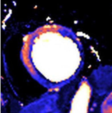
MRI utilizes magnetic fields to produce detailed cross-sectional images of tissue structures, providing very good soft tissue contrast. Contrast between tissues in the breast (fat, glandular tissue, lesions, etc.) depends on the mobility and magnetic environment of the hydrogen atoms in water and fat that contribute to the measured signal that determines the brightness of tissues in the image. In the breast, this results in images showing predominantly parenchyma and fat, and lesions, if they are present. A paramagnetic small molecular gadolinium-based contrast agent is injected intravenously to provide reliable detection of cancers and other lesions. Thus, contrast enhanced MRI has been shown to have a high sensitivity for detecting breast cancer in high-risk asymptomatic and symptomatic women, although reports of specificity have been more variable.2, [3], [4], [5], [6], [7]–8 This high signal from enhancing lesions can be difficult to separate from fat, leading to the use of subtraction images or fat suppression, or both, to assess disease. Because parenchymal tissue also enhances, but generally more slowly than malignant lesions, and also because contrast can wash out rapidly from some tumors, it is important to look at images at an early time point after contrast injection (typically 1 to 3 minutes). MRI examinations may involve examining images at one time point or, more often, will collect a preinjection image with sequential sets of images after contrast injection (dynamic contrast-enhanced [DCE]-MRI). Both the appearance of lesions and, where available, the uptake and washout pattern can be used to identify malignant disease and discriminate it from benign conditions.

These techniques, which have been widely employed for assessing symptomatic disease, have recently been shown to provide good sensitivity as a screening tool for breast cancer in women at increased risk based on family history.9, [10], [11], [12], [13]–14 The approach requires appropriate techniques and equipment, together with experienced staff. Higher quality images are produced by dedicated breast MRI coils, rather than body, chest, or abdominal coils.

IDENTIFICATION OF WOMEN WITH A HIGH RISK OF BREAST CANCER

Three approaches are available for identifying women with a high risk of breast cancer: family history assessment, genetic testing, and review of clinical history. All contribute to identifying women who are candidates for breast MRI screening.

Family History

Although a high proportion of women in the general population have at least one relative with breast cancer, for the majority of these women, this “family history” either does not increase risk at all (ie, the cancer was sporadic) or is associated with, at most, a doubling of lifetime risk (due to either shared environmental risk factors or an inherited gene of low penetrance). Only 1% to 2% of women have a family history suggestive of the inheritance of an autosomal dominant, high-penetrance gene conferring up to an 80% lifetime risk of breast cancer. In some families, there is also a high risk of ovarian cancer. Features of the family history which suggest the cancers may be due to such a high-penetrance gene include 2 or more close (generally first- or second-degree) relatives with breast or ovarian cancer; breast cancer occurring before age 50 years (premenopausal) in a close relative; a family history of both breast and ovarian cancer; one or more relatives with 2 cancers (breast and ovarian cancer or 2 independent breast cancers); and male relatives with breast cancer.15, [16], [17]–18

Two breast/ovarian cancer susceptibility genes, BRCA1 and BRCA2, have been identified.19,20 Inherited mutations in these genes can be found in approximately 50% of families in which an inherited risk is strongly suspected based on the frequency and age of onset of breast cancer cases, and in most families in which there is a much higher than expected incidence of both breast and ovarian cancer.

Several models can assist clinicians to estimate breast cancer risk or the likelihood that a BRCA mutation is present (Online Supplemental Material). The Gail, Claus, and Tyrer-Cusick models estimate breast cancer risk based on family history, sometimes in combination with other risk factors, such as reproductive history or prior breast biopsies.16,21, [22]–23 Although risk prediction is generally similar for the different models, an individual woman’s risk estimate may vary with different models.21,24,25

Two decision models have been developed to estimate the likelihood that a BRCA mutation is present, BRCAPRO18,26 and the Breast and Ovarian Analysis of Disease Incidence and Carrier Estimation Algorithm (BOADICEA)27; the BOADICEA model also provides estimates of breast cancer risk (Online Supplemental Material).

Genetic Testing

The prevalence of BRCA mutations is estimated to be between 1/500 and 1/1,000 in the general population28; however, in women of Jewish ethnicity, the prevalence is 1/50.29,30 Women with cancer-predisposing mutations in either BRCA1 or BRCA2 have an increased risk of both breast and ovarian cancer. From population-based studies, women with BRCA1 mutations are estimated to have a 65% risk by age 70 years for developing breast cancer (95% confidence interval [CI], 44% to 78%); the corresponding risk for BRCA2 mutations is 45% (95% CI, 31% to 56%).31 Risks estimated from cancer-prone families seen in referral centers are higher, with limit of risk in the 85% to 90% range.31 These mutations follow an autosomal dominant pattern of transmission, which means that the sister, mother, or daughter of a woman with a BRCA mutation has a 50% chance of having the same mutation.

The benefits and risks of genetic testing are beyond the scope of this article, but are reviewed in the American Society of Clinical Oncology policy statement update on genetic testing for cancer susceptibility.32 Genetic testing for a BRCA1 or BRCA2 mutation is generally offered to adult members of families with a known BRCA mutation, or to women with at least a 10% likelihood of carrying such a mutation, based on either validated family history criteria or one of the above-mentioned models. If a woman from a family in which a BRCA mutation has been previously identified does not have that mutation, one can generally safely conclude that her breast cancer risk is no higher than it would have been if she did not have a family history of breast cancer. However, in a high-risk family without a known mutation, failure to find a mutation in a particular member does not reduce her risk estimate.

A high risk of breast cancer also occurs with mutations in the TP53 gene (Li-Fraumeni syndrome) and the PTEN gene (Cowden and Bannayan-Riley-Ruvalcaba syndromes).33 Accurate prevalence figures are not available, but these conditions appear to be very rare.34,35

Clinical Indicators of Risk

Some clinical factors are associated with substantial breast cancer risk. Among women with Hodgkin disease, increased breast cancer risk has been consistently and significantly associated with mantle field radiation treatment. In several studies of women treated between 1955 and 1995, risk was inversely related to age at treatment in patients diagnosed between the ages of 10 to 30 years, with only slight or no increased risk when diagnosis was before age 10 years or after age 30 years.36, [37], [38], [39], [40]–41 Risk following treatment with radiation and chemotherapy was half that of treatment with radiation alone in two studies,39,42 which may reflect the effect of chemotherapy on earlier onset of menopause; risk was equivalent in a third study.43 Risk of breast cancer significantly increased 15 to 30 years after radiation therapy.41 More recently, treatment approaches have used lower doses of radiation and limited-field radiotherapy. In one study, which compared patients who received radiation therapy in 1966 to 1974 and 1975 to 1985, treatment in the later timeframe was not related to increased risk of breast cancer after a median follow up of 13 years, whereas patients treated between 1966 and 1974 were at increased risk, suggesting that Hodgkin disease survivors treated with current approaches will not face substantially increased breast cancer risk.44

Lobular carcinoma in situ (LCIS) and atypical lobular hyperplasia (ALH), together described as lobular neoplasia, are associated with substantially increased risk of subsequent breast cancer, with lifetime risk estimates ranging from 10% to 20%.45 This equates to a continuous risk of about 0.5% to 1.0% per year. The invasive cancers may be ipsilateral or contralateral, are usually invasive lobular cancers, and more than 50% of these diagnoses occur more than 15 years after the original diagnosis of LCIS. Similar findings have been reported by Fisher et al,46 describing a 12-year update of 180 women with LCIS who were treated with local excision alone and followed by the National Surgical Adjuvant Breast Project (NSABP), as well as Li et al, who described the risk of invasive breast cancer among 4,490 LCIS patients using Surveillance, Epidemiology, and End Results (SEER) data between 1988 to 2001.47

A typical ductal hyperplasia (ADH) is part of the continuum of ductal proliferative breast diseases ranging from usual ductal hyperplasia to ductal carcinoma in situ (DCIS). The literature review by Arpino et al45 suggests a 4- to 5-fold increased risk of invasive breast cancer (compared with a 6- to 10-fold risk with LCIS) at a median follow up of 17 years, which is doubled if the woman has an associated family history of breast cancer. It is unclear, however, what percentage of the women with this family history and ADH are at this significantly increased risk because they are carriers of a BRCA1 or 2 gene mutation.

Mammographic density has been shown to be a strong independent risk factor for the development of breast cancer.48, [49], [50]–51 In several studies, women with the most breast density were found to have a 4- to 6-fold increased risk of breast cancer, compared with women with the least dense breasts.52, [53], [54], [55]–56 For example, women with 75% or higher mammographic density had a more than five-fold increased risk of breast cancer, compared with women with less than 1% density.57 In addition, it has been shown that malignant tumors of the breast are more likely to arise in the areas of greatest mammographic density, compared with the more fatty areas of the breast.58

The absolute risk of contralateral breast cancer in women with a personal history of breast cancer is estimated to be 0.5% to 1% per year, or 5% to 10% during the 10 years following diagnosis, significantly higher than that of the general population.59 Hormone therapy and/or chemotherapy for the primary cancer is likely to subsequently lower the risk of contralateral breast cancer.

EVIDENCE AND RATIONALE

Evidence of Efficacy from MRI Screening Studies

In the mid to late 1990s, at least 6 prospective, nonrandomized studies were initiated in The Netherlands, the United Kingdom (UK), Canada, Germany, the United States (US), and Italy to determine the benefit of adding annual MRI to (film) mammography for women at increased risk of breast cancer. Some of the studies included ultrasound and/or clinical breast examination, as well. Despite substantial differences in patient population (age, risk, etc.) and MRI technique, all reported significantly higher sensitivity for MRI compared with mammography (or any of the other modalities). All studies that included more than one round of screening reported interval cancer rates below 10%. Participants in each of these 6 studies had either a documented BRCA1 or BRCA2 mutation or a very strong family history of breast cancer. Some of the studies included women with a prior personal history of breast cancer.

Kriege et al screened 1,909 unaffected women aged 25 to 70 years with an estimated 15% or higher lifetime risk of breast cancer (19% proven to have a BRCA mutation) at 6 centers across The Netherlands.9 After a median of 3 rounds of screening, 50 breast cancers (44 invasive) were diagnosed. Eighty percent of the invasive cancers were detected by MRI, compared with 33% by mammography. However, mammography outperformed MRI for detecting DCIS. Of the invasive cancers, 43% were 1 cm or smaller in diameter, and 33% had spread to axillary lymph nodes. The specificity of MRI was 90%, compared with 95% for mammography.

Leach et al screened 649 unaffected women aged 35 to 49 years who had at least a 25% lifetime risk of breast cancer (19% proven to have a BRCA mutation) at 22 centers in the UK.11 After a median of 3 rounds of screening, 35 cancers (29 invasive) were diagnosed. Sensitivity of MRI was 77%, compared with 40% for mammography, with specificities of 81% and 93%, respectively. MRI was most sensitive and mammography least sensitive for women with BRCA1 mutations. Forty-five percent of the cancers were 1 cm or less in size, and 14% had spread to axillary lymph nodes. There were two interval cancers.

Warner et al screened 236 women aged 25 to 65 years with a BRCA mutation at a single center in Toronto for up to 3 years and detected 22 cancers (16 invasive).14 Sensitivity of MRI was 77%, compared with 36% for mammography, with 50% of the cancers 1 cm or smaller, and 13% were node positive. There was one interval cancer. Specificity was 95% for MRI and 99.8% for mammography.

Kuhl et al screened 529 women aged 30 years and older with a lifetime breast cancer risk of at least 20% at a single center in Bonn for a mean of 5 years.10 They detected 43 cancers (34 invasive), with 1 interval cancer. The sensitivity of MRI was 91%, compared with 33% for mammography. The node positive rate was 16%. Specificity of both MRI and mammography was 97%.

The International Breast MRI Consortium screened 390 women aged 25 years and older with more than a 25% lifetime risk of breast cancer at 13 centers (predominantly in the US) on a single occasion.12 Four cancers were found by MRI, and only one of these by mammography. However, because the patients were not followed after screening, the false-negative rate could not be determined. MRI specificity was 95%, compared with 98% for mammography.

In a study in Italy with 9 participating centers, Sardanelli et al screened 278 women aged 25 years and older; 27% carried a BRCA mutation or had a first-degree relative with a BRCA mutation.13 After a median of 1.4 rounds of screening, 18 cancers (14 invasive) were found. MRI sensitivity was 94%, compared with 59% for mammography, 65% for ultrasound, and 50% for clinical breast examination. MRI specificity was 99%.

Overall, studies have found high sensitivity for MRI, ranging from 71% to 100% versus 16% to 40% for mammography in these high-risk populations. Three studies included ultrasound, which had sensitivity similar to mammography. The Canadian, Dutch, and UK studies9,11,14 reported similar sensitivity (71% to 77%) within CIs for MRI, although the single-center study from Germany10 reported a higher sensitivity, which may reflect the concentration of radiological practice and higher patient volume per radiologist at a single center. There is evidence of a learning curve for radiologists conducting MRI breast screening, with the number of lesions investigated falling with experience.60 The three multicenter studies reflect the likely initial effectiveness of this modality in a population context, and it is expected that, with training and advances in technology, sensitivity will increase further.

Table 2 provides a summary of these six screening studies.

Table TABLE 2. Published Breast MRI Screening Study Results
The Netherlands Canada United Kingdom Germany United States Italy
  1. n/a = not applicable.
No. of centers 6 1 22 1 13 9
No. of women 1,909 236 649 529 390 105
Age range 25–70 25–65 35–49 ≥30 ≥25 ≥25
No. of cancers 50 22 35 43 4 8
Sensitivity (%)
   MRI 80 77 77 91 100 100
   Mammogram 33 36 40 33 25 16
   Ultrasound n/a 33 n/a 40 n/a 16
Specificity (%)
   MRI 90 95 81 97 95 99
   Mammogram 95 >99 93 97 98 0
   Ultrasound n/a 96 n/a 91 n/a 0

Most of the available data are based on screening women at high risk due to family history and/or genetic mutations. More recently, smaller studies have provided information on the potential benefit of MRI screening for women with clinical factors that put them at increased risk. Preliminary data were obtained from one retrospective study, in which Port et al61 reviewed the screening results of 252 women with biopsy-confirmed LCIS and 126 women with atypical hyperplasia (either ductal or lobular), of whom half were screened with annual mammography and biennial clinical exams and half were also screened with MRI. The women who were screened with MRI were younger and more likely to have a strong family history. MRI screening offered a small advantage to patients with LCIS, but not atypical hyperplasia, and also resulted in increased biopsies: 6 cancers were detected by MRI in 5 women with LCIS (4% of patients undergoing MRI), and none were detected in women with atypical hyperplasia. Biopsies were recommended for 25% of MRI screened patients; 13% of biopsies had a cancer detected. All of the cancers in women screened with MRI were Stage 0 to I, whereas all of the cancers in women who were not screened with MRI were Stage I to II. Cancer was detected on the first MRI in 4 of 5 patients. The sensitivity of MRI was 75%, the specificity was 92%, and the positive predictive value was 13%.

Technological Limitations and Potential Harms Associated with MRI Screening

Although the efficacy of breast MRI has been demonstrated, it does not achieve perfect sensitivity or specificity in women undergoing screening, and as such, the issue of adverse consequences for women who do, but especially those who do not, have breast cancer is important to address. As with mammography and other screening tests, false negatives after MRI screening can be attributed to inherent technological limitations of MRI, patient characteristics, quality assurance failures, and human error; false positives also can be attributed to these factors, as well as heightened medical-legal concerns over the consequence of missed cancers. A patient’s desire for definitive findings in the presence of a low-suspicion lesion may also contribute to a higher rate of benign biopsies. The consequences of all these factors include missed cancers, with potentially worse prognosis, as well as anxiety and potential harms associated with interventions for benign lesions.

The specificity of MRI is significantly lower than that of mammography in all studies to date, resulting in more recalls and biopsies. Call-back rates for additional imaging ranged from 8% to 17% in the MRI screening studies, and biopsy rates ranged from 3% to 15%.9, [10], [11], [12], [13]–14 However, several researchers have reported that recall rates decreased in subsequent rounds of screening: prevalence screens had the highest false-positive rates, which subsequently dropped to less than 10%.9,62,63 Most call backs can be resolved without biopsy. The call-back and biopsy rates of MRI are higher than for mammography in high-risk populations; while the increased sensitivity of MRI leads to a higher call-back rate, it also leads to a higher number of cancers detected. The proportion of biopsies that are cancerous (positive predictive value) is 20% to 40%.9, [10], [11], [12], [13]–14 Since false-positive results appear to be common, more data are needed on factors associated with lower specificity rates.

Table 3 compares the likelihood of detection and follow-up tests for women who underwent screening MRI and mammography in two screening studies (Dutch and UK). The study populations differed, with the Dutch study having a wider age group and lower risk category, compared with the UK study.9,11 This affected both the prevalence of cancer and the pick-up rate by modality in the two studies. These results, drawn from two trials, demonstrate the relatively high recall rate in the high-risk population, as well as the fact that MRI is a relatively new technique. Despite the high number of recalls, because of the high cancer rate, the rate of benign surgical biopsy in the UK study per cancer detected was similar to that experienced in the population-based national breast screening service. Recalls will inevitably lead to additional investigations, many of which will not demonstrate that cancer is present.

Table TABLE 3. Rates of Detection and Follow-up Tests for Screening MRI Compared with Mammography
MRI Mammography
The Netherlands United Kingdom The Netherlands United Kingdom
Positives 13.7% 19.7% 6.0% 7.2%
Recalls 10.84% 10.7% 5.4% 3.9%
Biopsies 2.93% 3.08% 1.3% 1.33%
Cancers 1.04% 1.44% 0.46% 0.69%
False negatives 0.23% 0.43% 0.81% 1.52%

Given the high rate of cancer combined with the risk of false-positive scans in a high-risk population undergoing MRI-based screening, the psychological health of these women merits study. In a subgroup of 611 women in the UK study, 89% reported that they definitely intended to return for further screening, and only 1% definitely intended not to return. However, 4% found breast MRI “extremely distressing,” and 47% reported still having intrusive thoughts about the examination 6 weeks afterward.64

In a sample of 357 women from the Dutch study, psychological distress remained within normal limits throughout screening for the group as a whole. However, elevated breast cancer-specific distress related to screening was found in excessive (at least once per week) breast self-examiners, risk overestimators, and women closely involved in the breast cancer case of a sister. At least 35% of the total sample belonged to one of these subgroups. It was recommended that patients in one of these vulnerable subgroups be approached for additional psychological support.65

In a small sample of women from the Toronto study followed over a course of 2 years, there was no evidence of any effect on global anxiety, depression, or breast cancer-related anxiety.66 In another sample of 57 women, almost 50% had elevated baseline general and/or breast cancer-specific anxiety, but in 77% of cases this was attributed by the patients to life events, including relatives with cancer. A nonsignificant increase in general anxiety and breast cancer-related anxiety, compared with baseline, was found in the subset of women recalled for further imaging or biopsies.67 Follow-up time is still insufficient to determine whether anxiety scores return to baseline once the work up has been completed.

There is a special responsibility to alert patients to this technology, with its potential strengths and harms, and to be encouraging, while allowing for shared decision making. The interplay between risks, benefits, limitations, and harms is complicated by the fact that individual women likely will weigh these differently depending on their age, values, perception of risk, and their understanding of the issues. Steps should be taken to reduce anxiety associated with screening and the waiting time to diagnosis, and conscientious efforts should be made to inform women about the likelihood of both false-negative and false-positive findings. How information is conveyed to the patient greatly influences the patient’s response: it is important that providers not convey an undue sense of anxiety about a positive MRI finding. While the high rate of biopsies and further investigations is acceptable in women with a high risk of breast cancer, the number of such investigations in women at lower risk will be much higher than would be appropriate, leading to the need to counsel women in lower risk categories that MRI screening is not advisable and that the harms are believed to outweigh the benefits. Such advice needs to be based on considerations of family history, genetic mutation status, other risk factors, age, and mammographic breast density.

There are substantial concerns about costs of and limited access to high-quality MRI breast screening services for women with familial risk. In addition, MRI-guided biopsies are not widely available. With many communities not providing MRI screening and with MRI-guided biopsies not widely available, it is recognized that these recommendations may generate concerns in high-risk women who may have limited access to this technology.

The ability of MRI to detect breast cancer (both invasive and in situ disease) is directly related to high-quality imaging, particularly the signal-to-noise ratio, as well as spatial resolution of the MR image. In order to detect early breast cancer (ie, small invasive cancers, as well as DCIS), simultaneous imaging of both breasts with high spatial resolution is favored. High spatial resolution imaging should be performed with a breast coil on a high field magnet with thin slices and high matrix (approximately 1 mm in-plane resolution). These technical parameters are considered to be the minimal requirements to perform an adequate breast MRI study. The ability to perform MRI-guided biopsy is absolutely essential to offering screening MRI, as many cancers (particularly early cancers) will be identified only on MRI. The American College of Radiology (ACR) is currently developing an accreditation process for performing breast MRI, and, in addition to the performance of high spatial resolution images, the ability to perform MRI intervention (ie, needle localization and/or biopsy) will be essential in order to obtain accreditation by this group. Accreditation will be voluntary and not mandatory. This guideline will likely be available in 2007.

There is a learning curve with respect to interpretation for radiologists. Published trial sites that experience a high volume of cases are experienced, but community practice groups have reported call-back rates over 50% in the majority of the studies that are interpreted. Experience and familiarity with patterns of enhancement, normal and possibly abnormal, are thought to decrease recall rates and increase positive biopsy rates. The ACR accreditation process will stipulate a minimum number of exams that must be read for training purposes and a minimum number for ongoing accreditation. Sites performing breast MRI are encouraged to audit their call-back rates, biopsy rates, and positive biopsy rates.

Cost-effectiveness

Only limited data are available on the cost-effectiveness of breast MRI screening. One recent study modeled cost-effectiveness for adding MRI to mammography screening for women of different age groups who carry a BRCA1 or BRCA2 mutation.68 The authors concluded that the cost per quality-adjusted life year (QALY) saved for annual MRI plus film mammography, compared with annual film mammography alone, varied by age and was more favorable in carriers of a mutation in BRCA1 than BRCA2 because BRCA1 mutations confer higher cancer risk, and higher risk of more aggressive cancers, than BRCA2 mutations.31 Estimated cost per QALY for women aged 35 to 54 years was $55,420 for women with a BRCA1 mutation and $130,695 for women with a BRCA2 mutation. Cost-effectiveness was increased when the sensitivity of mammography was lower, such as in women with very dense breasts on mammography: estimated costs per QALY were $41,183 for women with a BRCA1 mutation and $98,454 for women with a BRCA2 mutation with dense breast tissue. The most important determinants of cost-effectiveness were breast cancer risk, mammography sensitivity, MRI cost, and quality of life gains from MRI.

An evaluation of the cost-effectiveness of the UK study69 has determined that the incremental cost per cancer detected for women at approximately 50% risk of carrying a BRCA gene mutation was $50,911 for MRI combined with mammography over mammography alone. For known mutation carriers, the incremental cost per cancer detected decreased to $27,544 for MRI combined with mammography, compared with mammography alone. Analysis supporting the introduction of targeted MRI screening in the UK for high-risk women70 identified the incremental cost of combined screening per QALY in 40- to 49-year-old women as $14,005 for a BRCA1 carrier with a 31% 10-year riskthe group in which MRI screening is seen to be most effective; $53,320 for women with a 12% 10-year risk; and $96,379 for women with a 6% 10-year risk. For the 30- to 39-year-old age range, the incremental costs per QALY are $24,275 for a BRCA1 carrier with an 11% 10-year risk and $70,054 for a women with a 5% 10-year risk. Based on these estimates, which are based on costs within the UK National Health Service, MRI screening will be offered to women at familial risk aged 30 to 39 years at a 10-year risk greater than 8%, and to women at familial risk aged 40 to 49 years at a 10-year risk greater than 20%, or greater than 12% when mammography has shown a dense breast pattern.

Evidence Supporting Benefit of MRI Screening Among Women in Different Risk Categories

The guideline recommendations were based on consideration of (1) estimates of level of risk for women in various categories and (2) the extent to which risk groups have been included in MRI studies, or to which subgroup-specific evidence is available. Because of the high false-positive rate of MRI screening, and because women at higher risk of breast cancer are much more likely to benefit than women at lower risk, screening should be recommended only to women who have a high prior probability of breast cancer. There is growing evidence that breast cancer in women with specific mutations may have biological and histological features that differ from sporadic cancers. This may result in observed variations in the sensitivity of MRI relative to mammography in detecting cancer in women with a BRCA mutation and those at high familial risk, but without mutations in these genes.11

Women at Increased Risk Based on Family History

The threshold for defining a woman as having significantly elevated risk of breast cancer is based on expert opinion. Any woman with a BRCA1 or BRCA2 mutation should be considered at high risk. The panel has not restricted its recommendations only to women with BRCA mutations because BRCA testing is not always available or informative, and other risk indicators identify additional subsets of women with increased breast cancer risk. If mutation testing is not available, has been done and is noninformative, or if a woman chooses not to undergo testing, pedigree characteristics suggesting high risk may be considered. Very careful family history analysis is required, using tools such as BRCAPRO.18,26 Risk assessment is likely to offer the greatest potential benefit for women under the age of 40 years. Table 4 provides examples of women with a family history indicative of moderate and high risk. The online supplemental material provides guidance for accessing and using risk assessment models.

Table TABLE 4. Breast Cancer Risks for Hypothetical Patients, Based on 3 Risk Models
Family History BRCAPRO*,18 Claus16 Tyrer-Cuzick23
  1. C = breast cancer.
  2. OC = ovarian cancer.
  3. * BRCAPRO (1.4–2) Breast cancer risk calculated to age 85 years.
  4. †Breast cancer risk calculated to age 79 years.
  5. ‡Breast cancer risk calculated for lifetime. Other personal characteristics included in the Tyrer-Cuzick risk model for each case were age at menarche = 12; age at first birth = 28; height = 1.37 meters (5 feet, 4 inches); weight = 61 kg (134 lbs); woman has never used hormone replacement therapy (HRT); no atypical hyperplasia or lobular carcinoma in situ (LCIS).
35-year-old woman
   Mother BC 33
   Maternal aunt BC 42 19% 36% 28%
35-year-old woman
   Paternal aunt BC 29, OC 49
   Paternal grandmother BC 35 23% 24% 32%
35-year-old woman
   Paternal aunt BC 29
   Paternal grandmother BC 35 18% 24% 31%
35-year-old woman
   Mother BC 51
   Maternal aunt BC 60 13% 18% 23%
35-year-old woman of Jewish ancestry
   Mother BC 51
   Maternal aunt BC 60 18% 18% 28%

Women at Increased Risk Based on Clinical Factors

Additional factors that increase the risk of breast cancer, and thus may warrant earlier or more frequent screening, include previous treatment with chest irradiation (eg, for Hodgkin disease), a personal history of LCIS or ADH, mammographically dense breasts, and a personal history of breast cancer, as discussed above. There are little data to assess the benefit of MRI screening in women with these risk factors. Women at increased risk or who are concerned about their risk may find it helpful to have their provider clarify the bases for MRI screening recommendations, as well as areas of uncertainty. For some women, mammography may be as effective as for women at average risk, and MRI screening may have little added benefit. In contrast, mammography is less effective in women with very dense breasts, and MRI screening may offer added benefit.

Women who have received radiation treatment to the chest, such as for Hodgkin disease, compose a well-defined group that is at high risk. Although evidence of the efficacy of MRI screening in this group is lacking, it is expected that MRI screening might offer similar benefit as for women with a strong family history, particularly at younger ages and within 30 years of treatment. Because of the high risk of secondary breast cancer in this group, MRI screening is recommended based on expert consensus opinion.

While lifetime risk of breast cancer for women diagnosed with LCIS may exceed 20%, the risk of invasive breast cancer is continuous and only moderate for risk in the 12 years following local excision.46 Only one MRI screening study has included a select group of women with LCIS,61 which showed a small benefit over mammography alone in detecting cancer. This benefit was not seen in patients with atypical hyperplasia. MRI use should be decided on a case-by-case basis, based on factors such as age, family history, characteristics of the biopsy sample, breast density, and patient preference.

Although there have been several trials reported looking at the accuracy and positive predictive value of MRI and mammography in women with high breast density, all of these trials have been conducted in women with known or highly-suspected malignancies within the breast.71, [72], [73]–74 To this point, there has been no Phase III randomized trial reported that has shown a reduction in either mortality or in the size of diagnosed breast cancer when comparing breast MRI with mammography in women with high mammographic density.

Scant data are available for MRI screening of women with a personal history of breast cancer. In one study, MRI detected more cancers in women who had both a personal history and a family history, compared with women at high risk based on family history alone.75 While women with a previous diagnosis of breast cancer are at increased risk of a second diagnosis, the ACS panel concluded that the estimated absolute lifetime risk of 10% does not justify a recommendation for MRI screening at the present time.

Limitations of Evidence from MRI Studies and Research Needs

Assiduous attempts were made to base recommendations on solid evidence. However, outcome data from screening MRI studies are not sufficient to form a solid basis for many of the recommendations. It was therefore necessary to rely on available inferential evidence and expert opinion to provide the guidance needed for patients and their health care providers.

Although the literature shows very good evidence for greater sensitivity of MRI than mammography and good evidence for a stage shift toward earlier, more favorable tumor stages by MRI in defined groups of women at increased risk, there are still no data on recurrence or survival rates, and therefore, lead-time bias is still a concern. Further, a large randomized, mortality endpoint study is unlikely to take place, and it will be necessary in the foreseeable future to rely on evidence of stage of disease and types of cancers. In the absence of randomized trials, recurrence and survival data will come from observational study designs.

The age at which screening should be initiated for women at high risk is not well established. The argument for early screening is based on the cumulative risk of breast cancer in women with BRCA1 mutations and a strong family history of early breast cancer, which is estimated to be 3% by age 30 years and 19% by age 40 years.76 Population-based data also indicate that risk for early breast cancer is increased by a family history of early breast cancer.16 Based on these observations, some experts have suggested that breast cancer screening begin 5 to 10 years before the earliest previous breast cancer in the family. In 1997, an expert panel suggested that screening be initiated at some time between the ages of 25 and 35 years for women with a BRCA1 or BRCA2 mutation.77 Because these recommendations were based on limited observational data, the decision regarding when to initiate screening should be based on shared decision making, taking into consideration individual circumstances and preferences. No data are available related to the effectiveness of screening women beyond age 69 years with MRI and mammography versus mammography alone; most of the current data are based on screening in younger women, and thus, similar investigations are needed in older age cohorts. For most women at high risk, screening with MRI and mammography should begin at age 30 years and continue for as long as a woman is in good health.1

Most of the available data are based on annual MRI screening; there is a lack of evidence regarding shorter or longer screening intervals. Further, while good data are available for the first screening exam (ie, the “prevalent screen”), considerably less data are available from subsequent screening exams (ie, “incidence screens”), and the available data include relatively short follow-up times. Most studies of annual MRI have shown few interval cancers, certainly fewer than with mammography. Given the probably shorter duration of the detectable preclinical phase, or sojourn time, in women with BRCA mutations, MRI has demonstrated superiority to mammography in this regard. Therefore, to the best of our knowledge, MRI should be performed annually. However, in view of data suggesting that tumor doubling time in women with an inherited risk decreases with age,78 it is conceivable that older women can safely be screened less frequently than younger women. The available evidence is limited, and additional research regarding optimal screening interval by age and risk status is needed.

Some experts recommend staggering MRI screening and mammography screening every 6 months. The potential advantage of this approach is that it may reduce the rate of interval cancers. Other experts recommend MRI and mammography at the same time or within a short time period. This approach allows for the results of both screening tests to be interpreted together and reported to the patient at the same time. All of the clinical trials screened participants with both MRI and mammography at the same time. There is no evidence to support one approach over the other. For the majority of women at high risk, it is critical that MRI screening be provided in addition to, not instead of, mammography, as the sensitivity and cancer yield of MRI and mammography combined is greater than for MRI alone. However, where there is a concern about raised radiation sensitivity, it may be advisable to employ MRI alone despite the overall lower sensitivity.

In order to pursue answers to some of the unresolved questions related to the use of MRI and mammography to screen women at increased risk, it is important to develop creative strategies related to data gathering and study design. Multicenter studies can result in greater efficiency in accumulating sufficiently large enough data sets in this subgroup of women. Conventional study designs with randomization may prove difficult given the potential advantage of adding MRI to mammography in higher-risk groups, and thus, design strategies that utilize surrogate markers and historic controls may prove both more practical and feasible. To move forward, we encourage the development of a simple, common data collection protocol to capture information from the growing number of centers that offer MRI and formal systems to collect outcome data. Because many insurers presently cover MRI screening for high-risk women, it may be economical to do prospective surveillance studies since screening costs are covered by third parties. A common surveillance protocol could permit pooling of data, much like presently is done within the framework of the National Cancer Institute’s Breast Cancer Surveillance Consortium, a collaborative network of seven mammography registries in the United States with linkages to tumor and/or pathology registries that was organized to study the delivery and quality of breast cancer screening and related patient outcomes in the United States.79 We also encourage seeking opportunities for broad international research collaboration on study questions of common interest.

Several further clinical trials of screening women at increased risk of breast cancer are underway, including an international study of MRI and ultrasound in conjunction with the International Breast MRI Consortium and Cancer Genetics Network, and the American College of Radiology Imaging Network (ACRIN) 666 screening trial of mammography compared with ultrasound. An amendment to the ACRIN trial, 6666, will screen patients with one round of MRI.

CONCLUSION

Often no available screening modality is uniquely ideal. For breast MRI, there is an increasing body of observational data showing that screening can identify cancer in patients of specific risk groups, ie, high-risk patients facing a lifetime risk of ∼20–25% or greater related to family history as estimated by one or more of the different risk models. We have specified a range of risk because estimates from the risk models vary and because each of the risk models is imperfect. Furthermore, these models likely will continue to be refined over time; therefore, these risk estimates for different family history profiles are likely to change. Thus, when estimating patient risk it is important to always be certain that the most current model is being used. In addition to family history, clinical factors as described earlier may be a relevant factor in individualized decisions about MRI screening when family history alone does not predict a risk of approximately 20–25%.

Several studies have demonstrated the ability of MRI screening to detect cancer with early-stage tumors that are associated with better outcomes. While survival or mortality data are not available, MRI has higher sensitivity and finds smaller tumors, compared with mammography, and the types of cancers found with MRI are the types that contribute to reduced mortality. It is reasonable to extrapolate that detection of noninvasive (DCIS) and small invasive cancers will lead to mortality benefit.

The guideline recommendations for MRI screening as an adjunct to mammography for women at increased risk of breast cancer take into account the available evidence on efficacy and effectiveness of MRI screening, estimates of level of risk for women in various categories based on both family history and clinical factors, and expert consensus opinion where evidence for certain risk groups is lacking. All of these groups of women should be offered clinical trials of MRI screening, if available. Women should be informed about the benefits, limitations, and potential harms of MRI screening, including the likelihood of false-positive findings. Recommendations are conditional on an acceptable level of quality of MRI screening, which should be performed by experienced providers in facilities that provide MRI-guided biopsy for the follow up of any suspicious results.