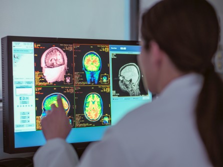
AI Could Speed Up and Equalize Medical Imaging


Doctors rely on imaging technologies that let them see inside the human body to diagnose diseases ranging from pneumonia to cancer. But access to these technologies is rife with inequities: low-income countries average just 1.9 radiologists per million people (the US has about one radiologist per 10,000 people). And within the US, patients in rural areas often have delays in imaging — and as a result, delays in disease diagnosis — because of a lack of equipment and physician shortages.

Fellow Sarah Hooper from the Fannie and John Hertz Foundation is helping develop a solution to both reduce diagnostic imaging costs and address shortages and delays in radiology: integrate automated, machine learning algorithms into the medical imaging workflow. Machine learning algorithms have the potential to transform medical imaging, from changing the way we build imaging systems to altering how images are analyzed and what information we can extract from them. During graduate school at Stanford, Hooper developed ways to improve the automated processing of cardiac MRIs. Now, as a research scientist at the National Heart, Lung, and Blood Institute’s (NHLBI) Imaging AI program, she’s working to integrate machine learning across a variety of imaging platforms.

“You can insert machine learning into any point of the imaging pipeline to make things more efficient,” says Hooper. “One program might make it easier to acquire images so you don’t need as much expertise to operate a machine; another might automate parts of the analysis workflow so that a radiologist can spend five minutes looking at an image instead of twenty. That all translates into patients getting results faster.”

As an undergraduate, Hooper worked with Drs Rebecca Richards-Kortum and Maria Oden at Rice University’s Institute for Global Health Technologies, studying how to reduce the cost of healthcare technologies for use in resource-limited settings. At the same time, during the classes required by her electrical engineering degree, she learned about the burgeoning field of machine learning — the branch of artificial intelligence (AI) that tries to teach computers to learn from experience the way humans do.

“At the time, there wasn’t much overlap in the resource-limited healthcare technology world and the machine learning world,” Hooper recalls. “But it was clear that these two worlds were going to coincide and that there was a lot of potential at their intersection.”

Hooper saw particular promise with automating medical imaging — radiology costs are high, the acquisition and analysis of scans is technically advanced, and imaging is critical to patient care. Hooper pursued her graduate degree as a Hertz Fellow in electrical engineering at Stanford University with the goal of working at this intersection and found mentors in both radiology and computer science.

“I had a very interdisciplinary mentorship team, and that was really enabled by my Hertz Fellowship,” Hooper says. “It enabled me to approach people from diverse fields and say ‘I’m looking for your expertise and I already have funding.’”

Over the next five years, Hooper developed algorithms that could learn to identify and label parts of medical images more easily than previous technologies. Specifically, she focused on cardiac MRIs —high-resolution images of the heart that doctors use to assess cardiac health. In standard image analysis pipelines, cardiologists often draw lines around different parts of the heart in many successive images to calculate key metrics about cardiac health, such as how much blood the heart is pumping over time. This “segmentation” of images into different parts by hand is time-consuming and can be inaccurate.

Hooper wanted a computer algorithm to carry out the segmentation. But machine learning algorithms must be trained: a computer is given data that humans have already analyzed to find patterns and learn how to automate the analysis. Previous machine learning algorithms that might have worked for cardiac MRI segmentation required a huge amount of training data from cardiologists before they could work on their own.

“The key technical challenge was figuring out how to do this without requiring weeks or months of clinicians’ time,” Hooper says.

Hooper successfully wrote new algorithms that could learn how to segment cardiac MRIs using far fewer already-analyzed images: about 100 instead of the 16,000 or more required previously. Researchers at the NHLBI, with whom Hooper collaborated, integrated these algorithms into a larger machine learning platform that is now being used by clinicians to analyze cardiac MRIs more easily.

Today, Hooper is herself a research scientist at the NHLBI’s Imaging AI program. She’s working more broadly across different imaging technologies, building an image analysis platform to help researchers and clinicians who want to segment many different kinds of images — from scans of the human body to pictures of cells growing in a dish or mouse organs being used to study disease.

“There might be pathology slides and we want to identify how many cancer cells there are on the slide, or we might want to calculate the size of a liver based on a CT scan,” Hooper explains. “For all these different images, the technical challenge of using machine learning to identify parts of an image is largely the same.”

Integrated with the image analysis platform, Hooper and the Imaging AI team are implementing additional machine learning algorithms to speed up image acquisition and improve image reconstruction. Ultimately, this platform will support machine learning algorithms throughout the imaging pipeline.

AI can predict the effectiveness of neoadjuvant chemotherapy in breast cancer patients


Engineers at the University of Waterloo have developed artificial intelligence (AI) technology to predict if women with breast cancer would benefit from chemotherapy prior to surgery.

The new AI algorithm, part of the open-source Cancer-Net initiative led by Dr. Alexander Wong, could help unsuitable candidates avoid the serious side effects of chemotherapy and pave the way for better surgical outcomes for those who are suitable.

“Determining the right treatment for a given breast cancer patient is very difficult right now, and it is crucial to avoid unnecessary side effects from using treatments that are unlikely to have real benefit for that patient,” said Wong, a professor of systems design engineering.

“An AI system that can help predict if a patient is likely to respond well to a given treatment gives doctors the tool needed to prescribe the best personalized treatment for a patient to improve recovery and survival.”

In a project led by Amy Tai, a graduate student with the Vision and Image Processing (VIP) Lab, the AI software was trained with images of breast cancer made with a new magnetic image resonance modality, invented by Wong and his team, called synthetic correlated diffusion imaging (CDI).

With knowledge gleaned from CDI images of old breast cancer cases and information on their outcomes, the AI can predict if pre-operative chemotherapy treatment would benefit new patients based on their CDI images.

Known as neoadjuvant chemotherapy, the pre-surgical treatment can shrink tumors to make surgery possible or easier and reduce the need for major surgery such as mastectomies.

I’m quite optimistic about this technology as deep-learning AI has the potential to see and discover patterns that relate to whether a patient will benefit from a given treatment.”

Dr. Alexander Wong, Director of the VIP Lab and the Canada Research Chair in Artificial Intelligence and Medical Imaging

A paper on the project, Cancer-Net BCa: Breast Cancer Pathologic Complete Response Prediction using Volumetric Deep Radiomic Features from Synthetic Correlated Diffusion Imaging, was recently presented at Med-NeurIPS as part of NeurIPS 2022, a major international conference on AI.

The new AI algorithm and the complete dataset of CDI images of breast cancer have been made publicly available through the Cancer-Net initiative so other researchers can help advance the field.

Artificial Intelligence in Medical Imaging—Learning From Past Mistakes in Mammography


Artificial intelligence (AI) in medical imaging shows promise in improving health care efficiency and outcomes. However, automated AI disease detection is not a new concept. Pressure to adopt such emerging technologies is reminiscent of the rapid rise of adjunct computer-aided detection (CAD) tools in mammography 2 decades ago. The CAD cues in mammography mark image features suspicious for breast cancer, while new deep learning AI algorithms similarly mark suspicious image features and provide scores for cancer risk. Despite the intended outcomes of these tools, their use in clinical practice often generates unexpected results. Without a more robust approach to the evaluation and implementation of AI, given the unabated adoption of emergent technology in clinical practice, we have not learned from our past mistakes in the field of mammography.

In 1998, CAD received US Food & Drug Administration (FDA) clearance as an adjunct tool for mammography. Within a few years, the Centers for Medicare & Medicaid Services approved its reimbursement. Nearly a decade after FDA clearance, a seminal article found that CAD did not improve mammography accuracy after dissemination into routine clinical practice.1 Results of initial CAD reader studies were summarized in simple receiver operating characteristic (ROC) curve figures comparing results with CAD vs control. While early reader studies showed improved accuracy with CAD, after FDA clearance and clinical adoption, results were reversed with lower accuracy with CAD.1

By 2016, more than 92% of US imaging facilities used CAD for mammography interpretation despite further research confirming that CAD did not improve radiologist accuracy over 2 decades of use in clinical practice.2,3 The CAD tools are associated with increased false-positive rates, leading to overdiagnosis of ductal carcinoma in situ and unnecessary diagnostic testing. In 2018, Medicare ceased add-on payments for CAD but not before the widespread embrace of CAD had resulted in more than $400 million per year in unnecessary health care expenditures.2 The premature adoption of CAD is consistent with the embrace of emergent technology before its association with patient outcomes is fully understood. As AI algorithms are increasingly receiving FDA clearance and becoming commercially available with ROC curves similar to what we observed prior to CAD clearance and adoption, how can we prevent history from repeating itself?

First, we must remember that there are complex interactions between a computer algorithm output and the interpreting physician. While much research is being done in the development of the AI algorithms and tools, the extent to which physicians may be influenced by the many types and timings of computer cues when interpreting remains unknown. Automation bias, or the tendency of humans to defer to a presumably more accurate computer algorithm, likely affects physician judgment negatively if presented prior to a physician’s independent assessment. In the case of CAD, 2 to 4 markings were shown to radiologists per screening mammogram–as breast cancer is present in about 5 per 1000 screening mammograms, almost all of these markings are false positives. Yet, radiologists do not want to miss cancer, thus leading to higher rates of additional testing, resulting in false-positive results and benign biopsies.4 Before widespread adoption of new AI tools for medical imaging, we need to evaluate the different user interfaces between AI and human interpreters and better understand how and when AI outputs should be presented. Ideally, we need prospective studies incorporating AI into routine clinical workflow.

Second, reimbursement of AI technologies needs to be incumbent on improved patient outcomes, not just improved technical performance in artificial settings. Currently, FDA clearance requires small reader studies and a demonstration of noninferiority to existing technologies (eg, CAD). Newer AI technologies need to demonstrate disease detection that matters. For example, the use of AI in mammography should correspond with increased detection of invasive breast cancers with poor prognostic markers and decreased interval cancer rates. To demonstrate improved patient outcomes, AI technologies need to be evaluated in large population-based, real-world screening settings with longitudinal data collection and linkage to regional cancer registries. If more benefits than harms are identified, then we need to confirm that these results are consistent across diverse populations and settings to ensure health equity. Given the rapid pace of innovation and the many years often needed to adequately study important outcomes, we suggest coverage with evidence development, whereby payment is contingent on evidence generation and outcomes are reviewed on a periodic basis.

Third, we need to embrace revisions to the FDA clearance process for AI algorithms to encourage continued technological improvement. The benefit of deep learning is the ability of machines to continuously improve their algorithms over time. Unfortunately, current FDA review for AI tools in medical imaging is only provided for static unchanging software tools. The consequence is a loss of incentive for continuously improving deep learning algorithms. The FDA is drafting regulatory frameworks for AI-based software as a medical device that pivots from approving static algorithms to oversight over the total product lifecycle including postmarket evaluation.5 The details are yet to be finalized, but will likely require the development of robust data sharing infrastructure necessary for continuous monitoring. One potential avenue for more vigorous, continuous evaluation of AI algorithms is to create prospectively collected imaging data sets that keep up with other temporal trends in medical imaging and are representative of target populations. For instance, the Breast Cancer Surveillance Consortium collects longitudinal data linked to long-term cancer outcomes data permitting prospective image collection, including from the most up-to-date manufacturers and digital breast tomosynthesis (3 dimensional mammograms) technologies. Then, updated algorithms can be independently validated on these representative, population-based imaging examination cohorts continuously.

Fourth, we need to address the impact of AI on medical-legal risk and responsibility in medical imaging interpretation. A great promise of AI is that sophisticated algorithms could eventually interpret images by themselves and free some of physicians’ time to concentrate on more complex tasks. However, truly independent AI is not currently possible because radiologists continue to be the responsible legal parties for accurate imaging interpretation. For example, the recall rate of mammography screening is heavily influenced by medical-legal risk, with missed breast cancer as one of the leading causes of medical malpractice cases in the US.6 Fear of being sued is likely a major reason that American radiologists call back twice as many women after screening mammograms as other countries, even though the cancer detection rate is the same in American and European populations.7,8 Physicians will remain unwilling to bypass inspecting images unless malpractice concerns are addressed. With the Mammography Quality Standards Act now requiring direct patient disclosure of additional risk information (eg, breast density), the appropriate use of supplemental technologies will likely make missed breast cancer even more of a malpractice lawsuit target. One potential solution is to amend the Mammography Quality Standards Act regarding cancer screening liability to better define standards for who can interpret mammograms, how AI can be used for interpretation, and provide guidance on capping limits to AI-related malpractice payouts. Without better guidance on individual party responsibilities for missed cancer, AI creates a new network of who, or what, is legally liable in complex and prolonged, multiparty malpractice lawsuits. Without national legislation addressing the medical and legal aspects of using AI, adoption will slow and we risk opportunities for predatory legal action.

We stand at the precipice of widespread adoption of AI-directed tools in many areas of medicine beyond mammography, and the harms vs benefits hang in the balance for patients and physicians. We need to learn from our past embrace of emerging computer support tools and make conscientious changes to our approaches in reimbursement, regulatory review, malpractice mitigation, and surveillance after FDA clearance. Inaction now risks repeating past mistakes.

Radiation in Medicine: Medical Imaging Procedures


Medical imaging tests are non-invasive procedures that allow doctors to diagnose diseases and injuries without being intrusive. Some of these tests involve exposure to ionizing radiation, which can present risks to patients. However, if patients understand the benefits and risks, they can make the best decisions about choosing a particular medical imaging procedure.

Man getting a CAT scan

Most people have had one or more medical imaging tests. Imaging procedures are medical tests that allow doctors to see inside the body in order to diagnose, treat, and monitor health conditions. Doctors often use medical imaging procedures to determine the best treatment options for patients. The type of imaging procedure that your doctor may suggest will depend on your health concern and the part of the body that is being examined. Some common examples of imaging tests include:

  • X-rays (including dental x-rays, chest x-rays, spine x-rays)
  • CT or CAT (computed tomography) scans
  • Fluoroscopy

If your doctor suggests x-rays or other medical imaging tests, you should consider the following:

  • Medical imaging tests should be performed only when necessary.
  • The U.S. Food and Drug Administration (FDA) recommends discussing the benefits and risks of medical imaging procedures with your doctor.

Benefits and Risks of Medical Imaging Procedures That Use Ionizing Radiation

Medical imaging tests can help doctors:

  • Obtain a better view of organs, blood vessels, tissues and bones.
  • Determine whether surgery is a good treatment option.
  • Guide medical procedures involving placement of catheters, stents, or other devices inside the body, locate tumors for treatment and locate blood clots or other blockages.
  • Guide joint replacement options and treatment of fractures.

As in many areas of medicine, there are risks associated with the use of medical imaging which uses ionizing radiation to create images of the body. Risks from exposure to ionizing radiation include:

  • A small increase in the likelihood that a person exposed to radiation will develop cancer later in life.
  • Health effects that could occur after a large acute exposure to ionizing radiation such as skin reddening and hair loss.
  • Possible allergic reactions associated with a contrast dye injected into the veins to better see body structures being examined.

How can you reduce your exposure to diagnostic ionizing radiation?

In the case of x-rays or other tests involving exposure to ionizing radiation, doctors and radiation experts can help reduce your exposure to and risk of harm from diagnostic ionizing radiation by:

  • Checking to see if you have had a similar test done recently that can provide them with the background information they need.
  • Checking to see if a test that does not use ionizing radiation can provide similar information.
  • Making certain the least possible amount of radiation needed to obtain a good quality image is used for your procedure.
  • Providing protective lead shielding to prevent exposing other areas of the body to radiation.

What are the risks of medical imaging procedures for pregnant women?

Talk to your physician about the potential risks and benefits from the medical procedures. In many cases, the risk of an x-ray procedure to the mother and the unborn child is very small compared to the benefit of finding out about the medical condition of the mother or the child.

However, small risks should not be taken if they’re unnecessary. You can reduce risks from medical imaging procedures by telling your doctor if you are, or think you might be, pregnant whenever an abdominal x-ray is suggested by your doctor. Other options suggested by FDA that may be considered are as follows:

  • If you are pregnant, the doctor may decide that it would be best to cancel the medical imaging procedure, to postpone it, or to modify it to reduce the amount of radiation.
  • Depending on your medical needs, and realizing that the risk is very small, the doctor may feel that it is best to proceed with using a medical imaging procedure as planned.

In any case, you should feel free to discuss the decision with your doctor. For more information on medical imaging and pregnancy, please see X-rays, Pregnancy and You. Also, for more information on radiation safety in adult medical imaging, please visit the Image Wisely website.

Are there special considerations for children?

It is important that x-rays and other imaging procedures performed on children use the lowest exposure setting needed to obtain a good clinical image. TheImage Gently Alliance, part of the Alliance for Radiation in Pediatric Imaging, suggests the following for imaging of children:

  • Use imaging examinations when the medical benefit outweighs the risk.
  • Use the most appropriate imaging techniques, matched to the size of the child.
  • Use alternative imaging methods (such as ultrasound or MRI) when possible.

For more information about medical imaging procedures that do not use ionizing radiation, please see Radiation in Medicine: Medical Imaging Procedures.

The FDA also provides information for parents, patients, and healthcare providers to address concerns about the benefits and risks of medical imaging procedures for children.

Medical Imaging: Phase-Contrast X-Ray Imaging .


Making blood vessels visible without contrast agents; differentiating tumors more clearly from healthy tissues — and all with a low radiation dose and large energy savings. These are the objectives of a new generation of X-ray systems from the Siemens laboratories.

What happened on November 8, 1895, late one Friday evening in the Physics Institute of the University of Würzburg can doubtless be described as one of the most revolutionary developments in the history of medicine. Wilhelm Conrad Röntgen discovered a “new type of radiation” that seemed able to penetrate matter with ease, and he quickly recognized how useful this type of radioscopy  would be for medicine. Two days before Christmas, he succeeded in making the first “X-ray photograph”: it was an image of his wife’s hand, in which not only the wedding ring but also the bones were clearly visible. The fact that Röntgen was awarded the first Nobel Prize in Physics in 1901 was only a logical consequence of his extraordinary achievement.

And it wasn’t long before the first commercial products appeared. On March 24, 1896, just three months after Röntgen’s discovery, the company Siemens&Halske obtained a patent for a new X-ray tube that was “especially suited to transillumination of the entire body of adult persons.” And to this day, Siemens has remained faithful to diagnostic radiology. The company offers a whole range of solutions, from mobile devices to fully digital systems to CT scanners for 3D images.

The Shortcomings of Current X-ray Systems

Over 90 percent of all medical imaging examinations worldwide now rely on X-rays. But the technology is still based on the fundamental principle that was used 120 years ago: electrons that are generated in a cathode and accelerated to high energies collide with a fixed anode — usually made of the heavy metal tungsten — and thereby release X-rays. The X-rays, in turn, are absorbed to a greater degree by bone than by soft tissue. The bones therefore appear dark in an X-ray image, and the soft tissues appear light.

From left to right: Prof. Alessandro Olivo (UCL), Prof. Dr. Oliver Heid (Siemens CT) and Dr. Paul Diemoz (UCL) inspect an “apertured” mask, a phase-contrast component. These are low-aspect ratio structures, which are inexpensive to build and can be scaled up to large imaging areas.

Despite the success of this technique in medical engineering, it does have a few drawbacks. For example, the electrons that collide with the anode produce mainly heat. No more than one percent of the energy is converted to X-rays — a huge waste of energy. There are also many applications, such as tumor diagnostics, in which physicians want to be able to distinguish among various soft tissues more easily. But if  contrast is increased, the patient is exposed to a higher dose of X-ray radiation — which should be avoided, because high radiation doses can damage body tissue. In X-ray examinations involving cardiovascular diseases, on the other hand, contrast agents are often needed in order for the angiography systems to be able to make blood vessels visible in X-ray light — but nearly one out of ten patients suffers allergic reactions to these substances, which can lead to shock and kidney failure. A technique that uses smaller quantities of contrast agent, or even none at all, would therefore be beneficial to millions of people.

On the Horizon: A New Revolution in Medical Diagnostics

“The technology that we’re currently developing at Siemens could help us overcome all these challenges,” says Prof. Oliver Heid, head of the Global Technology Field of Healthcare Technology and Concepts at Siemens Corporate Technology. Heid is a medical doctor and holder of approximately 300 patents in a large variety of fields, from high-frequency technology to superconductivity, materials science, accelerators and software solutions. “We’re in the process of completely rethinking everything and changing everything: the method by which X-rays are generated as well as the technique used for detecting them. If everything goes well with our next-generation X-ray system, it will be another revolution in medical diagnostics,” says Dr. Heinrich Kolem, CEO for Angiography and Interventional X-Ray Systems at Siemens Healthcare.

graphic: Siemens next generation X-ray tube

Siemens’ next generation X-ray tubes will be completely different from today’s. Electrons will no longer come from a hot cathode, but from a cold cathode ring made of nanostructured carbon — and the X-ray light will be generated in a thin jet of liquid metal, rather than at a solid anode.

This multi-year R&D project, which is scheduled to run until 2017, brings together just the right innovators: alongside Heid and Kolem, they also include the team of Components and Vacuum Technology at Siemens Healthcare lead by its CEO Dr. Peter Molnar, researchers from Siemens Corporate Technology in Russia, external partners from institutions such as Oxford University, as well as Prof. Alessandro Olivo of University College London, whose contribution to the development team includes both scientific expertise and insights from clinical practice. Molnar, whose business unit produces approximately 22,000 X-ray tubes per year for CT machines, angiography systems, and X-ray equipment from Siemens, underscores the value of this cooperation. “Our shared objective is to commercialize the new system in a competitive form and successfully launch it on the market. Only then does a good idea become a true innovation,” he says.

Substantially Higher Energy Densities with Significantly Reduced Energy Demand

What is being changed exactly? It starts with the cathode. Here, the team is no longer using 2,000-degree Celsius filaments to emit electrons. Instead, they are using a ring-shaped “cold cathode” of nanostructured carbon that operates at a high voltage and at room temperature. The advantage of this approach is that it uses less energy than the previous cathodes.

The electrons no longer collide with a fixed target of tungsten, but with a new device invented by Siemens researchers that they’ve named LiMA, which stands for “liquid metal jet alloy” target. In other words, the electron target is a jet of liquid metal as thin as a human hair. The metal consists of 95 percent lithium and 5 percent heavy elements such as bismuth or lanthanum. The latter produces short wavelength X-rays, the former acts as a coolant. The energy of electrons leaving the liquid-metal-jet anode can potentially be reclaimed and fed back into the energy cycle. The result is that the X-ray tube requires less than half the electricity and cooling of previous devices, which greatly reduces total energy demand.

graphic: How a wave front detector works graphic: How a wave front detector works

A wavefront sensor consists of millions of concave metal or silicon lenses that create a matrix of focal points on the detector. The refraction of the X-ray waves in the object — a tumor, for example — can be determined from the shift of these focal points.

Significantly more important, however, is the fact that the tube can achieve a much higher energy density at the target. At the same light intensity, the focus of the new X-ray source is 400 times smaller than in conventional X-ray tubes – “at the focal point, this X-ray radiation is four billion times brighter than the sun on the surface of the earth,” says Heid, “which results in a 20-fold higher imaging resolution.”

Twenty Times the Resolution of Today’s Systems

That, in turn, is the prerequisite for an entirely new imaging technique, one that scientists around the world have been working on for years: phase-contrast X-ray imaging. Whereas conventional radiography simply records whether X-rays penetrate a certain tissue or not, phase-contrast imaging measures the effect that passing through bodily tissue has on the wave phase – i.e. the sequence of wave crest and trough. This same physical phenomenon can be seen in the light effects on the bottom of a water-filled swimming pool on a sunny day. This phase shift is highly revealing, since it varies depending on the refractive power of the tissue through which the radiation passes. The approach described here would make it possible to distinguish different soft tissues, in particular fat from water or iron levels in blood, which is essential for being able to easily differentiate a tumor in an early stage of growth from healthy tissue.

The researchers discuss the advantages of the new x-ray-system in UCL's X-Ray Phase Contrast Laboratory.
The researchers discuss the advantages of the new X-ray-system at UCL’s X-Ray Phase Contrast Laboratory.

“To be able to measure these phase shifts, we’re also working on a completely new component on the detector side,” says Dr. Andreas Geisler, project manager for the new X-ray system on Heid’s team. To this end, a wavefront sensor of the kind used in optics or astronomy, for example, is to be used for the first time for X-ray light. The sensor consists of millions of concave metallic or silicon lenses that generate a matrix of focal points on the detector. Through the displacement of these focal points, the refraction in the object can be calculated. This is not possible today with conventional detectors alone.

“So not only will these next-generation X-ray systems be very efficient to operate, they will also do a good job of registering contrasts among soft tissues at a relatively low radiation dose,” says Geisler. Blood vessels could be made visible in this way without contrast agents; tumors could be more clearly recognized thanks to the 20-fold higher resolution and phase-contrast X-ray imaging; and the new technology would be ideal for minimally invasive surgery too. “We want to guide and navigate catheters using magnetic fields, for example, and know at any time via the X-ray imaging where exactly they are located in the body,” says Heinrich Kolem. That isn’t possible with conventional X-ray tubes, because they are sensitive to magnetic fields – “the next-generation X-ray systems won’t have this drawback, and at the same time, they’ll be able to provide images that are more useful diagnostically.”

Routine Urinalysis Not Helpful After Blunt Abdominal Trauma.


Routine urinalysis after blunt abdominal trauma won’t help find urogenital injury, Dutch researchers say.

“With the advancements made in CT scanning, there is now much greater accuracy in the detection (or ruling out) of injury to the urogenital system,” Dr. J. Carel Goslings from Academic Medical Center in Amsterdam told Reuters Health.

“In this study,” Dr. Goslings added, “we found the value of the routine performance of urinalysis in patients with a blunt trauma mechanism to be limited.”

The retrospective study involved 1815 patients. Most patients — 1031, or 57% — also had imaging studies, according to a paper online September 16th in Emergency Medicine Journal.

Among the patients who had imaging studies done, 795 (77%) had no hematuria, 220 (21%) had microscopic hematuria, and 16 (2%) had macroscopic hematuria.

Of the 220 patients with microscopic hematuria, eight had abnormal urogenital imaging studies, but only three of the eight had clinical consequences. Another eight patients with microscopic hematuria did have clinical consequences despite normal-looking imaging.

There were 332 patients who had urine collected but no imaging studies. In this group, 278 patients (84%) had no hematuria. In the 54 patients (16%) who did have microscopic hematuria, there were no clinical consequences, according to the authors.

Two hundred sixty-eight patients (15%) had urogenital imaging but no urinalysis. Only 10 had abnormal findings; four of the 10 had clinical consequences.

Ten percent of patients had neither urine collection nor imaging.

“The potential danger of performing urinalysis without imaging is to miss clinically relevant injuries (e.g., bleeding sites in the kidney parenchyma), which can only be shown by imaging,” the authors wrote. “Bypassing urinalysis and going straight for imaging…results in clinical consequences in 1.5% of the patients (4 out of 268). This is comparable to the percentage of clinical consequences in the patients who receive both urinalysis and imaging (2%; 22 out of 1031).”

They added, “The remaining 0.5% difference in clinical consequences consists of relatively minor consequences such as additional imaging and re-evaluation at the outpatient department, and this indicates little added value of the performance of urinalysis.”

Dr. Goslings told Reuters Health that the researchers “advise omitting this investigation as a routine part of the assessment of trauma patients, given that (good) imaging facilities are available in the hospital.”

But in specific circumstances, urinalysis might still be appropriate. Repeating by email some points from the paper, Dr. Goslings wrote, “In particular, patients with specific trauma mechanisms (e.g., fall from height, fall from horse or direct blow to the flank) or patients with a suspicion of pelvic (ring) injuries or thoracolumbar spinal cord injuries might benefit from urinalysis.”

“Future studies should focus on identifying the subgroups of patients in whom urinalysis is helpful,” Dr. Goslings added.

Targeted molecular imaging in oncology.


Improvement of scintigraphic tumor imaging is extensively determined by the development of more tumor specific radiopharmaceuticals. Thus, to improve the differential diagnosis, prognosis, planning and monitoring of cancer treatment, several functional pharmaceuticals have been developed. Application of molecular targets for cancer imaging, therapy and prevention using generator-produced isotopes is the major focus of ongoing research projects. Radionuclide imaging modalities (positron emission tomography, PET; single photon emission computed tomography,

SPECT) are diagnostic cross-sectional imaging techniques that map the location and concentration of radionuclide-labeled radiotracers.

99mTc- and 68Galabeled agents using ethylenedicysteine (EC) as a chelator were synthesized and their potential uses to assess tumor targets were evaluated.

99mTc (t1/2 = 6 hr, 140 keV) is used for SPECT and 68Ga (t1/2 = 68 min, 511 keV) for PET. Molecular targets labeled with Tc-99m and Ga-68 can be utilized for prediction of therapeutic response, monitoring tumor response to treatment and differential diagnosis. Molecular targets for oncological research in (1) cell apoptosis, (2) gene and nucleic acid-based approach, (3) angiogenesis (4) tumor hypoxia,and (5) metabolic imaging are discussed. Numerous imaging ligands in these categories have been developed and evaluated in animals and humans. Molecular targets were imaged and their potential to redirect optimal cancer diagnosis and therapeutics were demonstrated.

Source: http://www.jsnm.org