Lung cancer: screening with low-dose CT sharply improves long-term survival


Two annual repeat low-dose CT scans in a woman who was 60 years old at baseline...

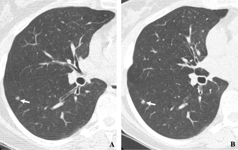
Two annual repeat low-dose CT scans in a woman who was 60 years old at baseline enrollment in 1999. At baseline enrollment, she was currently smoking and had a 30-pack-year smoking history. No nodules were identified on baseline low-dose CT scans. On the sixth annual low-dose CT scan (B), a right lower lobe solid nodule (arrow) measuring 4.5 mm in maximum diameter was identified. The nodule could be identified in retrospect on the prior annual CT scan (arrow in A), when it measured 2.0 mm in maximum diameter. Estimated tumor volume doubling time was 161 days. Lobectomy was performed 2 months later, and diagnosis of stage 1aN0M0 moderately differentiated adenocarcinoma measuring 6.0 mm in maximum diameter was made. Expert pathologic panel review (22) of the pathologic specimen updated the diagnosis to adenocarcinoma with mixed subtype (80% acinar, 20% bronchoalveolar carcinoma components) with 5 mm of invasion.

Diagnosing early-stage lung cancer with low-dose CT screening dramatically improves the long-term survival rate of cancer patients, according to a large-scale, 20-year international study.

The research was published in Radiology, a journal of the Radiological Society of North America (RSNA). The results show that patients diagnosed with lung cancer by low-dose CT screening have a 20-year survival rate of 81%. If diagnosed in the earliest Stage I, long-term survival was 95%. 

“It is the first time that 20-year survival rates from annual screening have been reported,” said the study’s lead author, Claudia Henschke, Ph.D., M.D., professor of radiology and director of the Early Lung and Cardiac Action Program at the Icahn School of Medicine at Mount Sinai in New York. “This 20-year survival rate of 81% is the estimated cure rate of all participants with lung cancers diagnosed by annual screening. This is a huge benefit compared to waiting for a diagnosis that, in usual care, is symptom-prompted.” 

Lung cancer can be cured if you enroll in an annual screening program using a well-defined protocol and comprehensive management systemClaudia Henschke

Lung cancer is the leading cause of cancer death. According to the American Lung Association, the average lung cancer five-year survival rate is 18.6%. Only 16% of lung cancers are diagnosed at an early stage, and more than half of people with lung cancer die within one year of being diagnosed. While treatments of more advanced-stage cancers with targeted therapy and immunotherapy have come a long way, the best tool in the fight against cancer deaths is early diagnosis through low-dose CT screening before symptoms appear. 

Since 1992, Dr. Henschke and colleagues have been studying the effectiveness of low-dose CT screening for lung cancer. This led to the creation of the International Early Lung Cancer Action Program (I-ELCAP) which has enrolled more than 89,000 participants in over 80 institutions worldwide. This international program will continue its collaborations in 2024 with clinical and government organizations to roll out lung screening programs for underserved and low-income countries on two additional continents. 

portrait of claudia henschke

In 2006, the researchers identified a 10-year survival rate of 80% for the patients whose cancer was identified by CT screening. For this study, they looked at 20-year survival rates. “We were excited to see that the estimated cure rate we reported in 2006 has persisted after 20 years of follow-up,” Dr. Henschke said. The new study also found that among the 1,257 I-ELCAP participants diagnosed with lung cancer, 81% had Stage I disease. A Stage I lung cancer is a very small tumor that has not spread to any lymph nodes. In this study, the long-term survival rate for Stage I cancers was 87%. 

The findings demonstrate the importance of low-dose CT screening for early detection of lung cancer. The researchers also included participants who have smoked less than 10 pack-years, including those who had never smoked cigarettes but had passive exposure to cigarette smoke. “The less than 10 pack-years of smoking includes people who have never smoked,” Dr. Henschke said. “In the United States, some 25% of lung cancers are diagnosed in people who have never smoked.” 

The results from this study show that after 20 years, patients diagnosed with lung cancer at an early stage via CT screening have significantly better outcomes. By treating the cancer when it is small, patients can be effectively cured in the long term. “Lung cancer can be cured if you enroll in an annual screening program using a well-defined protocol and comprehensive management system,” Dr. Henschke said. “It is important to return for annual screening.” 

The U.S. Preventive Services Task Force recommends annual lung cancer screening with low-dose CT in adults aged 50 to 80 years who have a 20 pack-year smoking history and currently smoke or have quit within the past 15 years. 

Urinary detection of lung cancer in mice via noninvasive pulmonary protease profiling


Noninvasive nanoparticles for lung cancer

Previously developed nanoparticle technology has been shown to detect the hallmark protease activity of many cancers, amplifying it into a urinary readout. Now, Kirkpatrick et al. optimize protease activity–based nanosensors for the detection of lung cancer. Intratracheal instillation of nanosensors enabled detection of localized lung adenocarcinoma in two immunocompetent, autochthonous mouse models. The sensors distinguished between lung cancer and lung inflammation, and did not detect protease activity in a colorectal cancer xenograft model. Further work will need to confirm the approach for human lung cancer and other lung cancer subtypes and to formulate the nanosensors for intrapulmonary delivery in patients.

Abstract

Lung cancer is the leading cause of cancer-related death, and patients most commonly present with incurable advanced-stage disease. U.S. national guidelines recommend screening for high-risk patients with low-dose computed tomography, but this approach has limitations including high false-positive rates. Activity-based nanosensors can detect dysregulated proteases in vivo and release a reporter to provide a urinary readout of disease activity. Here, we demonstrate the translational potential of activity-based nanosensors for lung cancer by coupling nanosensor multiplexing with intrapulmonary delivery and machine learning to detect localized disease in two immunocompetent genetically engineered mouse models. The design of our multiplexed panel of sensors was informed by comparative transcriptomic analysis of human and mouse lung adenocarcinoma datasets and in vitro cleavage assays with recombinant candidate proteases. Intrapulmonary administration of the nanosensors to a Kras– and Trp53-mutant lung adenocarcinoma mouse model confirmed the role of metalloproteases in lung cancer and enabled accurate detection of localized disease, with 100% specificity and 81% sensitivity. Furthermore, this approach generalized to an alternative autochthonous model of lung adenocarcinoma, where it detected cancer with 100% specificity and 95% sensitivity and was not confounded by lipopolysaccharide-driven lung inflammation. These results encourage the clinical development of activity-based nanosensors for the detection of lung cancer.

INTRODUCTION

Lung cancer is the most common cause of cancer-related death (25.3% of cancer deaths in the United States), with dismal 18.6% 5-year survival rates (1). Underlying this high mortality is the fact that 57% of patients with lung cancer have distant spread of disease at the time of diagnosis (1). Because patients with regional or localized disease have 6- to 13-fold higher 5-year survival rates than patients with distant metastases (1), substantial effort has been dedicated to early detection of lung cancer. In the United States, screening with low-dose computed tomography (LDCT) is recommended in high-risk patients [adults aged 55 to 80 years with a 30 pack-year smoking history (2)] and enabled a relative reduction in mortality of 20% when compared to chest radiography in the National Lung Screening Trial (NLST) (3). However, in addition to expense (4) and risks associated with radiation exposure (5), LDCT suffers from high false-positive rates (3), leading to a considerable burden of complications incurred during unnecessary follow-up procedures. Transthoracic needle biopsy, for example, is associated with a 15% rate of pneumothorax and a 6.6% rate of pneumothorax requiring chest drainage (6). Overall, the risk of dying or suffering a major complication in an LDCT-screened patient with a benign nodule is 4.1 and 4.5 per 10,000, respectively (5). As a result of these limitations, screening by LDCT has not been widely adopted outside of the United States (7), and there is an urgent need to develop diagnostic tests that increase the effectiveness of lung cancer screening.

Great strides in molecular diagnostics have yielded promising approaches that may be used in conjunction with or as an alternative to LDCT for lung cancer screening. Circulating tumor DNA (ctDNA) has emerged as a promising tool for noninvasive molecular profiling of lung cancer (8, 9). However, the presence of ctDNA scales with tumor burden, and there are thus fundamental sensitivity limits for early-stage disease (8, 10). In patients with a suspicious nodule identified by LDCT, transcriptional profiling of bronchial brushings can enhance the diagnostic sensitivity of bronchoscopy alone (11), leveraging the “field of injury” that results from smoking and other environmental exposures. However, as with any invasive procedure, bronchoscopy carries the risk of attendant complications such as pneumothorax (3, 5).

Rather than relying on imaging or the detection of endogenous biomarkers in circulation, we have developed a class of “activity-based nanosensors” that monitor for a disease state by detecting and amplifying the activity of aberrant proteases and that function as urinary reporters (1219). Protease activity is dysregulated in cancer, and proteases across all catalytic classes play a direct role in tumorigenesis (20, 21). Activity-based nanosensors leverage dysregulated protease activity to overcome the insensitivity of previous biomarker assays, amplifying disease-associated signals generated in the tumor microenvironment and providing a concentrated urine-based readout. We have previously explored the sensitivity of this approach using mathematical modeling (22) and cell transplant models (16). However, to drive accurate diagnosis in a heterogeneous disease, a diagnostic must also be highly specific. Here, we explored the potential to attain both sensitive and specific lung cancer detection by multiplexing 14 activity-based nanosensors in two immunocompetent, autochthonous mouse models driven by either Kras/Trp53 (KP) mutations or Eml4-Alk (EA) fusion. Clinically, activity-based nanosensors may have utility as an alternative to invasive follow-up procedures in patients with positive LDCT findings.

DISCUSSION

In this work, we present an advance toward clinical translation of a new class of biomarkers, activity-based nanosensors. We found that such multiplexed nanosensors, when delivered by intratracheal instillation, performed with specificity of 100% and sensitivity up to 95% for detection of localized disease in two autochthonous LUAD models representing Kras/Trp53 and Alk-mutant disease. Furthermore, we found that LPS-induced lung inflammation did not result in false positives. Our approach overcomes the intrinsic sensitivity limitation of blood-based diagnostic assays for localized disease by profiling disease activity directly within the tumor microenvironment and providing multiple steps of signal amplification (22). Using intrapulmonary delivery, we further ensured that virtually all nanosensors reached the lung and bypassed nonspecific activation in off-target organs.

This study represents a step toward clinical implementation of activity-based nanosensors for lung cancer testing, validating the efficacy of the tool in two autochthonous, immunocompetent models of localized LUAD. The use of genetically engineered mouse models offered several advantages over cell transplant models, including the ability to explore stage-specific differences, as well as proteolytic contributions from immune cells. Activity-based nanosensors detected disease as early as 7.5 weeks after initiating the KP model, when only grade 1 AAH and grade 2 adenomas are present (24). Furthermore, although metalloprotease-sensitive nanosensors were, as expected, preferentially cleaved in KP mice at both 7.5 and 10.5 weeks, the activation of PP11 (a serine protease-sensitive substrate) in KP10.5wk mice could point to an unexpected role of serine protease activity in tumor progression at this disease stage. One hypothesis is that tumor-infiltrating immune cells, which secrete a multitude of serine proteases (37), may contribute to nanosensor cleavage in KP10.5wk mice. Neutrophils are known to infiltrate KP tumors around 10 weeks after tumor induction (38). The potential capacity of activity-based nanosensors to measure immune-mediated protease activity (18) raises the prospect of rapid, noninvasive, and longitudinal immunotherapy response monitoring.

Here, we report improved sensitivity of activity-based nanosensors relative to previous work by our group, as well as existing and emerging blood-based diagnostics for cancer. We found that our nanosensors could detect tumors in KP7.5wk mice, whose total tumor volumes were, on average, only 2.78 mm3, more than an order of magnitude smaller than our most sensitive method to date (36 mm3 in an ovarian cancer model) (16). By comparison, in the LS174T colorectal cancer xenograft model, ctDNA is detectable when tumor volumes reach 1000 mm3 (39), carcinoembryonic antigen is detectable around 135 to 330 mm3 (12, 39), and intravenously administered activity-based nanosensors have previously been shown to detect disease in this model around 130 mm3 (12). Last, in the autochthonous KrasG12D-mutant “K” lung cancer model, ctDNA bearing the KrasG12D mutation was only detectable when average tumor volumes were 7.1 mm3 (40), even with collection of 2.5% of the total mouse blood volume, scaling to 125 ml in humans.

In the NLST, 96.4% of positive LDCT findings were false positives (3, 5), and many of these patients went on to suffer major complications during invasive follow-up procedures (4, 5). Therefore, there is a need to develop noninvasive diagnostic methods that can distinguish between lung cancer and benign lung disease. Here, we demonstrated the specificity of activity-based nanosensors for lung cancer, rather than benign lung inflammation, through multiplexing and machine learning. Although fewer than half of the 14 reporters were differentially enriched in the urine of KP mice and healthy controls, several more had diagnostic power in EA mice, and others were informative in the classification of malignant versus inflammatory disease. As a result, we found that a pretrained random forest classifier could distinguish between lung cancer-bearing mice (regardless of subtype) and benign disease controls. Although a clinical study would be necessary to directly assess the effectiveness of activity-based nanosensors in the setting of LDCT lung cancer screening, our results suggest that activity-based nanosensors may complement LDCT for discrimination of malignant lesions from benign disease.

Although this work represents a step toward translation of activity-based nanosensors for lung cancer detection, there are limitations that must be addressed before clinical implementation. In this work, we demonstrated the sensitivity and specificity of intrapulmonary activity-based nanosensors for localized lung cancer in two genetically engineered mouse models of LUAD. Although the advantages of such models over xenograft models in recapitulating human disease are numerous (41), mouse models cannot fully capture the native oncogenic properties or heterogeneity found in human lung cancer, and further in vivo validation is needed to confirm the generalizability of activity-based nanosensors to other lung cancer subtypes. Similarly, although activity-based nanosensors can discriminate between lung cancer and LPS-driven lung inflammation, it is possible that clinical lung cancer testing may be confounded by other benign lung disease etiologies or chronic exposure to tobacco smoke. Because of the inherent limitations of mouse models, clinical trials will be necessary to fully validate the robustness of activity-based nanosensors in detecting lung cancer and distinguishing malignant from benign and extrapulmonary disease in humans. Last, the intrapulmonary delivery methods presented here must be optimized before clinical translation. Here, we delivered activity-based nanosensors by intratracheal instillation and demonstrated their stability after aerosolization. However, a clinically relevant intrapulmonary delivery method such as dry powder inhalation or nebulization will be required for clinical implementation.

In summary, intrapulmonary activity-based nanosensors perform with high sensitivity and specificity for detection of localized lung cancer in autochthonous mouse models via a noninvasive urine test. To engineer these nanosensors, we leveraged analysis of LUAD gene expression datasets to nominate candidate proteases, screened these proteases in vitro against a panel of peptide substrates, and directly delivered nanosensors carrying these substrates into the lungs of mice. Activity-based nanosensors may have clinical utility as a rapid, safe, and cost-effective follow-up to LDCT, reducing the number of patients referred for invasive testing. With further optimization and validation studies, activity-based nanosensors may one day provide an accurate, noninvasive, and radiation-free strategy for lung cancer testing.

How a Simple Urine Test Could Reveal Early-Stage Lung Cancer


Lung cancer is the deadliest cancer in the world, largely because so many patients are diagnosed late.

Screening more patients could help, yet screening rates remain critically low. In the United States, only about 6% of eligible people get screened , according to the American Lung Association. Contrast that with screening rates for breast, cervical, and colorectal cancer, which all top 70%.

But what if lung cancer detection was as simple as taking a puff on an inhaler and following up with a urine test?

Researchers at the Massachusetts Institute of Technology (MIT), Cambridge, Massachusetts, have developed nanosensors that target lung cancer proteins and can be delivered via inhaler or nebulizer, according to research published this month in Science Advances. If the sensors spot these proteins, they produce a signal in the urine that can be detected with a paper test strip.

“It’s a more complex version of a pregnancy test, but it’s very simple to use,” said Qian Zhong, PhD, an MIT researcher and co-lead author of the study.

Currently, the only recommended screening test for lung cancer is low-dose CT. But not everyone has easy access to screening facilities, said the other co-lead author Edward Tan, PhD, a former MIT postdoc and currently a scientist at the biotech company Prime Medicine, Cambridge, Massachusetts.

“Our focus is to provide an alternative for the early detection of lung cancer that does not rely on resource-intensive infrastructure,” said Dr. Tan. “Most developing countries don’t have such resources” — and residents in some parts of the United States don’t have easy access, either, he said.

How It Works

The sensors are polymer nanoparticles coated in DNA barcodes, short DNA sequences that are unique and easy to identify. The researchers engineered the particles to be targeted by protease enzymes linked to stage I lung adenocarcinoma. Upon contact, the proteases cleave off the barcodes, which make their way into the bloodstream and are excreted in urine. A test strip can detect them, revealing results about 20 minutes from the time it’s dipped.

The researchers tested this system in mice genetically engineered to develop human-like lung tumors. Using aerosol nebulizers, they delivered 20 sensors to mice with the equivalent of stage I or II cancer. Using a machine learning algorithm, they identified the four most accurate sensors. With 100% specificity, those four sensors exhibited sensitivity of 84.6%.

“One advantage of using inhalation is that it’s noninvasive, and another advantage is that it distributes across the lung quite homogeneously,” said Dr. Tan. The time from inhalation to detection is also relatively fast — in mice, the whole process took about 2 hours, and Dr. Zhong speculated that it would not be much longer in humans.

Other Applications and Challenges

An injectable version of this technology, also developed at MIT, has already been tested in a phase 1 clinical trial for diagnosing liver cancer and nonalcoholic steatohepatitis. The injection also works in tandem with a urine test, the researchers showed in 2021. According to Tan, his research group (led by Sangeeta Bhatia, MD, PhD) was the first to describe this type of technology to screen for diseases.

The lab is also working toward using inhalable sensors to distinguish between viral, bacterial, and fungal pneumonia. And the technology could also be used to diagnose other lung conditions like asthma and chronic obstructive pulmonary disease, Dr. Tan said.

The tech is certainly “innovative,” remarked Gaetano Rocco, MD, a thoracic surgeon and lung cancer researcher at Memorial Sloan Kettering Cancer Center, Basking Ridge, New Jersey, who was not involved in the study.

Still, challenges may arise when applying it to people. Many factors are involved in regulating fluid volume, potentially interfering with the ability to detect the compounds in the urine, Rocco said. Diet, hydration, drug interference, renal function, and some chronic diseases could all limit effectiveness.

Another challenge: Human cancer can be more heterogeneous (containing different kinds of cancer cells), so four sensors may not be enough, Zhong said. He and colleagues are beginning to analyze human biopsy samples to see whether the same sensors that worked in mice would also work in humans. If all goes well, they hope to do studies on humans or nonhuman primates.

Perioperative Nivolumab and Chemotherapy in Stage III NSCLC


The addition of nivolumab to platinum-based chemotherapy improved pathologic complete response rates in patients with resectable stage IIIA or IIIB NSCLC.

Approximately 20% of patients with non–small-cell lung cancer (NSCLC) have stage III disease. Although therapeutic intent is curative for patients with locally advanced disease, historically, treatment outcomes have been poor, and there is lack of consensus on the most appropriate management.

In the industry-sponsored, open-label, multicenter, phase 2 NADIM II trial, 86 treatment-naive patients with resectable stage IIIA or IIIB NSCLC were randomized 2:1 to receive either neoadjuvant nivolumab and paclitaxel plus carboplatin (experimental group) or paclitaxel plus carboplatin alone (control group), followed by surgery. Patients in the experimental group who had R0 resections received adjuvant nivolumab for 6 months.

Pathologic complete response (pCR), the primary endpoint, occurred in 37% of patients in the experimental group compared with 7% in the control group (relative risk, 5.34; 95% CI 1.34–21.23; P=0.02). Progression-free survival at 24 months was 67.2% in the experimental group and 40.9% in the control group (hazard ratio for disease progression, disease recurrence, or death, 0.47; 95% CI, 0.25–0.88).

During neoadjuvant treatment, grade 3 or 4 adverse events occurred in 19% of patients in the experimental group compared with 10% in the control group, most commonly febrile neutropenia (5%) and diarrhea (4%). A higher percentage of patients in the experimental group underwent surgery (93% vs. 69%); there were no delays in surgery due to adverse events. All patients who attained pCR were free from progression at the time of data cutoff.

Comment

In the NADIM II trial, patients with stage IIIA or stage IIIB NSCLC who were treated with neoadjuvant nivolumab and paclitaxel plus carboplatin achieved a higher rate of pCR and longer survival than those treated with chemotherapy alone. These findings add further support for a neoadjuvant chemo-immunotherapy strategy as demonstrated in CheckMate 816 (NEJM JW Oncol Hematol Apr 14 2022 and N Engl J Med 2022; 386:1973) and in KEYNOTE 677 (NEJM JW Oncol Hematol Jun 20 2023 and N Engl J Med 2023 Jun 3; [e-pub]) in a restricted population of patients with stage III NSCLC.

Olanzapine Improves Weight Gain in Patients with Advanced Cancer on Chemotherapy


Among patients with newly diagnosed advanced gastric, hepatopancreaticobiliary, or lung cancers starting chemotherapy, low-dose daily olanzapine improved appetite and weight gain.

Patients with advanced cancer often present with anorexia and weight loss. Poor nutritional status is associated with myriad adverse outcomes in this population. Medical management options have been limited, although corticosteroids and progesterone analogs (e.g., megestrol acetate) are commonly used. These investigators evaluated olanzapine — an antipsychotic agent that increases appetite and decreases nausea — for the treatment of chemotherapy-related anorexia in patients with locally advanced or metastatic gastric, hepatopancreaticobiliary, or lung cancer.

In the randomized, double-blind study, 124 adult patients received olanzapine (2.5 mg daily) or placebo for 12 weeks, starting on the first day of the first chemotherapy cycle. A significantly higher percentage of patients in the olanzapine arm than the placebo arm had weight gain >5% (60% vs. 9%; P<.001) and improved appetite (43% vs. 13%; P<.001) — the primary outcomes. The olanzapine arm also had significantly improved quality of life and nutritional status, as well as less chemotoxicity ≥ grade 3 (12% vs. 37%; P=.002). Adverse events attributable to olanzapine included mild, limited drowsiness.

Comment

This study fills an important gap in the literature, helping clinicians better support patients with advanced cancer who have cancer-associated weight loss and anorexia.

As noted in a recent ASCO Guideline Rapid Recommendation Update on cancer cachexia, daily low-dose olanzapine should be offered to patients with advanced cancer to support appetite and weight gain, along with nutritional support (J Clin Oncol 2023; 41:4178). For patients unable to tolerate olanzapine, a short-term trial of a corticosteroid or progesterone analog may be considered. Although the Update applies to all adult patients with advanced cancer, the majority of evidence derives from patients with gastrointestinal or lung malignancies and those receiving cytotoxic chemotherapy.

The use of mirtazapine for cancer-associated weight loss and anorexia should be limited, as a recent study found it to be no better than placebo (J Pain Symptom Manage 2021; 62:1207). Importantly, there are no FDA-approved medications for cancer cachexia.

Patritumab Deruxtecan Granted Priority Review in the U.S. for Certain Patients with Previously Treated Locally Advanced or Metastatic EGFR-Mutated Non-Small Cell Lung Cancer


Daiichi Sankyo (TSE: 4568) and Merck, known as MSD outside of the United States and Canada, (NYSE: MEK) announced today that the U.S. Food and Drug Administration (FDA) has accepted and granted Priority Review to the Biologics License Application (BLA) for patritumab deruxtecan (HER3-DXd) for the treatment of adult patients with locally advanced or metastatic EGFR-mutated non-small cell lung cancer (NSCLC) previously treated with two or more systemic therapies.

The Prescription Drug User Fee Act (PDUFA) date, the FDA action date for their regulatory decision, is June 26, 2024. The Priority Review follows receipt of Breakthrough Therapy Designation granted by the FDA in December 2021.

The FDA grants Priority Review to applications for medicines that, if approved, would offer significant improvements over available options by demonstrating safety or efficacy improvements, preventing serious conditions or enhancing patient compliance. The BLA is being reviewed under the Real-Time Oncology Review (RTOR) program, an initiative of the FDA which is designed to bring safe and effective cancer treatments to patients as early as possible. RTOR allows the FDA to review components of an application before submission of the complete application.

Patritumab deruxtecan is a specifically engineered potential first-in-class HER3 directed DXd antibody drug conjugate (ADC) discovered by Daiichi Sankyo and being jointly developed and commercialized by Daiichi Sankyo and Merck.

The BLA is based on the primary results from the HERTHENA-Lung01 pivotal phase 2 trial and data results presented at the IASLC 2023 World Conference on Lung Cancer (#WCLC23), which were simultaneously published in the Journal of Clinical Oncology.

In HERTHENA-Lung01, patritumab deruxtecan was studied in 225 patients with EGFR-mutated locally advanced or metastatic NSCLC following disease progression with an EGFR TKI and platinum-based chemotherapy, which demonstrated an objective response rate (ORR) of 29.8% (95% CI: 23.9-36.2), including one complete response and 66 partial responses. The median duration of response was 6.4 months (95% CI: 4.9-7.8). The safety profile of patritumab deruxtecan observed in HERTHENA-Lung01 was consistent with previous phase 1 clinical trials in NSCLC with a treatment discontinuation rate of 7.1% due to treatment-emergent adverse events (TEAEs). Grade 3 or higher TEAEs occurred in 64.9% of patients. The most common (≥5%) grade 3 or higher TEAEs were thrombocytopenia (21%), neutropenia (19%), anemia (14%), leukopenia (10%), fatigue (6%), hypokalemia (5%) and asthenia (5%). Twelve patients (5.3%) had confirmed treatment-related interstitial lung disease (ILD) as determined by an independent adjudication committee. One grade 5 ILD event was observed.

“The FDA’s prioritization of the BLA submission reflects the strength of the data from HERTHENA-Lung01 and emphasizes the need to provide new options to patients with locally advanced or metastatic EGFR-mutated non-small cell lung cancer previously treated with two or more systemic therapies,” said Ken Takeshita, MD, Global Head, R&D, Daiichi Sankyo. “If approved, patritumab deruxtecan could become the first HER3 directed medicine approved in the US and the second DXd antibody drug conjugate approved from Daiichi Sankyo’s oncology pipeline.”

“The acceptance of the BLA submission of patritumab deruxtecan marks an important step in potentially bringing this new medicine to previously treated patients with EGFR-mutated non-small cell lung cancer who often experience recurrence and have few remaining treatment options,” said Marjorie Green, MD, Senior Vice President and Head of Late-Stage Oncology, Global Clinical Development, Merck Research Laboratories. “Today is the first of many important milestones from our collaboration with Daiichi Sankyo, as we work together to bring new and potentially first-in-class antibody drug conjugates to people living with cancer.”

About HERTHENA-Lung01

HERTHENA-Lung01 is a global, multicenter, open-label, two-arm phase 2 trial evaluating the safety and efficacy of patritumab deruxtecan in patients with EGFR-mutated locally advanced or metastatic NSCLC following disease progression with an EGFR TKI and platinum-based chemotherapy. Patients were randomized 1:1 to receive 5.6 mg/kg (n=225) or an uptitration regimen (n=50). The uptitration arm was discontinued as the dose of 5.6 mg/kg of patritumab deruxtecan was selected following a risk-benefit analysis conducted from the phase 1 trial assessing the doses in a similar patient population.

The primary endpoint of HERTHENA-Lung01 was ORR as assessed by blinded independent central review (BICR). Secondary endpoints included duration of response, progression-free survival (PFS), disease control rate, and time to response – all assessed by both BICR and investigator assessment – as well as investigator-assessed ORR, overall survival (OS), safety and tolerability.

HERTHENA-Lung01 enrolled patients in Asia, Europe, North America and Oceania. For more information about the trial, visit ClinicalTrials.gov.

About EGFR-Mutated Non-Small Cell Lung Cancer

Lung cancer is the second most common cancer and the leading cause of cancer-related deaths worldwide. NSCLC accounts for approximately 85% of all lung cancers – 55% having distant spread at diagnosis – with EGFR mutations occurring in 14% to 38% of all NSCLC tumors worldwide.

About HER3

HER3 is a member of the EGFR family of receptor tyrosine kinases. It is estimated that about 83% of primary NSCLC tumors and 90% of advanced EGFR-mutated tumors express HER3 after prior EGFR TKI treatment. There is currently no HER3 directed therapy approved for the treatment of any cancer.

About Patritumab Deruxtecan

Patritumab deruxtecan (HER3-DXd) is an investigational HER3 directed ADC. Designed using Daiichi Sankyo’s proprietary DXd ADC technology, patritumab deruxtecan is composed of a fully human anti-HER3 IgG1 monoclonal antibody attached to a number of topoisomerase I inhibitor payloads (an exatecan derivative, DXd) via tetrapeptide-based cleavable linkers.

Patritumab deruxtecan was granted Breakthrough Therapy Designation by the U.S. Food and Drug Administration in December 2021 for the treatment of patients with EGFR-mutated locally advanced or metastatic NSCLC with disease progression on or after treatment with a third-generation TKI and platinum-based therapies.

Patritumab deruxtecan is currently being evaluated as both a monotherapy and in combination with other therapies in a global development program, which includes HERTHENA-Lung02, a phase 3 trial versus platinum-based chemotherapy in patients with EGFR-mutated locally advanced or metastatic NSCLC following disease progression on or after treatment with a third-generation EGFR TKI; a phase 1 trial in combination with osimertinib in EGFR-mutated locally advanced or metastatic NSCLC; and a phase 1 trial in previously treated patients with advanced NSCLC. A phase 1/2 trial in HER3 expressing metastatic breast cancer also has been completed.

About the Daiichi Sankyo and Merck Collaboration

Daiichi Sankyo and Merck entered into a global collaboration in October 2023 to jointly develop and commercialize patritumab deruxtecan (HER3-DXd), ifinatamab deruxtecan (I-DXd) and raludotatug deruxtecan (R-DXd), except in Japan where Daiichi Sankyo will maintain exclusive rights. Daiichi Sankyo will be solely responsible for manufacturing and supply.

About the DXd ADC Portfolio of Daiichi Sankyo

The DXd ADC portfolio of Daiichi Sankyo currently consists of six ADCs in clinical development across multiple types of cancer. ENHERTU, a HER2 directed ADC, and datopotamab deruxtecan (Dato-DXd), a TROP2 directed ADC, are being jointly developed and commercialized globally with AstraZeneca. Patritumab deruxtecan (HER3-DXd), a HER3 directed ADC, ifinatamab deruxtecan (I-DXd), a B7-H3 directed ADC, and raludotatug deruxtecan (R-DXd), a CDH6 directed ADC, are being jointly developed and commercialized globally with Merck. DS-3939, a TA-MUC1 directed ADC, is being developed by Daiichi Sankyo.

Designed using Daiichi Sankyo’s proprietary DXd ADC technology to target and deliver a cytotoxic payload inside cancer cells that express a specific cell surface antigen, each ADC consists of a monoclonal antibody attached to a number of topoisomerase I inhibitor payloads (an exatecan derivative, DXd) via tetrapeptide-based cleavable linkers.

Datopotamab deruxtecan, ifinatamab deruxtecan, patritumab deruxtecan, raludotatug deruxtecan and DS-3939 are investigational medicines that have not been approved for any indication in any country. Safety and efficacy have not been established.

What Are the Risk Factors for Lung Cancer?


Research has found several risk factors that may increase your chances of getting lung cancer.

Smoking

Benefits of Quitting Smoking over Time

https://www.youtube-nocookie.com/embed/KX82NJMguvA?autohide=0&enablejsapi=1&playerapiid=3407&modestbranding=1&rel=0&origin=https://www.cdc.gov&showinfo=0//www.youtube-nocookie.com/embed/KX82NJMguvA?autohide=0&enablejsapi=1&playerapiid=3407&modestbranding=1&rel=0&origin=https://www.cdc.gov&showinfo=0&wmode=opaque?enablejsapi=1

Audio DescriptionLow Resolution Video

This animated infographic shows the positive changes the body experiences over time after quitting smoking.

Cigarette smoking is the number one risk factor for lung cancer. In the United States, cigarette smoking is linked to about 80% to 90% of lung cancer deaths. Using other tobacco products such as cigars or pipes also increases the risk for lung cancer. Tobacco smoke is a toxic mix of more than 7,000 chemicals. Many are poisons. At least 70 are known to cause cancer in people or animals.

People who smoke cigarettes are 15 to 30 times more likely to get lung cancer or die from lung cancer than people who do not smoke. Even smoking a few cigarettes a day or smoking occasionally increases the risk of lung cancer. The more years a person smokes and the more cigarettes smoked each day, the more risk goes up.

People who quit smoking have a lower risk of lung cancer than if they had continued to smoke, but their risk is higher than the risk for people who never smoked. Quitting smoking at any age can lower the risk of lung cancer.

Cigarette smoking can cause cancer almost anywhere in the body. Cigarette smoking causes cancer of the mouth and throat, esophagus, stomach, colon, rectum, liver, pancreas, voicebox (larynx), lung, trachea, bronchus, kidney and renal pelvis, urinary bladder, and cervix, and causes acute myeloid leukemia.

Secondhand Smoke

Smoke from other people’s cigarettes, pipes, or cigars (secondhand smoke) also causes lung cancer. In the United States, one out of four people who don’t smoke, including 14 million children, were exposed to secondhand smoke during 2013 to 2014.

Radon

Are You At Risk for Radon?

https://www.youtube-nocookie.com/embed/bXrVoKxvW0k?autohide=0&enablejsapi=1&playerapiid=3407&modestbranding=1&rel=0&origin=https://www.cdc.gov&showinfo=0//www.youtube-nocookie.com/embed/bXrVoKxvW0k?autohide=0&enablejsapi=1&playerapiid=3407&modestbranding=1&rel=0&origin=https://www.cdc.gov&showinfo=0&wmode=opaque?enablejsapi=1

Low Resolution Video

This video explains what radon is, how it can enter your home and cause lung cancer, and how to fix a radon problem if needed.

After smoking, radon is the second leading cause of lung cancer in the United States. Radon is a naturally occurring gas that forms in rocks, soil, and water. It cannot be seen, tasted, or smelled. When radon gets into homes or buildings through cracks or holes, it can get trapped and build up in the air inside. People who live or work in these homes and buildings breathe in high radon levels. Over long periods of time, radon can cause lung cancer.

The U.S. Environmental Protection Agency (EPA) estimates that radon causes about 21,000 lung cancer deaths each year. The risk of lung cancer from radon exposure is higher for people who smoke than for people who don’t smoke. However, the EPA estimates that more than 10% of radon-related lung cancer deaths occur among people who have never smoked cigarettes. Nearly one out of every 15 homes in the United States has high radon levels. Learn how to test your home for radon and reduce the radon level if it is high.

Other Substances

Examples of substances found at some workplaces that increase risk include asbestos, arsenic, diesel exhaust, and some forms of silica and chromium. For many of these substances, the risk of getting lung cancer is even higher for those who smoke. Living in areas with higher levels of air pollution may increase the risk of getting lung cancer.

Personal or Family History of Lung Cancer

If you are a lung cancer survivor, there is a risk that you may develop another lung cancer, especially if you smoke. Your risk of lung cancer may be higher if your parents, brothers or sisters, or children have had lung cancer. This could be true because they also smoke, they live or work in the same place where they are exposed to radon and other substances that can cause lung cancer, or because of an inherited genetic mutation.

Radiation Therapy to the Chest

Cancer survivors who had radiation therapy to the chest are at higher risk of lung cancer.

Diet

Scientists are studying many different foods and dietary supplements to see whether they change the risk of getting lung cancer. There is much we still need to know. We do know that people who smoke and take beta-carotene supplements have increased risk of lung cancer. For more information, visit Lung Cancer Prevention.

Also, arsenic and radon in drinking water (primarily from private wells) can increase the risk of lung cancer.

Study Finds Higher Downstream Procedures and Complications With Lung Cancer Screening


Rates of downstream procedures and complications associated with lung cancer screening are substantially higher in routine clinical practice than previously observed in the National Lung Screening Trial (NLST). The study of more than 9,000 persons is published in Annals of Internal Medicine.

Lung cancer screening using low dose computed tomography (LDCT) reduces lung cancer mortality and can help catch lung cancer earlier in high-risk patients. As with any cancer screening exam, lung cancer screening can also lead to downstream procedures, complications, and other potential harms. The rates of these harms and how often they may occur in clinical practice are unclear and may deviate from the NLST.

With support from the National Cancer Institute, researchers from the Perelman School of Medicine at the University of Pennsylvania in collaboration with researchers across the Population-based Research to Optimize the Screening Process (PROSPR) network studied healthcare data for 9,266 persons screened for lung cancer across 5 U.S. health care systems between 2014 and 2018 to identify rates of downstream procedures and complications associated with screening. The authors found that among all screened patients, 15.9% had a baseline LDCT showing abnormalities. Of those patients presenting abnormalities, 9.5% were diagnosed with lung cancer within 12 months. Of all patients, 31.9% underwent downstream imaging and 2.8% underwent downstream procedures. In patients undergoing invasive procedures after abnormal findings, complication rates were substantially higher than those in NLST. According to the authors, their findings highlight the need for practice-based strategies to assess and improve variations in the quality of care and to prioritize LCS among those patients most likely to receive a net benefit from screening in relation to potential complications and other harms.

Surprising Signs You Might Have Lung Cancer


Is It Lung Cancer?

Is It Lung Cancer?

1/15

In its early stages, lung cancer doesn’t typically have symptoms you can see or feel. Later, it often causes coughing, wheezing, and chest pain. But there are other, lesser-known effects that can show up, too — in places you may not expect. (Of course, lung cancer isn’t the only thing that can cause these symptoms.)

Fatter Fingertips

Fatter Fingertips

2/15

Some lung tumors make hormone-like chemicals. One of them pushes more blood and fluid to the tissues in your fingertips, so they look thicker or larger than usual. The skin next to your nails may seem shiny, or your nails may curve more than usual when you look at them from the side. It’s not common, but finger clubbing is strongly linked to lung cancer: Around 80% of people who have it also have the disease.

Tummy Troubles

Tummy Troubles

3/15

One or two of every 10 people with cancer develop high calcium levels, a condition called hypercalcemia. Too much calcium in your blood can give you belly aches and make you queasy or constipated. You may not feel like eating and be really thirsty. Another hormone-like substance that some tumors make will mess with your kidneys, causing cramps and nausea.

Mental Health Issues

Mental Health Issues

4/15

In a Danish study, the odds of being diagnosed with small-cell lung cancer were higher for people who had seen a mental health professional for the first time in the past year for illnesses such as anxiety, depression, and dementia. It might be because of how the cancer affects your immune system or hormones, or that it can spread to the brain. High calcium levels related to cancer can also cause confusion, muddled thinking, and depression.

Back or Shoulder Pain

Back or Shoulder Pain

5/15

A Pancoast tumor is a type of lung cancer that grows in the upper part of your lung and spreads to your ribs, vertebrae in your spine, nerves, and blood vessels. Because of where these tumors grow, they rarely affect your respiratory system. They’re more likely to make your shoulder blade, upper back, and arm hurt instead.

Fatigue

Fatigue

6/15

A low red blood cell count, or anemia, is a very common effect of lung cancer. Anemia can make you really tired because your body’s tissues aren’t getting enough oxygen. And generally speaking, cancer cells like to feed off the nutrients you need to power through a day. So when you have the disease, you may feel like you’re dragging.

Being Off-Balance

Being Off-Balance

7/15

Small-cell lung cancers may tell your immune system to attack your nervous system, which can in turn affect how your muscles work. It may be hard to stand up when you’re sitting, or you might feel unsteady. You could be dizzy from anemia or from a backup in your superior vena cava, the large vein that moves blood from your head to your heart, if it’s crowded by a tumor in the upper right lung.

Weight Change

Weight Change

8/15

Some people with small-cell lung cancer get Cushing’s syndrome. The cancer may tell your body to make a hormone called ACTH, which raises the level of cortisol. This leads to fluid retention and weight gain. (You may bruise really easily and feel drowsy, too.)

On the other hand, hypercalcemia and SIADH, a hormone problem that affects your kidneys, tend to make you lose your appetite, so you may start to drop pounds without trying.

Eye Problems

Eye Problems

9/15

Pancoast tumors can also affect the nerves to your eyes and part of your face. This is called Horner syndrome. Symptoms include a small pupil in one of your eyes with a droopy eyelid. You also won’t be able to sweat as well on that side of your face.

Small-cell lung cancer that turns your immune system against your nervous system can show up as trouble seeing.

Swollen Breasts in Men

Swollen Breasts in Men

10/15

It’s rare for lung cancer to be the cause of gynecomastia, but it’s possible. Large-cell lung cancer can disrupt your hormone balance and cause tenderness and swelling in male breast tissue.

Headaches

Headaches

11/15

A tumor in the right place can squeeze your superior vena cava, narrowing it so it’s harder for blood to get through. The backed-up blood can make your head pound. You could even pass out. High calcium levels often give you splitting headaches, too.

New headaches or changes in your headache pattern are a reason to see your doctor.

Heart Problems

Heart Problems

12/15

Both hypercalcemia and anemia can cause symptoms such as a fast or irregular heartbeat. If your heart issues are from hypercalcemia, chances are it’s severe, and you could have a heart attack or go into a coma. Severe anemia can also cause chest pains and shortness of breath.

Puffy Face, Neck, or Arms

Puffy Face, Neck, or Arms

13/15

When your superior vena cava is choked off by a tumor, blood from the upper parts of your body doesn’t have anywhere else to go. Your neck, arms, and face may swell from the extra fluid that’s waiting to get through. You may also get a bluish-red skin color on your chest.

Weakness and Achiness

Weakness and Achiness

14/15

When lung cancer spreads (metastasizes), cells often travel to your bones through the bloodstream and form new tumors, or lesions. These lesions typically damage your bones, making them more fragile and painful. A mineral imbalance from hypercalcemia or SIADH can make you weak and hurt all over. If the cancer affects your nervous system, it could weaken muscles so that you have trouble speaking or swallowing.

Blood Clots: DVT and PE

Blood Clots: DVT and PE

15/15

Someone with lung cancer is more likely to get a blood clot in their legs or arms (called deep vein thrombosis) and lungs (pulmonary embolism). We don’t know exactly why. The cancer could cause inflammation that triggers the clotting process, or chemicals from the tumor itself might cause clots. Your doctor may do tests if you have a blood clot and other symptoms of cancer, too, like unexplained weight loss.

Adjuvant Immunotherapy Approved for Some Patients with Lung Cancer


The Food and Drug Administration (FDA) has approved the immunotherapy drug atezolizumab (Tecentriq) as an additional, or adjuvant, treatment after surgery and chemotherapy for some patients with non-small cell lung cancer (NSCLC).

Atezolizumab (Tecentriq) is approved as an additional treatment for some patients with stage II to IIIA (pictured) non-small cell lung cancer.

The approval marks the first time an immunotherapy has been cleared as an adjuvant treatment for people with lung cancer.

Under the October 15 approval, patients must have stage II to IIIA NSCLC, which means that their cancer has only spread “locally,” that is, nearby to the tumor. In addition, their tumors have to express the protein PD-L1 on 1% or more of their tumor cells, which must be determined by an FDA-approved test. FDA also approved a companion diagnostic test called Ventana PD-L1 (AP263) to identify patients who are candidates for adjuvant treatment with atezolizumab.

The approval is based on results from the IMpower010 clinical trial, which included more than 1,000 patients with NSCLC who had had their tumors removed surgically. The patients all received adjuvant chemotherapy before being randomly assigned to receive atezolizumab or best supportive care.

The group that received adjuvant atezolizumab lived about 7 months longer without dying, experiencing a recurrence of their cancer, or developing a new lung cancer (disease-free survival) than the group that received supportive care.

The improvement was more pronounced among patients whose tumors had elevated levels of PD-L1, researchers reported in The Lancet on September 20. PD-L1 is the most widely used biomarker to guide the use of immunotherapy drugs called immune checkpoint inhibitors, including atezolizumab.

Treatment with atezolizumab after surgery and adjuvant chemotherapy offers “a promising treatment option” for some patients with early-stage NSCLC, the study authors wrote.

“This is the first phase 3 clinical trial to demonstrate a benefit from immunotherapy for patients with early-stage NSCLC,” said Enriqueta Felip, M.D., Ph.D., of the Vall d’Hebron University Hospital in Barcelona, who led the study.

Hoffman-LaRoche, the maker of atezolizumab, sponsored the trial.

“This well-done study clearly demonstrates the value of adjuvant immunotherapy when given after adjuvant chemotherapy in patients with early-stage lung cancer,” said Fred Hirsch, M.D., Ph.D., executive director of the Center for Thoracic Oncology at the Tisch Cancer Institute at Mount Sinai.

In an interview before the FDA approval was announced, Dr. Hirsch, who was not involved in the research, added, “My strong opinion is that the findings will lead to a change in clinical practice quickly.”

An “Important Step Forward” for Lung Cancer

Patients receive adjuvant treatments to reduce the risk of cancer coming back after primary treatment, such as surgery. Adjuvant treatments may include chemotherapy, radiation therapy, hormone therapy, and targeted therapy. Many patients with early-stage NSCLC, for example, receive adjuvant chemotherapy after surgery. In many patients, however, the cancer eventually comes back.

IMpower 010 is just one of several phase 3 clinical trials evaluating immune checkpoint inhibitors as adjuvant treatments for NSCLC following surgery. The NCI-sponsored ALCHEMIST trial (also called ANVIL) is evaluating adjuvant nivolumab (Opdivo), and the PEARLS study is testing pembrolizumab (Keytruda).

As the first of these trials to report results, the IMpower010 study “is an important step forward,” Justin Gainor, M.D., of Massachusetts General Hospital, wrote in an accompanying editorial in The Lancet that appeared before the FDA approval was announced.

Atezolizumab and several other immune checkpoint inhibitors are already approved as initial treatments for people with metastatic NSCLC.

Results of the IMpower010 Trial

IMpower010 trial participants had lung cancers ranging from stage IB through IIIA. However, the data reported in The Lancet, which formed the basis for the FDA approval, focused mainly on those with stage II through IIIA disease specifically.

In these patients, the tumors have only spread locally and not metastasized to distant locations in the body. In stage IIIA, for example, the cancer has spread to lymph nodes on the same side of the chest as the primary, or original, tumor.

Most patients in the trial received standard adjuvant chemotherapy, as planned. Those assigned to the atezolizumab group then went on to receive it every 3 weeks, for up to a year.

After a median follow up of nearly 3 years, more participants in the atezolizumab group than the best supportive care group were alive without any evidence of their cancer returning or of a new primary NSCLC developing.

The improvement in 3-year disease-free survival with immunotherapy was even greater when looking at just those patients whose tumors expressed PD-L1 on 1% or more of tumor cells. That’s consistent with many other studies of immune checkpoint inhibitors, which have shown that patients with advanced cancer whose tumor cells express PD-L1 tend to benefit more from the treatment than patients whose tumor cells don’t express it.

When all patients with phase II-IIIA disease were considered together, regardless of PD-L1 expression level, the median disease-free survival was 42.3 months for the patients in the atezolizumab group and 35.3 months for the supportive care group: a 21% reduction in risk of a disease-free survival event (i.e., return of their cancer or discovery of a new primary lung cancer).

Three-Year Disease-Free Survival Rate
 All patientsPD-L1 on ≥1% of tumor cells
Atezolizumab group56%60%
Supportive care group49%48%

For those patients whose tumors expressed PD-L1 on 1% or more of tumor cells (slightly more than half of the patients in the study overall), the median disease-free survival was 35.3 months in the supportive care group but, because too few patients had experienced a worsening of their cancer, has not yet been reached in the atezolizumab group. That translates to a 34% reduction in the risk of a disease-free survival event.

The researchers also looked specifically at patients whose tumors expressed PD-L1 on at least 50% of tumor cells and found that the benefit of atezolizumab was greatest in such patients. Among this group, people in the atezolizumab group were almost 60% less likely to experience a disease-free survival event.

Longer follow-up will be needed to show if the use of adjuvant atezolizumab following adjuvant chemotherapy and surgery for lung cancer helps patients to live longer, the researchers wrote.

The trial did not uncover any previously unknown side effects of atezolizumab. The most common atezolizumab-related side effects included hypothyroidism, itchy skin, and rash.

Overall, more side effects were reported in the atezolizumab group than in the other group. Approximately 18% of patients in atezolizumab group stopped taking the drug because of side effects, and 8 patients died due to the treatment, the researchers reported.

A New Idea that Could Be Here to Stay

Adjuvant atezolizumab should become a new standard treatment for the appropriate patients with NSCLC, Dr. Gainor wrote in his editorial. A limitation of the IMpower010 trial, he noted, was that the researchers could not yet determine whether adjuvant atezolizumab helped patients live longer than patients who did not receive the treatment.

Nonetheless, he continued, other studies of patients with NSCLC have suggested that prolonging the control of the disease might be “clinically meaningful.” This knowledge, together with the results of IMpower010, supports use of adjuvant atezolizumab in the appropriate patients with NSCLC, he concluded.

Stephen Liu, M.D., director of thoracic oncology at Georgetown Lombardi Comprehensive Cancer Center, agreed, writing last month on Twitter that the study results were “practice changing.”Exit Disclaimer Dr. Liu stressed, however, that more work is needed to clarify which patients are likely to benefit most from adjuvant immunotherapy.

The available data, Dr. Hirsch noted, suggest that patients whose tumors express high levels of PD-L1 may benefit the most.

“The [IMpower010 trial] paves the way for the use of immunotherapy in the adjuvant setting for treatment of lung cancer,” he said.