Strokes are the second leading cause of death in the world, with more than 140,000 people dying – every year – from a stroke in the U.S. alone. To put the magnitude of the situation in perspective, someone, in the U.S., has a stroke about every 40 seconds! But, as you’ll soon see, your vitamin C levels can have a huge impact on your risk.
In fact, there is research published in the American Journal of Clinical Nutrition and Stroke: A Journal of Cerebral Circulation which found “vitamin C to be exceptionally helpful in stroke prevention.” Basically, an elevated blood plasma level of vitamin C seems to have a protective effect on the arteries.
Vitamin C levels linked to tissue health and stroke prevention
For example, stroke researcher Stéphane Vannier, MD and her colleagues recently found that plasma vitamin C concentrations lower than 11 micromoles per liter can lead to hemorrhagic effects. Sixty-five people who suffered hemorrhagic stroke all showed depleted vitamin C levels; the control group, who did not suffer stroke, had normal C levels.
Vitamin C levels in the body already have an established relationship to tissue strength and integrity; and we know that vitamin C deficiency can cause bleeding gums and the disease scurvy. Researchers speculate a deterioration in tissue strength could be contributing to the bleeding in the brain that accompanies hemorrhagic stroke.
A 1995 study detailed in the British Medical Journal showed elderly persons with low levels of vitamin C had greater risk of all types of stroke; plus a 2008 University of Cambridge study found people with high C levels reduced their risk of stroke by 42 percent.
Experts say: ‘The benefits of vitamin C are undeniable’
’Vitamin C is the world’s best natural antibiotic, antiviral, antitoxin and antihistamine… Let the greats be given their due. The importance of vitamin C cannot be overemphasized.’ – Andrew W. Saul, PhD
“Man’s body was designed to function best with high blood and cellular levels of vitamin C – synthesized as needed by the liver. Due to an inborn error of metabolism, the vast majority of us no longer have the ability to make it, but that does not lessen our need for vitamin C or the benefits derived from it.” – Thomas E. Levy, MD, JD
In July 2013, researchers at Columbia University published a definitive study, looking at over 16,000 Americans, which showed that those with the highest concentration of vitamin C were 45% less likely to die of any cause.
Simple way to improve the health of your cardiovascular system
In addition to supporting tissue health, studies have shown that vitamin C helps to maintain blood vessel health, regulate blood pressure, stabilize blood sugar, and support the digestive system by helping to neutralize free radicals. It is also extremely beneficial to cardiovascular health. With confirmation that it is also protective against occurrences of stroke, the compelling list of reasons to take vitamin C just got longer.
You can optimize your vitamin C levels by eating lots of fresh (organic) vitamin C-rich foods, on a daily basis. The best foods to eat for vitamin C, beyond an orange, are yellow (or red) bell peppers, camu camu or other berries, broccoli and many of the dark, leafy green vegetables – to name a few.
Naturally, depending on your personal health issues, you may benefit from supplemental forms of IV, powder or liposomal vitamin C.
Oral vitamin C supplements offer consistent intake on a daily basis, but you’ll likely need to take more than the recommended daily allowance of 75 to 90 mg. Talk to an experienced healthcare provider to determine the right vitamin C dosage for you.
Another good idea would be to consider splitting up your daily oral dosage – to be taken at intervals throughout the day – to help maintain optimal levels. And, remember, many forms of vitamin C are derived from genetically modified corn, so like anything else, know your source before you consume it.
Stroke is a very common and serious medical problem. Ischemic strokes happen when a blood clot blocks blood flow to the brain, causing brain cells to die. A hemorrhagic stroke happens when a brain artery breaks and blood flows freely into the brain. Telling the difference between the two types of stroke is very important as their treatments are quite different, and the longer a stroke goes without treatment, the more brain cells die. Artificial intelligence (AI), a computerised form of learning, has allowed great progress in identifying situations or items in the technological world, but it has rarely been used in medicine. Recently, however, there have been a few reports. For example, AI has been used to detect bone fractures from X-ray images. For this project, AI was tested on computed tomography (CT) images of ischemic and hemorrhage strokes to see whether AI could tell the difference between the two types. The online AI program Google Teachable Machine was used for these experiments. First, multiple images of the two types of strokes were selected from freely-available online sources. Then the images were cleaned to focus on the brain only. The images were organised into categories: ischemic vs. hemorrhagic, large vs. small, and training vs. validation sets. The training set was used to teach the AI program, while the validation set was used to test it. AI correctly identified the type of stroke 77.4% of the time in the validation set. AI incorrectly identified the stroke type 22.6% of the time. AI did very well at telling the difference between the two stroke types, but for this approach to be useful in the hospital, the error rate would need to improve. If the AI program trained on more images, and its error rate went down, then it has potential to be used as a verification method for stroke diagnosis for doctors in the future.
Introduction
Every 40 seconds someone in the United States has a stroke.[1] Each year about 795,000 people in the US suffer a stroke, and 140,000 of those people die. Knowing the difference between stroke types is a life or death situation. There are two types of strokes: ischemic and hemorrhagic.[2] Ischemic strokes happen when an artery in the brain gets blocked, usually by a blood clot preventing blood from reaching a part of the brain. As blood carries oxygen and nutrients to the cells in the body; without these the brain cells die. This is the most common type of stroke, occurring in 87% of cases.[3] Hemorrhagic strokes happen when a blood artery in the brain bursts, causing blood to spill out into the brain.[2] This can be caused by high blood pressure and/or weak arteries.
Both ischemic and hemorrhagic strokes can be life threatening. Ischemic strokes are treated with a clot-busting drug called alteplase (also known as tissue plasminogen activator, or tPA).[2] However, if this drug was given to someone with a hemorrhagic stroke, it would make things much worse by increasing bleeding. Hemorrhagic strokes are often treated with medicine to lower blood pressure to stop bleeding. If this was done for an ischemic stroke patient, it could reduce the blood flow to the brain even more, making the stroke worse. Therefore, it is important to know the difference between these types of strokes as the wrong treatment for either could lead to a worse prognosis. Artificial intelligence (AI) could help with this.
Intelligence is the ability to take memories and do something useful with them. AI is the ability of a computer program to gain and apply knowledge, and to use this knowledge to make decisions.[4-6] To do this, AI needs inputs, which consists of data from many possible sources. The term AI was coined by John McCarthy in 1995 and implementations of AI include Siri on the Apple iPhone and Alexa on Amazon Echo. AI has also been used recently in the field of medicine. For example, AI has been used in studies to help doctors find dangerous infections[7] and identify bone fractures on X-ray images.[8] In the second of these studies, AI improved doctors’ ability to detect bone fractures. In this study, AI was applied to stroke.
Ischemic and hemorrhagic strokes can be distinguished by magnetic resonance imaging (MRI) or computerised tomography (CT) scans[9], two ways of forming images of the inside of the head or body. In this project, only CT scan images were used. Although a trained professional can tell the difference between an ischemic and hemorrhagic stroke on a CT scan, there may not always be someone immediately available with that knowledge, hence the potential of the use of AI. It could also be used as a verification method even when a trained doctor is available.
A CT is a scan that uses X-rays to look inside the head or body taking many pictures from different angles to create a 3D image.[9] CT scans are fast, painless, and accurate, and can reveal internal injuries and bleeding, as well as being used to determine what kind of stroke a person is having. In an ischemic stroke, the brain gets darker (hypodense) where the stroke occurred (Figure 1). In a hemorrhagic stroke, the blood that spills into the brain appears brighter than the rest of the brain (hyperdense).
Figure 1: Appearance of ischemic and hemorrhagic strokes on CT scan. An ischemic stroke[10] appears dark compared to the rest of the brain, while a hemorrhagic stroke[11] appears bright. In both cases above, the stroke appears on the left side of the brain.
The objective of this project is to inverstigate whether AI can determine the difference between the two types of strokes based on CT scan pictures. Based on the ability of AI to correctly identify pictures in common technology applications, the hypothesis or expected outcome is that AI will be able to tell the difference between CT scan images of ischemic and hemorrhagic strokes at least 75% of the time after training on >25 pictures of each stroke type.
Methods
In this project, the internet will be used to find CT images of stroke and brain hemorrhage. A subset of these will be input into an AI program while telling the program which type of stroke each image represents (AI training). Then the AI program will be tested on a new set of images to see whether it has learned the difference between these types of strokes (AI validation).
Variables and Constants
The independent variable is the number of CT images of each brain stroke and brain hemorrhage used to train the AI program. The dependent variable is AI’s accuracy at telling the difference between new CT scan images of brain stroke and brain hemorrhage. Some constants include: the same kind of image (CT scan), all images in same orientation (axial; Figure 2), and the conditions (lighting, positioning of camera and image) in the room when showing the AI program the images for training and validation.
Figure 2: The three different imaging orientations.[12] The axial orientation was used for all experiments.
Selecting the Images
First, CT scan images were found through the use of the search engine “Google.com” by searching terms ‘CT stroke,’ and ‘CT large stroke’. This was then repeated while using the terms ‘CT hemorrhage’ to find images of CT scans of hemorrhagic stroke. Examples of each are in Figure 1. A neurologist helped to identify CT images, ensuring they were correctly identified.
Cleaning up the Images
Images were cleaned up by splitting any images that had multiple panels into separate images and removing anything outside the head using image processing software (Adobe Photoshop). The goal was to make the only differences the strokes themselves; not objects outside of the head or labels.
Organising the Images for training and validation
The images were organised into ischemic and hemorrhagic stroke by splitting them into separate folders. These folders were then each split into ‘big’ and ‘small’ based on the size of the stroke. The small strokes were less than approximately 20% of a brain hemisphere (one side of the brain). The big strokes were larger than this approximate cut off. These four sets were then divided into training and validation sets. The training set was 90% of the images (of each stroke type and size), and the validation set was 10% of the images. The training and validation sets of each stroke were visually compared to make sure that they looked similar. In the final training set, there were 127 images, 56 images of ischemic stroke (45 large and 11 small) and 71 images of hemorrhagic stroke (39 large and 32 small). In the final validation set there were 41 images, 20 images of ischemic stroke (18 large and 2 small) and 21 images of hemorrhagic stroke (10 large and 11 small).
Training AI
The free website for Google Teachable Machine (https://teachablemachine.withgoogle.com/) was used for these experiments. The training and testing was done on a laptop with a camera (to input the images). The images for training and testing were downloaded onto an iPad to display to the camera. The iPad and laptop were set up so that the input frame for the AI program was completely filled with the iPad display (Figure 3). To minimise error in the methodology, masking tape was used to carefully keep the computer and the iPad in the same position throughout the experiments. Training images on the iPad were shown one after the other to the AI program. First, the AI program was trained with ischemic stroke images, then with hemorrhagic stroke images. Next the AI program was tested with new images of each type of stroke type, and its confidence percentage was recorded for each, along with the minimum and maximum confidence displayed (this varied somewhat over time). Several screenshots of this process were recorded (Figure 4).
Figure 3: Setup of AI training and testing. Images were shown on the iPad, and the AI was trained on the laptop.
Results
AI correctly identified the two stroke types in the validation images the majority of the time, as shown in Table 1. AI’s few failures in identification were spread throughout, independent of the type of stroke or stroke size. When AI misclassified the stroke type, its confidence was generally lower than when it classified the stroke correctly. There was also a tendency when AI misclassified the stroke type for its confidence to fluctuate more than when it was correct, as shown in the range column. In one case of a large ischemic stroke validation image (image 5), AI could not reach a conclusion, reporting a confidence of 50% for each stroke type.
Table 1: Performance of AI on the validation set
Figure 4 shows examples of the performance of the trained AI program when tested on validation images. The top example shows AI correctly identifying a small ischemic stroke, with a confidence of 99%. In the middle, is an example of AI, with a confidence of 100%, correctly identifying a large hemorrhagic stroke. Occasionally, AI incorrectly classified a stroke. The bottom example is one such instance. In this example, AI incorrectly identified a hemorrhagic stroke as an ischemic one, with a confidence of 85%.
Figure 4: Examples of stroke classification from the validation set of images. The top two examples were correctly identified, and the bottom example was incorrectly identified.
Table 2 shows AI’s error rates on top and the correct rates on the bottom. The lowest percentage correct was 72.7% on small hemorrhagic strokes. The highest percentage correct was 100% on small ischemic strokes, but there were only two validation examples. It is difficult to distinguish whether this encouraging result is due to chance or is a result of significance. The performance of AI was otherwise similar in every stroke type and size. Overall, AI correctly identified the stroke type 77.4% of the time.
Table 2: The error rate and correct rate of the trained AI program. On average, AI was correct 77.4% of the time.
Discussion
This project is important because it shows that AI could be helpful in telling the difference between brain stroke and brain hemorrhage, a life or death decision. AI did very well in telling the difference between the two types of strokes, supporting the hypothesis that a correct rate of >75% could be achieved. AI did slightly better when injuries were large versus small, but the results were extremely close, and more examples would be needed to figure out if this is a true difference.
AI could be applicable to doctors as a verification method in the future. For example, following the reading of a stroke CT scan by a trained doctor, AI could be used to make an independent check. If AI disagrees with the doctor’s read, then the image could be passed on to another doctor for further confirmation. This might help to eliminate potential human errors and prevent patients from getting the wrong treatment. AI could instead be used in place of a doctor (as a first check). To do this, however, the error rate would have to be much lower. One way to work towards lowering the error rate, would be to give AI more images to train. In order to do this more images would be needed, not just from Google but from hospital and doctors’ databases.
If this study was repeated, there are some areas that could be improved. First, as AI is trained on the second type of stroke (hemorrhagic), it would be interesting to note its decisions on the images as it learns. This could show how many training images it takes before the percentage correct starts to improve. This could help to understand how much data is required for AI to learn. It would also be interesting to give the AI program a few training images of each type, then run the validation set (without learning), give a few more training images, run the validation set again, and repeat until it exceeds the goal accuracy. This would also help to understand how AI learns. There are also limitations related to the use of images from the internet. Hospital records may provide better reliability and may be more relevant than internet images. Such images were not available for this study, but could be used, providing patient privacy was protected. In the future, AI may be an important part of how doctors diagnose patients more often, or even help to suggest treatments.
Conclusion
In this project, AI was tested on how accurately it could tell the difference between CT scan images of brain stroke and brain hemorrhage. It is critical to know the difference between the two main types of brain strokes because the treatments for them are very different. This project is important because a computer program that can identify stroke type from CT scan images could save lives. Overall, AI did very well at identifying the type of brain stroke that was shown. AI correctly identified the type of stroke 77.4% of the time. This confirmed the study’s hypothesis, exceeding the goal of 75% correct. AI could be useful as a verification method for doctors in the future, catching errors to make sure patients get the correct medications. It could also be a first check in situations where a trained doctor is not available, providing the error rate could be lowered.
Acknowledgements
I would like to thank my family for encouraging me throughout the whole time I was doing this project. I would especially like to thank my dad for helping me, including providing identification of the type of stroke in the images used, as a neurologist. I would not have been able to do this without him.
References
“Stroke”, Centers for Disease Control and Prevention, last modified November 5, 2018, https://www.cdc.gov/stroke/.
Matthieu Komorowski, Leo A. Celi, Omar Badawi, Anthony C. Gordon and A. Aldo Faisal, “The Artificial Intelligence Clinician learns optimal treatment strategies for sepsis in intensive care,” Nature Medicine 24, no. 11 (November 2018): 1716–1720, https://doi.org/10.1038/s41591-018-0213-5.
Robert Lindsey, Aaron Daluiski, Sumit Chopra, Alexander Lachapelle, Michael Mozer, Serge Sicular, Douglas Hanel, et al., “Deep neural network improves fracture detection by clinicians,” PNAS 115, no. 45 (November 6, 2018): 11591-6, https://doi.org/10.1073/pnas.1806905115.
Stephan A. Mayer, Nikolai C. Brun, Kamilla Begtrup, Joseph Broderick, Stephen Davis, Michael N. Diringer, Brett E. Skolnick, Thorsten Steiner and Recombinant Activated Factor VII Intracerebral Hemorrhage Trial Investigators, “Recombinant activated factor VII for acute intracerebral hemorrhage,”The New England Journal of Medicine 352, no. 8 (February 24, 2005): 777-85, https://doi.org/10.1056/NEJMoa042991.
Ulf Nestler, Daniel Memia-Zolo, Nidal Salloum, Mehdi Mejdoubi, François Lengelle, Raoul Santiago, William Cécile, Remus Stegaru and Norbert Manzo, “Sinogenic Subdural Empyema in a Ten-Year-Old Boy with Sickle Cell Anemia,” Open Journal of Modern Neurosurgery 3, no. 4 (October 2013): 53-58, http://doi.org/10.4236/ojmn.2013.34012.
Sporadic arteriovenous malformations of the brain, which are morphologically abnormal connections between arteries and veins in the brain vasculature, are a leading cause of hemorrhagic stroke in young adults and children. The genetic cause of this rare focal disorder is unknown.
Methods
We analyzed tissue and blood samples from patients with arteriovenous malformations of the brain to detect somatic mutations. We performed exome DNA sequencing of tissue samples of arteriovenous malformations of the brain from 26 patients in the main study group and of paired blood samples from 17 of those patients. To confirm our findings, we performed droplet digital polymerase-chain-reaction (PCR) analysis of tissue samples from 39 patients in the main study group (21 with matching blood samples) and from 33 patients in an independent validation group. We interrogated the downstream signaling pathways, changes in gene expression, and cellular phenotype that were induced by activating KRAS mutations, which we had discovered in tissue samples.
Results
We detected somatic activating KRAS mutations in tissue samples from 45 of the 72 patients and in none of the 21 paired blood samples. In endothelial cell–enriched cultures derived from arteriovenous malformations of the brain, we detected KRAS mutations and observed that expression of mutant KRAS (KRASG12V) in endothelial cells in vitro induced increased ERK (extracellular signal-regulated kinase) activity, increased expression of genes related to angiogenesis and Notch signaling, and enhanced migratory behavior. These processes were reversed by inhibition of MAPK (mitogen-activated protein kinase)–ERK signaling.
Conclusions
We identified activating KRAS mutations in the majority of tissue samples of arteriovenous.
We’ve all heard that coffee is bad for you. Or is it good for you? Butter and eggs used to be bad for you, but now they’re good… or are they?
The mainstream media is constantly reporting on new scientific medical studies, dishing out more advice than a doctor on what (and what not) to consume to maintain our health. One month something will kill you, the next month it will save your life.
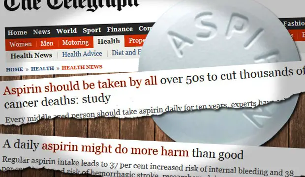
Take this recent example. A couple weeks ago, The Telegraph reported that, based on a new scientific study, all adults over the age of 50 should take aspirin every single day as a preventative measure to stave off cancer and heart disease. Everyone. Regardless of their previous warnings.
Are you over 50? Is your mom? Your grandma? Well according to The Telegraph’s reporting on this new study, you better be taking a daily aspirin every day for a decade. As The Telegraph reported:
“Middle-aged adults should take aspirin every day for ten years, according to scientists who found it could save more than 6,000 lives a year by preventing cancer and heart disease. Daily aspirin can prevent up to one third of cancers of the bowel, throat and stomach and can halve the risk of dying in some cases, according to the the largest, most comprehensive analysis of the drugs use.”
The article goes on to downplay the risks associated with taking a daily aspirin, stating: “It comes after previous research raised concerns about the side effects of aspirin, which include bleeding and ulcers. The new study found that while there was a small increased risk of a stroke, stomach bleeding and ulcers, the benefits of taking aspirin made it a ‘good bet’.”
A “small” increased risk of a stroke? Hm. That must be why the exact same media outlet reported back in December that “a daily aspirin may do more harm than good”, and noted that researchers at that time were claiming that regularly taking aspirin led to a 37 percent increased risk of internal bleeding and 38 percent increased risk of hemorrhagic stroke.
So which is it? Aspirin is good to stave off heart disease, but only if you want a nearly 40 percent increased risk of suffering a potentially deadly hemorrhagic stroke? How is that even remotely a “good bet”? Anyone??
Corporate Junk Science
So, listen to The Telegraph and go ahead and take aspirin every day for 10 years, you know… just in case. Never mind that it is already on record that drugs approved by the U.S. Food and Drug Administration kill 100,000 people every yearwho took the drugs as prescribed – this latest study claims it will save 6,000 lives. Notably, The Telegraph reporter doesn’t declare this little factoid about prescription death rates on the aspirin study until the very last line of her article: “Several of the authors of the analysis declared that they have worked for the pharmaceutical industry connected to aspirin but that the findings and conclusions in the study do not represent their respective organisations.
This is the other side of “science” — the bought-and-paid-for “science” conducted to paint the proper picture for whatever industry is funding it. Sadly though, most people do not read articles all the way to the end. Most casual mainstream media customers will read a headline and if it grabs them, they might skim the first few paragraphs, meaning they will seldom read the part where the industry itself is essentially revealed to be paying for the so-called “science” that propagates the false belief that these profitable products are good for them.
And this is just one example. This happens all the time, to the point that most people don’t know what’s healthy or not anymore, based on mainstream media standards. This isn’t to say that science gets everything wrong, obviously, but it illustrate that science can be paid for by corporate interests to say what those interests want it to say. And even a broken clock is right twice a day.
Writing on aspirin as a blood thinner in his book Health and Nutrition Secrets, Dr. Blaylock says that strokes are a huge concern with taking a daily aspirin, noting, “One of the largest stroke-prevention studies using aspirin, the Physician’s Health Study, involved 22,000 physicians and found that patients taking aspirin had twice as many brain hemorrhages as those who did not take aspirin.”
Dr. Blaylock went on to point out that that the problem with just taking a daily aspirin to prevent strokes and heart attacks is that it doesn’t do anything to strengthen the actual blood vessels themselves, something people who are concerned with heart issues should definitely take into consideration. Basically a weak blood vessel carrying thinned out blood is the perfect set up for a stroke. Makes sense.
Blaylock goes on to say that the herb Gingko Biloba not only thins the blood, but helps strengthen the collagen and elastin in blood vessel walls. In addition, flavonoids contained in Ginkgo biloba protect from free radical damage. These benefits are in addition to other numerous health effects of Ginkgo Biloba as reported by numerous other natural health outlets, including:
Enhances memory
Uplifts the spirit
Strengthens the eyes
Relaxes constricted blood vessels
Improves circulation
Acts as an energy restorative
Provides antioxidant boost
Relieves tension and anxiety
But Ginkgo biloba isn’t a manufactured chemical produced for a Big Pharma company in the past 100 years; it’s a plant that has been around for millions of years and has been used medicinally since long before aspirin was ever synthesized. But what does nature know? But by now, if we believe mainstream media’s medical advice, we should all be asking ourselves how anyone anywhere in the history of humanity managed to live to be over 60 before we could take aspirin every single day. Insanity.
This study examined dietary potassium effects on different stroke subtypes over 11 years in 90,137 postmenopausal women aged 50 to 79 years who had no stroke history at enrollment. Mean dietary potassium intake was 2,611 mg per day. The highest quartile of potassium intake was associated with lower incidence of ischemic and hemorrhagic stroke and total mortality. Comparing highest to lowest quartile of potassium intake with multivariate analyses yielded a hazard ratio of 0.90 (95% confidence interval [CI], 0.85-0.95) for all-cause mortality, 0.88 (95% CI, 0.79-0.98) for all stroke, and 0.84 (95% CI, 0.74-0.96) for ischemic stroke.
Stroke has devastating consequences for menopausal women, and treating modifiable risk factors should have a large payoff in reduction of morbidity and mortality. Dietary potassium intake has been associated with stroke risk in most but not all previous studies. In the largest study of menopausal women to date, using Women’s Health Initiative observational study data, the association between stroke and dietary potassium intake is confirmed. Interestingly, the association is strongest in nonhypertensive women. Hypertensive women with increasing potassium intake had lowered mortality but no lower incidence of stroke itself. The authors speculate that this may be because of a greater effect on arterial stiffness in the prehypertensive endothelial wall. Higher potassium intake was also associated with lowered all-cause mortality in all women, suggesting effects above and beyond the vascular system. The heterogeneity of “stroke” as a diagnosis was illustrated by the association with reduction in ischemic but not hemorrhagic stroke.
Of course, two major weaknesses of all such studies are our inability to infer causation from association in an observational study and reliance on the human brain to fill out food frequency forms. If potassium’s ability to reduce stoke in menopausal women was confirmed by a randomized clinical trial, public health efforts could be undertaken to increase the US population’s already lower-than-recommended potassium intake (only about 1/6 of these women met US Department of Agriculture potassium intake recommendations.) Such efforts would best take a simple macronutrient/whole foods approach, because counting dietary potassium intake itself would be difficult for patients. However, before considering increasing dietary potassium intake, special attention would have to be given to the potential for hyperkalemia in a population full of risk factors, such as chronic renal failure, and angiotensin-converting enzyme inhibitor use. Forget “an apple a day”—maybe “half a cantaloupe a day (with twice the potassium of a banana) makes the stroke go away” will be the new mantra for menopausal women.
A systematic review conducted by investigators at the National Center of Epidemiology, Carlos III Health Institute, in Madrid, Spain, showed that overall, after adjusting for potential confounders, 5 adjusted odds ratios (aORs) among selected studies showed an increased risk ranging from 2.0 to 19.7 for stroke or atherosclerosis among cocaine users, the authors, led by Luis Sordo, MD, report.
“This is the first systematic review to evaluate and synthesize the scientific evidence from epidemiological studies on the association between cocaine use and risk of stroke,” they write.
The study was published in the September issue of Drug and Alcohol Dependence.
Biologically Plausible
The authors note that the biological plausibility of cocaine as a cause of stroke is supported by preclinical evidence, and they point out that cocaine increases blood pressure, heart rate, and vasoconstriction, which, in turn reduce cerebral blood and oxygen and increase vascular resistance in the central nervous system.
“These effects may persist for hours due to the activity of different cocaine metabolites and, separately or in combination, might lead to ischemic or hemorrhagic stroke,” the investigators note.
“In addition, cocaine promotes thrombotic strokes causing hypercoagulable states and can produce arrhythmias that could lead to cardioembolic strokes,” they state.
The authors also note that although cocaine is generally considered to be a cause of stroke, no systematic review of the scientific evidence has ever been published.
The authors conducted a systematic review of all published epidemiologic evidence on the link between cocaine use and stroke. Of 996 articles that were reviewed, 9 were selected. These 9 consisted of 7 case-control studies and 2 cross-sectional studies.
Eight of the studies were from the United States, and 1 was from Australia. All of the studies were conducted in young adults, with ages ranging from 15 to 49 years.
The sample size of the case-control studies ranged from 291 to 1368 patients, and the 2 cross-sectional studies encompassed 822,332 patients.
Overall, after adjusting for potential confounders, 5 aORs among the 9 selected studies showed an increased risk for strokes or atherosclerosis among cocaine users. The odds ratios ranged from 2.0 to 19.7.
More specifically, an association between cocaine use and hemorrhagic stroke was found in 1 case-control study, with an aOR of 6.1 (95% confidence interval [CI], 3.3 – 11.8) and in 1 cross- sectional study (aOR = 2.33; 95% CI, 1.74 – 3.11).
The same cross-sectional study also found a positive link between cocaine use and ischemic stroke (aOR = 2.03; 95% CI, 1.48 – 2.79).
Another case-control study found cocaine use to be associated with stroke, but it did not distinguish between ischemic and hemorrhagic stroke. In that study, the aOR was 13.9 (95% CI, 2.8 – 69.4).
In the Australian case-control study, which was a forensic study comparing the presence of cerebrovascular atherosclerosis between deaths due to cocaine toxicity, opioid toxicity, and hanging, deaths associated with cocaine-positive toxicology showed a 14.3-fold increased risk (95% CI, 5.6 – 37) for atherosclerosis compared with opioid-related deaths, and a 4.6-fold increased risk (95% CI, 2.5 – 8.5) for atherosclerosis compared with deaths from hanging.
One case-control study found a statistically significant association between cocaine use and hemorrhagic stroke, but it did not provide an aOR.
Three case-control studies and 1 cross-sectional study failed to find any relationship between cocaine use and strokes.
Inadequate control for confounding was “not uncommon,” the authors state.
Need for More Research
“Controlling possible confounders is a methodological challenge in studying the association between cocaine use and strokes, given their potentially large number and difficulty of measurement,” the authors note.
They add that the review might be limited by publication bias, insofar as studies with negative results are less likely to be published.
The authors conclude that more research into the causal relationship between cocaine and stroke is warranted.
They also note that “confirmation and identification of the specific characteristics of this stroke risk factor will require a large, well-designed cohort study in the young-to-middle age population.”