Researchers Use fMRI to See Brain Activity Changes in Children With Anxiety After Treatment


In unmedicated children with anxiety disorders, researchers at the National Institutes of Health have found overactivation in many brain regions, including the frontal and parietal lobes and the amygdala. They also showed that treatment with cognitive behavioral therapy (CBT) led to improvements in clinical symptoms and brain functioning. The findings illuminate the brain mechanisms underlying the acute effects of CBT to treat one of the most common mental disorders. The study, published in the American Journal of Psychiatry, was led by researchers at NIH’s National Institute of Mental Health (NIMH). 

“The findings can help our understanding of how and for which children CBT works, a critical first step in personalizing anxiety care and improving clinical outcomes,” said senior author Melissa Brotman, PhD, Chief of the Neuroscience and Novel Therapeutics Unit in the NIMH Intramural Research Program.

Sixty-nine unmedicated children diagnosed with an anxiety disorder underwent 12 weeks of CBT following an established protocol. CBT, which involves changing dysfunctional thoughts and behaviors through gradual exposure to anxiety-provoking stimuli, is the current gold standard for treating anxiety disorders in children.

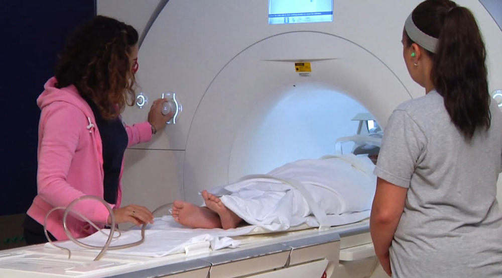
The researchers used clinician-rated measures to examine the change in children’s anxiety symptoms and clinical functioning from pre- to post-treatment. They also used task-based fMRI to look at whole-brain changes before and after treatment and compare those to brain activity in 62 similarly aged children without anxiety.

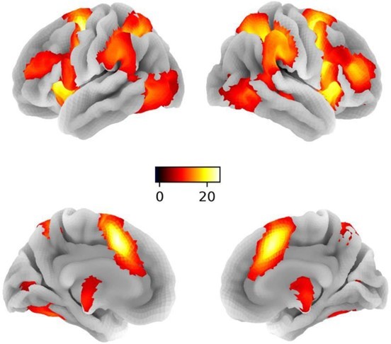
Children with anxiety showed greater activity in many brain regions, including cortical areas in the frontal and parietal lobes, which are important for cognitive and regulatory functions, such as attention and emotion regulation. The researchers also observed elevated activity in deeper limbic areas like the amygdala, which are essential for generating strong emotions, such as anxiety and fear.

Following three months of CBT treatment, children with anxiety showed a clinically significant decrease in anxiety symptoms and improved functioning. Increased activation seen before treatment in many frontal and parietal brain regions also improved after CBT, declining to levels equal to or lower than those of non-anxious children. According to the researchers, the reduced activation in these brain areas may reflect more efficient engagement of cognitive control networks following CBT.

However, eight brain regions, including the right amygdala, continued to show higher activity in anxious compared to non-anxious children after treatment. This persistent pattern of enhanced activation suggests some brain regions, particularly limbic areas that modulate responses to anxiety-provoking stimuli, may be less responsive to the acute effects of CBT. Changing activity in these regions may require a longer duration of CBT, additional forms of treatment, or directly targeting subcortical brain areas.

“Understanding the brain circuitry underpinning feelings of severe anxiety and determining which circuits normalize and which do not as anxiety symptoms improve with CBT is critical for advancing treatment and making it more effective for all children,” said first author Simone Haller, PhD, Director of Research and Analytics in the NIMH Neuroscience and Novel Therapeutics Unit.

In this study, all children with anxiety received CBT. For comparison purposes, the researchers also measured brain activity in a separate sample of 87 youth who were at high risk for anxiety based on their infant temperament (for example, showing a high sensitivity to new situations). Because these children were not diagnosed with an anxiety disorder, they had not received CBT treatment. Their brain scans were taken at 10 and 13 years.

In adolescents at temperamental risk for anxiety, higher brain activity was related to increased anxiety symptoms over time and matched the brain activity seen in children diagnosed with an anxiety disorder before treatment. This provides preliminary evidence that the brain changes in children with anxiety were driven by CBT and that they may offer a reliable neural marker of anxiety treatment.

Anxiety disorders are common in children and can cause them significant distress in social and academic situations. They are also chronic, with a strong link into adulthood when they become harder to treat. Despite the effectiveness of CBT, many children continue to show anxiety symptoms after treatment. Enhancing the therapy to treat anxiety more effectively during childhood can have short- and long-term benefits and prevent more serious problems later in life.

This study provide evidence—in a large group of unmedicated youth with anxiety disorders—of altered brain circuitry underlying treatment effects of CBT. The findings could, in time, be used to enhance treatment outcomes by targeting brain circuits linked to clinical improvement. This is particularly important for the subset of children who did not significantly improve after short-term CBT.

“The next step for this research is to understand which children are most likely to respond. Are there factors we can assess before treatment begins to make the most informed decisions about who should get which treatment and when? Answering these questions would further translate our research findings into clinical practice,” said Brotman.

fMRI-Based Modelling Gives Insight Into How Our Minds Control Impulses


A team of researchers from The University of Hong Kong and The University of Electronic Science and Technology of China has conclusively identified the right inferior frontal gyrus (rIFG) as a key input and causal regulator within the subcortical response inhibition nodes. This right-lateralized inhibitory control circuit, characterized by its significant intrinsic connectivity, highlights the crucial role of the rIFG in orchestrating top-down cortical-subcortical control, underscoring the intricate dynamics of brain function in response inhibition.

In this study, researchers employed dynamic causal modeling (DCM-PEB) and functional magnetic resonance imaging (fMRI) with a substantial sample size (n = 250) to explore inhibitory circuits in the brain, particularly focusing on the right inferior frontal gyrus (rIFG), caudate nucleus (rCau), globus pallidum (rGP), and thalamus (rThal). This approach treated the brain as a nonlinear dynamical system, enabling the estimation of directed causal influences among these nodes, influenced by task demands and biological variables. Findings revealed a high intrinsic connectivity within this neural circuit, with response inhibition notably enhancing causal projections from the rIFG to both rCau and rThal, particularly amplifying the regulatory role of the rIFG during such tasks. The study also uncovered that sex and performance metrics significantly affect the circuit’s functional architecture; for instance, women exhibited increased self-inhibition in the rThal and reduced modulation to the GP, while better inhibitory performance was linked to more robust communication from the rThal to the rIFG. Interestingly, these communication patterns were not mirrored in a left-lateralized model, highlighting a hemispheric asymmetry. The research indicates that different brain processes might mediate similar behavioral performances in response inhibition across genders, particularly in thalamic loops, with higher response inhibition accuracy associated with stronger information flow from the rThal to the rIFG.

These insights into the brain’s inhibitory control mechanisms have significant implications for understanding a range of mental and neurological disorders characterized by response inhibition deficits. The study’s findings could guide the development of targeted neuromodulation strategies and personalized interventions to address these deficits, enhancing the treatment and management of such conditions.

fMRI finds changes in brain connectivity can help diagnose and predict outcomes of mild TBI


A new study shows that patients with mild traumatic brain injury (mTBI), even without evidence of brain lesions, may exhibit changes in brain connectivity detectable at the time of the injury that can aid in diagnosis and predicting the effects on cognitive and behavioral performance at 6 months. Brain connectivity maps showed differences between patients with mTBI and healthy controls, including different patterns depending on the presence of brain lesions, as reported in an article in Journal of Neurotrauma, a peer-reviewed journal from Mary Ann Liebert, Inc., publishers. The article is available free on the Journal of Neurotrauma website until March 23, 2017.

The article entitled “Resting-State Functional Connectivity Alterations Associated with Six-Month Outcomes in Mild Traumatic Brain Injury” describes the prospective multicenter TRACK-TBI pilot study. Eva Palacios and coauthors from University of California, San Francisco, San Francisco General Hospital and Trauma Center, University of Texas, Austin, University of Pittsburgh Medical Center (PA), Virginia Commonwealth University (Richmond), Icahn School of Medicine at Mount Sinai (New York, NY), and Antwerp University Hospital (Edegem, Belgium) concluded that resting state functional magnetic resonance imaging (MRI) to assess brain connectivity and compare spatial maps of resting state brain networks can serve as a sensitive biomarker for early diagnosis of mTBI and later patient performance.

“While, as the authors acknowledge, they are not the first group to explore the utility of resting state functional MRI in probing the morbidity associated with mild traumatic brain injury, they do elegantly capitalize on the TRACK-TBI study population to critically evaluate functional connectivity in a patient population that is well characterized and followed by traditional imaging approaches,” says John T. Povlishock, PhD, Editor-in-Chief of Journal of Neurotrauma and Professor, Medical College of Virginia Campus of Virginia Commonwealth University, Richmond. “Their finding of altered patterns of functional connectivity even in that mild TBI patient population, revealing no CT/MRI abnormalities, is an extremely important observation, as is the fact that these same changes in functional connectivity portend the development of a persistent post-concussive syndrome.”

” INCEPTION STYLE ” Memory Implants Are Coming our Way


It may sound like something out of the latest Science-Fiction movie ” Inception “, but scientists have now discovered a way to implant subliminal messages into people’s brains without them even knowing it.  Although the thought of having an artificial memory pumped into our brain may sound a little daunting, the benefits that it could have for some people, including those with autism could be phenomenal.

During the research, subjects were asked to complete a series of tasks while lying underneath an fMRI machine and the instructions they received were to try and regulate their brain activity.  Afterward, the subjects were given scores on how well they apparently performed during the tests.  The results of the tests showed that when subjects unconsciously thought of the color red they received higher scores and when they were shown pictures of black and white stripes they somehow saw the color red!  Why is this?This is the result of subliminal message planting.  During the tests, while the subjects thought they were completing a series of tests, they were subconsciously trained to see the color red whenever they were shown pictures of black and white stripes.  The scientists found that this type of neurofeedback training is a great way to strengthen associative memories and to learn anywhere in the brain.

But, as well as enhancing memories, this type of technological process could also be used to delete bad experiences from the brain, which could be very useful in treating trauma victims. Or, it could also work by changing the way in which brain currently makes connections to help those with high-functioning autism or even depression. Although much more research is needed into this area, as well as some well-written rules, before we will see it commercially, this type of implant really could help millions of people one day.

Training the Brain’s Motivation Center


What biofeedback was to the 1970’s, neurofeedback could be to the 2020’s

fMRI images could someday allow us to control specific regions of our own brains 

My heart pounds as I sprint to the finish line. Thousands of spectators cheer as a sense of elation washes over me. I savor the feeling. But then, the image slowly fades away and my true surroundings come into focus. I am lying in a dark room with my head held firmly in place, inside an MRI scanner. While this might typically be unpleasant, I am a willing research study participant and am eagerly anticipating what comes next. I hold my breath as I stare at the bar on the computer screen representing my brain activity. Then the bar jumps. My fantasy of winning a race had caused the “motivation center” of my brain to surge with activity.

I am participating in a study about neurofeedback, a diverse and fascinating area of research that combines neuroscience and technology to monitor and modulate brain activity in real time. My colleagues, Katie Dickerson and Jeff MacInnes, in the Adcock Lab at Duke University, are studying whether people can train themselves to increase brain activity in a tiny region of the brain called the VTA. Notably, the VTA is thought to be involved in motivation—the desire to get something that you want. For example, if I told you that by buying a lottery ticket you would be guaranteed to win $1,000,000, you would probably be very motivated to buy the ticket and would have a spike in brain activity in this region of your brain. But while studies have shown that motivation for external rewards (like money) activate the VTA, until now, we didn’t know whether people could internally generate a motivational state that would activate this brain region.

To see if people can self-activate the VTA, my colleagues are using neurofeedback, which falls under the broader umbrella of biofeedback. Biofeedback uses technology to give people information about the functioning of their bodies so that they can try to change their physiology. Studies on using the technique to control heart rate, breathing rate, muscle tension, and skin temperature have been around since the 1960s, and these types of biofeedback are often used to help people reduce anxiety, chronic pain, or psychological disorders, with varying degrees of efficacy.

The latest iteration of biofeedback is neurofeedback, which provides a person with information on their brain activity milliseconds to seconds after it happens. Neurofeedback can use either an EEG machine, which records the electrical activity of the brain and is very fast, but not very specific, or it can use functional magnetic resonance imaging (fMRI), which records changes in blood flow in the brain and can better target specific brain regions, but is slower. The fMRI version of the technology has been around since as early as 1995 and its potential is slowly being realized. Studies on rtfMRI (the rt stands for “real-time”), have found that giving people feedback on their brain activity might be able to help them to control that activity. These studies both help us to better understand the functioning of the brain and can be used for clinical purposes. Perhaps the most common use to date has been for chronic pain, where people learn to decrease activation in regions of the brain that process pain perception.  [For more information about neurofeedback see “How Real-Time Brain Scanning Could Alleviate Pain,” by Heather Chapin and Sean Mackey; Scientific American Mind, March 1, 2013.]

The study I’m participating in is about using the technology to better understand the functioning of the VTA and its relationship with internally generated motivation, with potential for clinical applications down the road. In animal models, artificially affecting functioning of the VTA can affect how much an animal eats or drinks, and can even affect its emotional responses. If people could learn to activate their VTAs deliberately, it could have important applications to anything from helping someone stick to a diet to helping with psychological disorders.

Back in the fMRI, I lie on the uncomfortable scanner bed, looking up at the giant machine that reminds me of something I might encounter on a spaceship. Katie Dickerson’s voice comes over the intercom and gives me suggestions of ways to try and activate my VTA. She says I could try to think of phrases, like “you can do it!” or “increase that signal!” and says that it might help to think of the task as a fun game. So I think “I can do this!” but to my dismay the bar remains pretty flat. I think about winning $1,000,000, but don’t get much of a jump for that either. Then I picture myself running with a cheering crowd and music playing, and the bar goes through the roof.

For the study, participants were placed in one of four groups. All four groups started out by getting in the scanner and trying to activate their brains using motivation strategies, but first without receiving any feedback. After doing this for several minutes, people in the first group (like me) would try again, but this time would see a thermometer on the computer screen in front of them. When activity in the VTA went up, the bar on the thermometer would rise. When activity in the VTA went down, the thermometer would drop. The other groups either got feedback from a different brain region, got fake feedback, or were shown a visual distraction. These groups were used as comparisons to ensure that it really was the signal from the VTA that was being registered in the neurofeedback group. Afterward, all four groups tried one more time without the feedback (real or fake). At the end of the study, all participants were debriefed about their group assignment and the purpose of the study.

The results were published recently in the journal Neuron. It turns out that the strategies people tried initially did not activate their VTAs very much—the same experience I had. In other words, what people thought of as motivating did not match up with activity in what we consider to be the “motivation center” of the brain. How could that be? One possible explanation is that it can be difficult to get a sense of just how motivated we are to do something. Consider times when you might have thought you were highly motivated (“I know I am going to stick to my diet/exercise regimen this year”), and didn’t follow through. Another interpretation is that while we might have some sense of how motivated we are in a given moment, our subjective perceptions might not translate to VTA activation. There might not even be a clear feeling associated with the activation at all, explains MacInnes. That’s where the feedback came in.

The study found that, like me, people were better able to activate their VTAs, on average, once they got neurofeedback compared to people who got false feedback or no feedback. And the learning stuck—once people knew the strategies that worked for them, they were effective even once the feedback was taken away. Overall, different strategies worked for different people and some people in the control groups were still able to activate their VTAs even without the neurofeedback. The take-home message is that there is still a lot to learn.

Perhaps the biggest unanswered question is what could result from an ability to better activate one’s VTA. One possibility is that internally generated VTA activation could allow people to have the extra oomph to better meet their goals. So maybe when I need to do errands, but am really not in the mood, I can think about winning a race and it will give me the drive to go to the grocery store. For others, enhanced VTA activation might be able to help with studying. After all, studies have found VTA activation associated with better memory performance. And given other studies showing VTA signaling being related to eating and mood, it’s possible that it could help people with eating disorders or depression. Time will tell whether this method will be useful clinically, but for now I can say for certain that it can be a lot of fun getting to know your brain—and for me at least, it seems that having fun is one of the keys to activating my VTA.

Witnessing hateful people in pain modulates brain activity in regions associated with physical pain and reward.


How does witnessing a hateful person in pain compare to witnessing a likable person in pain? The current study compared the brain bases for how we perceive likable people in pain with those of viewing hateful people in pain. While social bonds are built through sharing the plight and pain of others in the name of empathy, viewing a hateful person in pain also has many potential ramifications. In this functional Magnetic Resonance Imaging (fMRI) study, Caucasian Jewish male participants viewed videos of (1) disliked, hateful, anti-Semitic individuals, and (2) liked, non-hateful, tolerant individuals in pain. The results showed that, compared with viewing liked people, viewing hateful people in pain elicited increased responses in regions associated with observation of physical pain (the insular cortex, the anterior cingulate cortex, and the somatosensory cortex), reward processing (the striatum), and frontal regions associated with emotion regulation. Functional connectivity analyses revealed connections between seed regions in the left anterior cingulate cortex and right insular cortex with reward regions, the amygdala, and frontal regions associated with emotion regulation. These data indicate that regions of the brain active while viewing someone in pain may be more active in response to the danger or threat posed by witnessing the pain of a hateful individual more so than the desire to empathize with a likable person’s pain.