An elderly epilepsy patient unexpectedly died during a brain scan, revealing bursts of activity associated with memory recall, meditation, and dreamin
:focal(2658x1810:2659x1811)/https://tf-cmsv2-smithsonianmag-media.s3.amazonaws.com/filer_public/64/0c/640c6ffb-8e94-49a1-b4bb-456811766639/image_from_ios.jpg)
New research is revealing what happens in the brain during our final moments of life. When scientists recorded the brainwaves of a dying man, he appeared to go through a sudden flash of memories seconds before and after his heart stopped beating. This first-of-its-kind study suggests we may experience a flood of memories when we die.
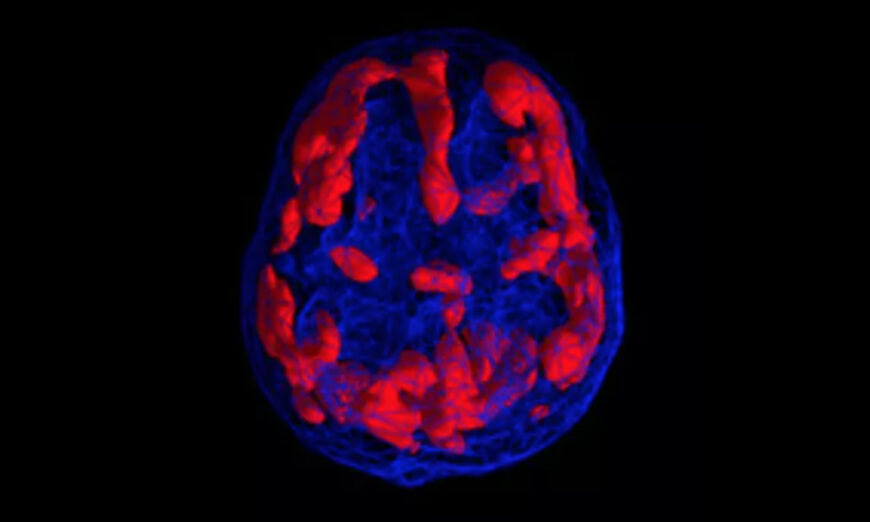
In the research published last week in Frontiers in Aging Neuroscience, doctors took brain scans of an 87-year-old Canadian patient with epilepsy. The team was performing a test that detects electrical activity in the brain, called an electroencephalogram (EEG), to learn more about what was happening during his seizures.
The elderly man had an unexpected heart attack and died during the procedure and, in accordance with the patient’s Do-Not-Resuscitate status, the doctors did not attempt any further treatment and the man soon passed away, reports Ed Cara for Gizmodo.
Because the EEG machine kept running, doctors got a glimpse into the man’s brain activity at the end of his life. Such scans had never before been captured on a dying individual.
“This is why it’s so rare, because you can’t plan this. No healthy human is going to go and have an EEG before they die, and in no sick patient are we going to know when they’re going to die to record these signals,” study author Ajmal Zemmar, a neurosurgeon at the University of Louisville in Kentucky, says to Insider’s Anna Medaris Miller.
For roughly 30 seconds before and after the man’s heart stopped beating, the scans showed increased acitivy in parts of the brain associated with memory recall, meditation, and dreaming. Different types of neural oscillations, also called brain waves, are involved in different brain functions. Researchers recorded both high-frequency gamma oscillations as well as slower-frequency theta, delta, alpha, and beta oscillations. Scientists say they were particularly intrigued by the presence of gamma waves, which suggest the man’s brain may have been replaying memories from throughout his life.
“Through generating oscillations involved in memory retrieval, the brain may be playing a last recall of important life events just before we die, similar to the ones reported in near-death experiences,” Zemmar says in a news release. And the patient’s brain activity didn’t immediately stop when he was declared dead. “Surprisingly, after the heart stops pumping blood into the brain, these oscillations keep going,” he tells Insider. “So that was extremely surprising for us to see.”
Because this phenomenon has only been observed in a single case so far, Zemmar and his colleagues caution against assuming brain activity is the same for all people upon death. The patient who died had epilepsy, which can alter gamma wave activity, per Live Science’s Harry Baker.
Despite the limitations of studying a single case, the results built on a 2013 study in rats that reported similar brain activity patterns before and after death, leading some to speculate that memory recall could be a universal experience of dying mammals.
“As a neurosurgeon, I deal with loss at times. It is indescribably difficult to deliver the news of death to distraught family members,” says Zemmar in a press release. “Something we may learn from this research is: although our loved ones have their eyes closed and are ready to leave us to rest, their brains may be replaying some of the nicest moments they experienced in their lives.”
An elderly epilepsy patient unexpectedly died during a brain scan, revealing bursts of activity associated with memory recall, meditation, and dreamin
New research is revealing what happens in the brain during our final moments of life. When scientists recorded the brainwaves of a dying man, he appeared to go through a sudden flash of memories seconds before and after his heart stopped beating. This first-of-its-kind study suggests we may experience a flood of memories when we die.
In the research published last week in Frontiers in Aging Neuroscience, doctors took brain scans of an 87-year-old Canadian patient with epilepsy. The team was performing a test that detects electrical activity in the brain, called an electroencephalogram (EEG), to learn more about what was happening during his seizures.
The elderly man had an unexpected heart attack and died during the procedure and, in accordance with the patient’s Do-Not-Resuscitate status, the doctors did not attempt any further treatment and the man soon passed away, reports Ed Cara for Gizmodo.
Because the EEG machine kept running, doctors got a glimpse into the man’s brain activity at the end of his life. Such scans had never before been captured on a dying individual.
“This is why it’s so rare, because you can’t plan this. No healthy human is going to go and have an EEG before they die, and in no sick patient are we going to know when they’re going to die to record these signals,” study author Ajmal Zemmar, a neurosurgeon at the University of Louisville in Kentucky, says to Insider’s Anna Medaris Miller.
For roughly 30 seconds before and after the man’s heart stopped beating, the scans showed increased acitivy in parts of the brain associated with memory recall, meditation, and dreaming. Different types of neural oscillations, also called brain waves, are involved in different brain functions. Researchers recorded both high-frequency gamma oscillations as well as slower-frequency theta, delta, alpha, and beta oscillations. Scientists say they were particularly intrigued by the presence of gamma waves, which suggest the man’s brain may have been replaying memories from throughout his life.
“Through generating oscillations involved in memory retrieval, the brain may be playing a last recall of important life events just before we die, similar to the ones reported in near-death experiences,” Zemmar says in a news release. And the patient’s brain activity didn’t immediately stop when he was declared dead. “Surprisingly, after the heart stops pumping blood into the brain, these oscillations keep going,” he tells Insider. “So that was extremely surprising for us to see.”
Because this phenomenon has only been observed in a single case so far, Zemmar and his colleagues caution against assuming brain activity is the same for all people upon death. The patient who died had epilepsy, which can alter gamma wave activity, per Live Science’s Harry Baker.
Despite the limitations of studying a single case, the results built on a 2013 study in rats that reported similar brain activity patterns before and after death, leading some to speculate that memory recall could be a universal experience of dying mammals.
“As a neurosurgeon, I deal with loss at times. It is indescribably difficult to deliver the news of death to distraught family members,” says Zemmar in a press release. “Something we may learn from this research is: although our loved ones have their eyes closed and are ready to leave us to rest, their brains may be replaying some of the nicest moments they experienced in their lives.”